Pterygopalatine ganglion

Last updated
Pterygopalatine ganglion
Gray779.png
Alveolar branches of superior maxillary nerve and pterygopalatine ganglion
Gray780.png
The pterygopalatine ganglion and its branches (pterygopalatine ganglion visible but not labeled, as large yellow ganglion in upper-right center)
Details
From Maxillary nerve and nerve of pterygoid canal
To Greater palatine nerve, lesser palatine nerve, posterior lateral nasal branches and nasopalatine nerve
Identifiers
Latin ganglion pterygopalatinum
TA98 A14.3.02.006
TA2 6665
FMA 6965
Anatomical terms of neuroanatomy

The pterygopalatine ganglion (aka Meckel's ganglion, nasal ganglion, or sphenopalatine ganglion) is a parasympathetic ganglion in the pterygopalatine fossa. It is one of four parasympathetic ganglia of the head and neck, (the others being the submandibular, otic, and ciliary ganglion).

Contents

It is innervated by the Vidian nerve (formed by the greater superficial petrosal nerve branch of the facial nerve and deep petrosal nerve) and maxillary division of the trigeminal nerve. Its postsynaptic axons project to the lacrimal glands and nasal mucosa. [1]

The flow of blood to the nasal mucosa, in particular the venous plexus of the conchae, is regulated by the pterygopalatine ganglion and heats or cools the air in the nose.

Structure

The pterygopalatine ganglion (of Meckel), the largest of the parasympathetic ganglia associated with the branches of the maxillary nerve, is deeply placed in the pterygopalatine fossa, close to the sphenopalatine foramen. It is triangular or heart-shaped, of a reddish-gray color, and is situated just below the maxillary nerve as it crosses the fossa.

The pterygopalatine ganglion supplies the lacrimal gland, paranasal sinuses, glands of the mucosa of the nasal cavity and pharynx, the gingiva, and the mucous membrane and glands of the hard palate. It communicates anteriorly with the nasopalatine nerve.

Roots

It receives a sensory, a parasympathetic, and a sympathetic root.

Sensory root

Its sensory root is derived from two sphenopalatine branches of the maxillary nerve of the trigeminal nerve; their fibers, for the most part, pass directly into the palatine nerves; a few, however, enter the ganglion, constituting its sensory root.

Parasympathetic root

Its parasympathetic root is derived from the nervus intermedius (a part of the facial nerve) through the greater petrosal nerve.

In the pterygopalatine ganglion, the preganglionic parasympathetic fibers from the greater petrosal branch of the facial nerve synapse with neurons whose postganglionic axons, vasodilator, and secretory fibers are distributed with the deep branches of the trigeminal nerve to the mucous membrane of the nose, soft palate, tonsils, uvula, roof of the mouth, upper lip and gums, and upper part of the pharynx. It also sends postganglionic parasympathetic fibers to the lacrimal nerve (a branch of the ophthalmic nerve, also part of the trigeminal nerve) via the zygomatic nerve, a branch of the maxillary nerve (from the trigeminal nerve), which then arrives at the lacrimal gland.

The nasal glands are innervated with secretomotor fibers from the nasal branches. Likewise, the palatine glands are innervated by the nasopalatine, greater palatine nerve and lesser palatine nerves. The pharyngeal nerve innervates pharyngeal glands. These are all branches of maxillary nerve.

Sympathetic root

The ganglion also consists of sympathetic efferent (postganglionic) fibers from the superior cervical ganglion. These fibers, from the superior cervical ganglion, travel through the carotid plexus, and then through the deep petrosal nerve. The deep petrosal nerve (carrying postganglionic sympathetics) joins with the greater petrosal nerve (carrying preganglionic parasympathetics) to form the nerve of the pterygoid canal, which passes through the pterygoid canal before entering the ganglion. The stellate ganglion is at the bottom of the cervical sympathetic chain. Fibers from the stellate ganglion pass up the chain to the superior cervical sympathetic ganglion and into and through the sphenopalatine ganglion.

Branches

Blockade and neuromodulation of the pterygopalatine (sphenopalatine) ganglion

Blockade of the ganglion with local anesthetic, clinically referred to as a 'sphenopalatine ganglion block' may be performed transcutaneously with a small needle, or topically via the nose with local anesthetic soaked swabs. The topical sphenopalatine ganglion block is used for treatment of persistent migraines and cluster headaches, demonstrating relief within 10–20 minutes. Sphenopalatine ganglion block has been used to treat post-dural-puncture headache, [2] though a 2020 trial comparing local anaesthetic sphenopalatine ganglion block to sham injection with saline failed to show difference in pain scores for those receiving local anaesthetic vs placebo, suggesting any efficacy is unrelated to local anaesthetic blockade. [3]

Self-administration of sphenopalatine ganglion blocks with cotton-tipped catheters and continual capillary feed is the most cost-effective method of treatment and has the benefit of allowing patients to avoid visits to physicians and emergency rooms. After initial visit self-administered sphenopalatine ganglion blocks are less than $1.00 per application. Frequent repeated administration of sphenopalatine ganglion blocks seems to increase effectiveness initially, after which decreased frequency is required. Self-administered sphenopalatine ganglion blocks can be used to treat acute pain symptoms, and prophylactically to reduce the onset of painful conditions and anxiety.

Self-administration of sphenopalatine ganglion blocks

Self-administered sphenopalatine ganglion blocks are extremely helpful for treatment of migraines, chronic daily headaches, anxiety and temporomandibular joint disorders. Neuromuscular dentistry and the role of the autonomic nervous system: Sphenopalatine ganglion blocks and neuromodulation. An International College of Cranio Mandibular Orthopedics (ICCMO) position paper They are also effective in about 13 of cases of essential hypertension.Bilateral sphenopalatine ganglion block reduces blood pressure in never treated patients with essential hypertension. A randomized controlled single-blinded study

The sphenopalatine ganglion block has been called "The Miracle Block" after publication of Albert Bengamin Gerber's book Miracles on Park Avenue, the story of octogenerian otorhinolargyngologist Dr. Milton Reder, whose entire medical practice was based on that procedure.[ citation needed ]

There are multiple new devices utilized for neuromodulation of the sphenopalatine ganglion. There is one non-invasive device, a myomonitor, an ultra low frequency transcutaneous electrical nerve stimulation that has been utilized safely for more than 50 years by neuromuscular dentists in the diagnosis and treatment of temporomandibular joint disorders and orofacial pain conditions. Spenopalatine ganglion neuromodulation (possibly via the vagal nerve) is a secondary effect making it extremely effective for TMD.Neuromuscular dentistry and the role of the autonomic nervous system: Sphenopalatine ganglion blocks and neuromodulation. An International College of Cranio Mandibular Orthopedics (ICCMO) position paper

Additional images

Related Research Articles

<span class="mw-page-title-main">Parasympathetic nervous system</span> Division of the autonomic nervous system

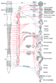
The parasympathetic nervous system (PSNS) is one of the three divisions of the autonomic nervous system, the others being the sympathetic nervous system and the enteric nervous system. The enteric nervous system is sometimes considered part of the autonomic nervous system, and sometimes considered an independent system.

<span class="mw-page-title-main">Facial nerve</span> Cranial nerve VII, for the face and tasting

The facial nerve, also known as the seventh cranial nerve, cranial nerve VII, or simply CN VII, is a cranial nerve that emerges from the pons of the brainstem, controls the muscles of facial expression, and functions in the conveyance of taste sensations from the anterior two-thirds of the tongue. The nerve typically travels from the pons through the facial canal in the temporal bone and exits the skull at the stylomastoid foramen. It arises from the brainstem from an area posterior to the cranial nerve VI and anterior to cranial nerve VIII.

Articles related to anatomy include:

<span class="mw-page-title-main">Glossopharyngeal nerve</span> Cranial nerve IX, for the tongue and pharynx

The glossopharyngeal nerve, also known as the ninth cranial nerve, cranial nerve IX, or simply CN IX, is a cranial nerve that exits the brainstem from the sides of the upper medulla, just anterior to the vagus nerve. Being a mixed nerve (sensorimotor), it carries afferent sensory and efferent motor information. The motor division of the glossopharyngeal nerve is derived from the basal plate of the embryonic medulla oblongata, whereas the sensory division originates from the cranial neural crest.

<span class="mw-page-title-main">Lacrimal gland</span> Exocrine gland, one for each eye, that secrete tears

The lacrimal glands are paired exocrine glands, one for each eye, found in most terrestrial vertebrates and some marine mammals, that secrete the aqueous layer of the tear film. In humans, they are situated in the upper lateral region of each orbit, in the lacrimal fossa of the orbit formed by the frontal bone. Inflammation of the lacrimal glands is called dacryoadenitis. The lacrimal gland produces tears which are secreted by the lacrimal ducts, and flow over the ocular surface, and then into canals that connect to the lacrimal sac. From that sac, the tears drain through the lacrimal duct into the nose.

<span class="mw-page-title-main">Otic ganglion</span> Parasympathetic ganglion of the head and neck

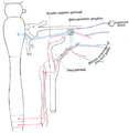
The otic ganglion is a small parasympathetic ganglion located immediately below the foramen ovale in the infratemporal fossa and on the medial surface of the mandibular nerve. It is functionally associated with the glossopharyngeal nerve and innervates the parotid gland for salivation.

<span class="mw-page-title-main">Greater petrosal nerve</span> Nerve in the skull

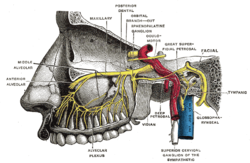
The greater petrosal nerve is a nerve of the head mainly containing pre-ganglionic parasympathetic fibres which ultimately synapse in the pterygopalatine ganglion. It branches from the facial nerve and is derived from the parasympathetic part of the nervus intermedius component of CN VII, with its cell bodies located in the superior salivary nucleus. In the connective tissue substance of the foramen lacerum, the greater petrosal nerve unites with the (sympathetic) deep petrosal nerve to form the nerve of the pterygoid canal which proceeds to the pterygopalatine ganglion.

<span class="mw-page-title-main">Maxillary nerve</span> Branch of the trigeminal nerve responsible for the mid-face

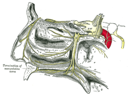
In neuroanatomy, the maxillary nerve (V2) is one of the three branches or divisions of the trigeminal nerve, the fifth (CN V) cranial nerve. It comprises the principal functions of sensation from the maxilla, nasal cavity, sinuses, the palate and subsequently that of the mid-face, and is intermediate, both in position and size, between the ophthalmic nerve and the mandibular nerve.

<span class="mw-page-title-main">Pterygopalatine fossa</span> Fossa in the skull

In human anatomy, the pterygopalatine fossa is a fossa in the skull. A human skull contains two pterygopalatine fossae—one on the left side, and another on the right side. Each fossa is a cone-shaped paired depression deep to the infratemporal fossa and posterior to the maxilla on each side of the skull, located between the pterygoid process and the maxillary tuberosity close to the apex of the orbit. It is the indented area medial to the pterygomaxillary fissure leading into the sphenopalatine foramen. It communicates with the nasal and oral cavities, infratemporal fossa, orbit, pharynx, and middle cranial fossa through eight foramina.

<span class="mw-page-title-main">Zygomatic nerve</span> Nerve of the face

The zygomatic nerve is a branch of the maxillary nerve. It arises in the pterygopalatine fossa and enters the orbit through the inferior orbital fissure before dividing into its two terminal branches: the zygomaticotemporal nerve and zygomaticofacial nerve.

<span class="mw-page-title-main">Submandibular ganglion</span>

The submandibular ganglion is part of the human autonomic nervous system. It is one of four parasympathetic ganglia of the head and neck..

<span class="mw-page-title-main">Parasympathetic ganglia</span> Autonomic ganglia of the parasympathetic nervous system

Parasympathetic ganglia are the autonomic ganglia of the parasympathetic nervous system. Most are small terminal ganglia or intramural ganglia, so named because they lie near or within (respectively) the organs they innervate. The exceptions are the four paired parasympathetic ganglia of the head and neck.

<span class="mw-page-title-main">Deep petrosal nerve</span>

The deep petrosal nerve is a post-ganglionic branch of the (sympathetic) internal carotid (nervous) plexus that enters the cranial cavity through the carotid canal, then passes perpendicular to the carotid canal in the cartilaginous substance which fills the foramen lacerum to unite with the (parasympathetic) greater petrosal nerve to form the nerve of pterygoid canal.

<span class="mw-page-title-main">Nerve of pterygoid canal</span>

The nerve of the pterygoid canal is formed by the union of the (parasympathetic) greater petrosal nerve and (sympathetic) deep petrosal nerve within the cartilaginous substance filling the foramen lacerum. From the foramen lacerum, the nerve of the pterygoid canal passes through the pterygoid canal to reach the pterygopalatine fossa, ending at the pterygopalatine ganglion.

<span class="mw-page-title-main">Salivatory nuclei</span>

The salivatory nuclei are two parasympathetic general visceral efferent cranial nerve nuclei - the superior salivatory nucleus and the inferior salivatory nucleus - that innervate the salivary glands. Both are located in the pontine tegmentum of the brainstem.

<span class="mw-page-title-main">Outline of human anatomy</span> Overview of and topical guide to human anatomy

The following outline is provided as an overview of and topical guide to human anatomy:

The autonomic nerve is a small nerve which carries postganglionic sympathetic and parasympathetic neurons from the zygomaticotemporal nerve; a branch of the maxillary nerve, to the lacrimal nerve; a branch of the ophthalmic nerve. These neurons derive from the superior cervical ganglion and the pterygopalatine ganglion respectively. They will travel to the lacrimal gland via the lacrimal nerve. Parasympathetic will induce lacrimation and vice versa.

<span class="mw-page-title-main">Outline of the human nervous system</span> Overview of and topical guide to the human nervous system

The following diagram is provided as an overview of and topical guide to the human nervous system:

<span class="mw-page-title-main">Roots of the ciliary ganglion</span>

The ciliary ganglion is a parasympathetic ganglion located just behind the eye in the posterior orbit. Three types of axons enter the ciliary ganglion but only the preganglionic parasympathetic axons synapse there. The entering axons are arranged into three roots of the ciliary ganglion, which join enter the posterior surface of the ganglion.

References

  1. Lundy, Jason A.; McNary, Thomas (2023), "Neuroanatomy, Pterygopalatine Ganglion", StatPearls, Treasure Island (FL): StatPearls Publishing, PMID   31424892 , retrieved 2023-12-07
  2. "Sphenopalatine Ganglion Block Postdural Puncture Headache « metajournal.com". www.metajournal.com. Retrieved 28 April 2023.
  3. Jespersen, Mads S.; Jaeger, Pia; Ægidius, Karen L.; Fabritius, Maria L.; Duch, Patricia; Rye, Ida; Afshari, Arash; Meyhoff, Christian S. (2020). "Sphenopalatine ganglion block for the treatment of postdural puncture headache: A randomised, blinded, clinical trial". British Journal of Anaesthesia. 124 (6): 739–747. doi: 10.1016/j.bja.2020.02.025 . PMID   32303377. S2CID   215809853.

PD-icon.svgThis article incorporates text in the public domain from page 891 of the 20th edition of Gray's Anatomy (1918)