Accessory nerve

Last updated

Accessory nerve
Brain human normal inferior view with labels en.svg
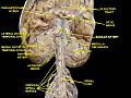
View of the human brain from below. The accessory nerve emerges from the medulla of the brainstem, and is visible at the bottom of the image in blue.
Details
Innervates Sternocleidomastoid muscle, trapezius muscle
Identifiers
Latin nervus accessorius
MeSH D000055
TA98 A14.2.01.184
A14.1.02.112
TA2 6352
FMA 6720
Anatomical terms of neuroanatomy

The accessory nerve, also known as the eleventh cranial nerve, cranial nerve XI, or simply CN XI, is a cranial nerve that supplies the sternocleidomastoid and trapezius muscles. It is classified as the eleventh of twelve pairs of cranial nerves because part of it was formerly believed to originate in the brain. The sternocleidomastoid muscle tilts and rotates the head, whereas the trapezius muscle, connecting to the scapula, acts to shrug the shoulder.

Contents

Traditional descriptions of the accessory nerve divide it into a spinal part and a cranial part. [1] The cranial component rapidly joins the vagus nerve, and there is ongoing debate about whether the cranial part should be considered part of the accessory nerve proper. [2] [1] Consequently, the term "accessory nerve" usually refers only to nerve supplying the sternocleidomastoid and trapezius muscles, also called the spinal accessory nerve. [3]

Strength testing of these muscles can be measured during a neurological examination to assess function of the spinal accessory nerve. Poor strength or limited movement are suggestive of damage, which can result from a variety of causes. Injury to the spinal accessory nerve is most commonly caused by medical procedures that involve the head and neck. [4] Injury can cause wasting of the shoulder muscles, winging of the scapula, and weakness of shoulder abduction and external rotation. [5]

The accessory nerve is derived from the basal plate of the embryonic spinal segments C1–C6.

Structure

The fibres of the spinal accessory nerve originate solely in neurons situated in the upper spinal cord, from where the spinal cord begins at the junction with the medulla oblongata, to the level of about C6. [1] [6] These fibres join together to form rootlets, roots, and finally the spinal accessory nerve itself. The formed nerve enters the skull through the foramen magnum, the large opening at the skull's base. [1] The nerve travels along the inner wall of the skull towards the jugular foramen. [1] Leaving the skull, the nerve travels through the jugular foramen with the glossopharyngeal and vagus nerves. [7] The spinal accessory nerve is notable for being the only cranial nerve to both enter and exit the skull. This is due to it being unique among the cranial nerves in having neurons in the spinal cord. [8]

After leaving the skull, the cranial component detaches from the spinal component. The spinal accessory nerve continues alone and heads backwards and downwards. In the neck, the accessory nerve crosses the internal jugular vein around the level of the posterior belly of digastric muscle. As it courses downwards, the nerve pierces through the sternocleidomastoid muscle (approximately 1cm above Erb's point) while sending it motor branches, then continues down until it reaches the trapezius muscle (entering at the junction of the middle and lower third of the anterior border of the trapezius) to provide motor innervation to its upper part. [9]

Nucleus

The fibres that form the spinal accessory nerve are formed by lower motor neurons located in the upper segments of the spinal cord. This cluster of neurons, called the spinal accessory nucleus, is located in the lateral aspect of the anterior horn of the spinal cord, and stretches from where the spinal cord begins (at the junction with the medulla) through to the level of about C6. [1] The lateral horn of high cervical segments appears to be continuous with the nucleus ambiguus of the medulla oblongata, from which the cranial component of the accessory nerve is derived. [8]

Variation

In the neck, the accessory nerve crosses the internal jugular vein around the level of the posterior belly of digastric muscle, in front of the vein in about 80% of people, and behind it in about 20%, [8] and in one reported case, piercing the vein. [10]

Traditionally, the accessory nerve is described as having a small cranial component that descends from the medulla and briefly connects with the spinal accessory component before branching off of the nerve to join the vagus nerve. [1] A study, published in 2007, of twelve subjects suggests that in the majority of individuals, this cranial component does not make any distinct connection to the spinal component; the roots of these distinct components were separated by a fibrous sheath in all but one subject. [6]

Development

The accessory nerve is derived from the basal plate of the embryonic spinal segments C1–C6. [11]

Function

The accessory nerve supplies the sternocleidomastoid and trapezius muscles 1610 Muscles Controlled by the Accessory Nerve-02.jpg
The accessory nerve supplies the sternocleidomastoid and trapezius muscles

The spinal component of the accessory nerve provides motor control of the sternocleidomastoid and trapezius muscles. [7] The trapezius muscle controls the action of shrugging the shoulders, and the sternocleidomastoid the action of turning the head. [7] Like most muscles, control of the trapezius muscle arises from the opposite side of the brain. [7] Contraction of the upper part of the trapezius muscle elevates the scapula. [12] The nerve fibres supplying sternocleidomastoid, however, are thought to change sides (Latin : decussate) twice. This means that the sternocleidomastoid is controlled by the brain on the same side of the body. Contraction of the stenocleidomastoid fibres turns the head to the opposite side, the net effect meaning that the head is turned to the side of the brain receiving visual information from that area. [7] The cranial component of the accessory nerve, on the other hand, provides motor control to the muscles of the soft palate, larynx and pharynx.

Classification

Among researchers there is disagreement regarding the terminology used to describe the type of information carried by the accessory nerve. As the trapezius and sternocleidomastoid muscles are derived from the pharyngeal arches, some researchers believe the spinal accessory nerve that innervates them must carry specific special visceral efferent (SVE) information. [13] This is in line with the observation that the spinal accessory nucleus appears to be continuous with the nucleus ambiguus of the medulla. Others consider the spinal accessory nerve to carry general somatic efferent (GSE) information. [14] Still others believe it is reasonable to conclude that the spinal accessory nerve contains both SVE and GSE components. [15]

Clinical significance

Examination

The accessory nerve is tested by evaluating the function of the trapezius and sternocleidomastoid muscles. [7] The trapezius muscle is tested by asking the patient to shrug their shoulders with and without resistance. The sternocleidomastoid muscle is tested by asking the patient to turn their head to the left or right against resistance. [7]

One-sided weakness of the trapezius may indicate injury to the nerve on the same side of an injury to the spinal accessory nerve on the same side (Latin: ipsilateral) of the body being assessed. [7] Weakness in head-turning suggests injury to the contralateral spinal accessory nerve: a weak leftward turn is indicative of a weak right sternocleidomastoid muscle (and thus right spinal accessory nerve injury), while a weak rightward turn is indicative of a weak left sternocleidomastoid muscle (and thus left spinal accessory nerve). [7]

Hence, weakness of shrug on one side and head-turning on the other side may indicate damage to the accessory nerve on the side of the shrug weakness, or damage along the nerve pathway at the other side of the brain. Causes of damage may include trauma, surgery, tumours, and compression at the jugular foramen. [7] Weakness in both muscles may point to a more general disease process such as amyotrophic lateral sclerosis, Guillain–Barré syndrome or poliomyelitis. [7]

Injury

Injury to the spinal accessory nerve commonly occurs during neck surgery, including neck dissection and lymph node excision. It can also occur as a result of blunt or penetrating trauma, and in some causes spontaneously. [16] [5] Damage at any point along the nerve's course will affect the function of the nerve. [9] The nerve is intentionally removed in "radical" neck dissections, which are attempts at exploring the neck surgically for the presence and extent of cancer. Attempts are made to spare it in other forms of less aggressive dissection. [5]

Injury to the accessory nerve can result in neck pain and weakness of the trapezius muscle. Symptoms will depend on at what point along its length the nerve was severed. [5] Injury to the nerve can result in shoulder girdle depression, atrophy, abnormal movement, a protruding scapula, and weakened abduction. [5] Weakness of the shoulder girdle can lead to traction injury of the brachial plexus. [9] Because diagnosis is difficult, electromyogram or nerve conduction studies may be needed to confirm a suspected injury. [5] Outcomes with surgical treatment appear to be better than conservative management, which entails physiotherapy and pain relief. [16] Surgical management includes neurolysis, nerve end-to-end suturing, and surgical replacement of affected trapezius muscle segments with other muscle groups, such as the Eden-Lange procedure. [16]

Damage to the nerve can cause torticollis. [17]

History

English anatomist Thomas Willis in 1664 first described the accessory nerve, choosing to use "accessory" (described in Latin as nervus accessorius) meaning in association with the vagus nerve. [18]

In 1848, Jones Quain described the nerve as the "spinal nerve accessory to the vagus", recognizing that while a minor component of the nerve joins with the larger vagus nerve, the majority of accessory nerve fibres originate in the spinal cord. [3] [19] In 1893 it was recognised that the heretofore named nerve fibres "accessory" to the vagus originated from the same nucleus in the medulla oblongata, and it came to pass that these fibres were increasingly viewed as part of the vagus nerve itself. [3] Consequently, the term "accessory nerve" was and is increasingly used to denote only fibres from the spinal cord; the fact that only the spinal portion could be tested clinically lent weight to this opinion. [3]

See also

Additional images

Related Research Articles

<span class="mw-page-title-main">Cranial nerves</span> Nerves that emerge directly from the brain and the brainstem

Cranial nerves are the nerves that emerge directly from the brain, of which there are conventionally considered twelve pairs. Cranial nerves relay information between the brain and parts of the body, primarily to and from regions of the head and neck, including the special senses of vision, taste, smell, and hearing.

<span class="mw-page-title-main">Vagus nerve</span> Main nerve of the parasympathetic nervous system

The vagus nerve, also known as the tenth cranial nerve, cranial nerve X, or simply CN X, is a cranial nerve that carries sensory fibers that create a pathway that interfaces with the parasympathetic control of the heart, lungs, and digestive tract. It comprises two nerves—the left and right vagus nerves—but they are typically referred to collectively as a single subsystem.

<span class="mw-page-title-main">Neck</span> Part of the body on many vertebrates that connects the head with the torso

The neck is the part of the body on many vertebrates that connects the head with the torso. The neck supports the weight of the head and protects the nerves that carry sensory and motor information from the brain down to the rest of the body. In addition, the neck is highly flexible and allows the head to turn and flex in all directions. The structures of the human neck are anatomically grouped into four compartments: vertebral, visceral and two vascular compartments. Within these compartments, the neck houses the cervical vertebrae and cervical part of the spinal cord, upper parts of the respiratory and digestive tracts, endocrine glands, nerves, arteries and veins. Muscles of the neck are described separately from the compartments. They bound the neck triangles.

<span class="mw-page-title-main">Trapezius</span> Muscle between the lower spine and the shoulder blade

The trapezius is a large paired trapezoid-shaped surface muscle that extends longitudinally from the occipital bone to the lower thoracic vertebrae of the spine and laterally to the spine of the scapula. It moves the scapula and supports the arm.

Articles related to anatomy include:

<span class="mw-page-title-main">Brachial plexus</span> Network of nerves

The brachial plexus is a network of nerves formed by the anterior rami of the lower four cervical nerves and first thoracic nerve. This plexus extends from the spinal cord, through the cervicoaxillary canal in the neck, over the first rib, and into the armpit, it supplies afferent and efferent nerve fibers to the chest, shoulder, arm, forearm, and hand.

<span class="mw-page-title-main">Glossopharyngeal nerve</span> Cranial nerve IX, for the tongue and pharynx

The glossopharyngeal nerve, also known as the ninth cranial nerve, cranial nerve IX, or simply CN IX, is a cranial nerve that exits the brainstem from the sides of the upper medulla, just anterior to the vagus nerve. Being a mixed nerve (sensorimotor), it carries afferent sensory and efferent motor information. The motor division of the glossopharyngeal nerve is derived from the basal plate of the embryonic medulla oblongata, whereas the sensory division originates from the cranial neural crest.

<span class="mw-page-title-main">Hypoglossal nerve</span> Cranial nerve XII, for the tongue

The hypoglossal nerve, also known as the twelfth cranial nerve, cranial nerve XII, or simply CN XII, is a cranial nerve that innervates all the extrinsic and intrinsic muscles of the tongue except for the palatoglossus, which is innervated by the vagus nerve. CN XII is a nerve with a sole motor function. The nerve arises from the hypoglossal nucleus in the medulla as a number of small rootlets, pass through the hypoglossal canal and down through the neck, and eventually passes up again over the tongue muscles it supplies into the tongue.

<span class="mw-page-title-main">Sternocleidomastoid muscle</span> Cervical muscle

The sternocleidomastoid muscle is one of the largest and most superficial cervical muscles. The primary actions of the muscle are rotation of the head to the opposite side and flexion of the neck. The sternocleidomastoid is innervated by the accessory nerve.

<span class="mw-page-title-main">Pyramidal tracts</span> The corticobulbar tract and the corticospinal tract

The pyramidal tracts include both the corticobulbar tract and the corticospinal tract. These are aggregations of efferent nerve fibers from the upper motor neurons that travel from the cerebral cortex and terminate either in the brainstem (corticobulbar) or spinal cord (corticospinal) and are involved in the control of motor functions of the body.

<span class="mw-page-title-main">Dura mater</span> Outermost layer of the protective tissues around the central nervous system (meninges)

In neuroanatomy, dura mater is a thick membrane made of dense irregular connective tissue that surrounds the brain and spinal cord. It is the outermost of the three layers of membrane called the meninges that protect the central nervous system. The other two meningeal layers are the arachnoid mater and the pia mater. It envelops the arachnoid mater, which is responsible for keeping in the cerebrospinal fluid. It is derived primarily from the neural crest cell population, with postnatal contributions of the paraxial mesoderm.

<span class="mw-page-title-main">Levator scapulae muscle</span> Slender skeletal muscle at the back and side of the neck

The levator scapulae is a slender skeletal muscle situated at the back and side of the neck. It originates from the transverse processes of the four uppermost cervical vertebrae; it inserts onto the upper portion of the medial border of the scapula. It is innervated by the cervical nerves C3-C4, and frequently also by the dorsal scapular nerve. As the Latin name suggests, its main function is to lift the scapula.

<span class="mw-page-title-main">Carotid sheath</span>

The carotid sheath is a condensation of the deep cervical fascia enveloping multiple vital neurovascular structures of the neck, including the common and internal carotid arteries, the internal jugular vein, the vagus nerve, and ansa cervicalis. The carotid sheath helps protects the structures contained therein.

<span class="mw-page-title-main">Occipital artery</span>

The occipital artery is a branch of the external carotid artery that provides arterial supply to the back of the scalp, sternocleidomastoid muscles, and deep muscles of the back and neck.

<span class="mw-page-title-main">Posterior triangle of the neck</span> Region of the neck

The posterior triangle is a region of the neck.

<span class="mw-page-title-main">Supraclavicular nerves</span>

The supraclavicular nerve is a cutaneous (sensory) nerve of the cervical plexus that arises from the third and fourth cervical (spinal) nerves. It emerges from beneath the posterior border of the sternocleidomastoid muscle, then split into multiple branches. Together, these innervate the skin over the shoulder.

<span class="mw-page-title-main">Accessory nerve disorder</span> Disorder caused due to injury to the spinal accessory nerve (11th cranial nerve or Cranial Nerve XI)

Accessory nerve disorder is an injury to the spinal accessory nerve which results in diminished or absent function of the sternocleidomastoid muscle and upper portion of the trapezius muscle.

<span class="mw-page-title-main">Outline of human anatomy</span> Overview of and topical guide to human anatomy

The following outline is provided as an overview of and topical guide to human anatomy:

<span class="mw-page-title-main">Spinal root of accessory nerve</span>

The spinal root of accessory nerve is firm in texture, and its fibers arise from the motor cells in the lateral part of the anterior column of the gray substance of the medulla spinalis as low as the fifth cervical nerve.

<span class="mw-page-title-main">Cranial root of accessory nerves</span> Portion of accessory nerve found in humans

The cranial root of accessory nerve is the smaller of the two portions of the accessory nerve. It is generally considered as a part of the vagus nerve and not part of the accessory nerve proper because the cranial component rapidly joins the vagus nerve and serves the same function as other vagal nerve fibers. Recently, the concept of a cranial root of the accessory nerve has been challenged by new neuroanatomical studies which found that an unambiguous cranial root was not present in the majority of the cases. However, a small study in 2007 followed by a substantially larger study published in 2012 both confirmed that the cranial root of the accessory nerve is commonly found in humans, matching traditional descriptions.

References

  1. 1 2 3 4 5 6 7 Gray's Anatomy 2008, p. 459.
  2. "Spinal Accessory Nerve". Structure of the Human body, Loyola University Medical Education Network. Archived from the original on 16 June 2007. Retrieved 17 June 2007.
  3. 1 2 3 4 Terence R. Anthoney (1993). Neuroanatomy and the Neurologic Exam: A Thesaurus of Synonyms, Similar-Sounding Non-Synonyms, and Terms of Variable Meanings. Boca Raton: CRC-Press. pp. 69–73. ISBN   0-8493-8631-4.
  4. London J, London NJ, Kay SP (1996). "Iatrogenic accessory nerve injury". Annals of the Royal College of Surgeons of England. 78 (2): 146–50. PMC   2502542 . PMID   8678450.
  5. 1 2 3 4 5 6 Kelley, Martin J.; Kane, Thomas E.; Leggin, Brian G. (February 2008). "Spinal Accessory Nerve Palsy: Associated Signs and Symptoms". Journal of Orthopaedic & Sports Physical Therapy. 38 (2): 78–86. doi:10.2519/jospt.2008.2454. PMID   18560187.
  6. 1 2 Ryan S, Blyth P, Duggan N, Wild M, Al-Ali S (2007). "Is the cranial accessory nerve really a portion of the accessory nerve? Anatomy of the cranial nerves in the jugular foramen". Anatomical Science International. 82 (1): 1–7. doi:10.1111/j.1447-073X.2006.00154.x. PMID   17370444. S2CID   25032538.
  7. 1 2 3 4 5 6 7 8 9 10 11 Talley, Nicholas (2014). Clinical Examination. Churchill Livingstone. pp. 424–5. ISBN   978-0-7295-4198-5.
  8. 1 2 3 Finsterer, Josef; Grisold, Wolfgang (2015). "Disorders of the lower cranial nerves". Journal of Neurosciences in Rural Practice. 6 (3): 377–91. doi: 10.4103/0976-3147.158768 . PMC   4481793 . PMID   26167022.
  9. 1 2 3 Gray's Anatomy 2008, p. 460.
  10. Saman M, Etebari P, Pakdaman MN, Urken ML (2010). "Anatomic relationship between the spinal accessory nerve and the jugular vein: a cadaveric study". Surgical and Radiologic Anatomy. 33 (2): 175–179. doi:10.1007/s00276-010-0737-y. PMID   20959982. S2CID   24202845.
  11. Skórzewska, A; Bruska, M; Woźniak, W (1994). "The development of the spinal accessory nerve in human embryos during 5th week (stages 14 and 15)". Folia Morphologica. 53 (3): 177–84. PMID   7883243.
  12. Kelley, Martin J.; Kane, Thomas E.; Leggin, Brian G. (February 2008). "Spinal Accessory Nerve Palsy: Associated Signs and Symptoms". Journal of Orthopaedic & Sports Physical Therapy. 38 (2): 78–86. doi:10.2519/jospt.2008.2454. PMID   18560187. The upper trapezius elevates, the middle trapezius retracts, and the lower trapezius depresses. In unison, the pri- mary function of the trapezius is to up- wardly rotate the scapula during shoulder elevation, forming a force couple with the serratus anterior
  13. William T. Mosenthal (1995). A Textbook of Neuroanatomy: With Atlas and Dissection Guide. Washington, DC: Taylor & Francis. p. 12. ISBN   1-85070-587-9.
  14. Duane E. Haines (2004). Neuroanatomy: an atlas of structures, sections, and systems . Hagerstown, MD: Lippincott Williams & Wilkins. ISBN   0-7817-4677-9.
  15. Joshi SS, Joshi SD (2001). "Muscle Dorso-Fascialis — A Case Report". Journal of the Anatomical Society of India. 50 (2): 159–160.
  16. 1 2 3 Martin, Ryan M.; Fish, David E. (2 November 2007). "Scapular winging: anatomical review, diagnosis, and treatments". Current Reviews in Musculoskeletal Medicine. 1 (1): 1–11. doi:10.1007/s12178-007-9000-5. PMC   2684151 . PMID   19468892.
  17. Tomczak, K (2013). "Torticollis". Journal of Child Neurology. 28 (3): 365–378. doi:10.1177/0883073812469294. PMID   23271760. S2CID   216099695.
  18. Davis, Matthew C.; Griessenauer, Christoph J.; Bosmia, Anand N.; Tubbs, R. Shane; Shoja, Mohammadali M. (January 2014). "The naming of the cranial nerves: A historical review". Clinical Anatomy. 27 (1): 14–19. doi:10.1002/ca.22345. PMID   24323823. S2CID   15242391.
  19. Jones Quain (1848). Richard Quain; William Sharpey (eds.). Elements of Anatomy. Vol. 2 (5th ed.). London: Taylor, Walton, and Maberly. p. 812.
Books