Facial canal

Last updated
Facial canal
Facial canal.png
Route of facial nerve, with facial canal labeled
Canalisnervifacialis.PNG
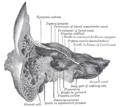
View of the inner wall of the tympanum. (Facial canal visible in upper left; promontory labeled at center)
Details
System Skeletal
Nerve Facial nerve (CN VII)
Identifiers
Latin canalis nervi facialis,
canalis facialis
TA98 A02.1.06.009
TA2 688
FMA 54952
Anatomical terminology

The facial canal (also known as the Fallopian canal) is a Z-shaped canal in the temporal bone of the skull. It extends between the internal acoustic meatus and stylomastoid foramen. It transmits the facial nerve (CN VII) (after which it is named).

Contents

Anatomy

The facial canal gives passage to the facial nerve (CN VII) (hence the name). [1] [ verification needed ][ better source needed ] Its proximal opening is at the internal auditory meatus; its distal opening is the stylomastoid foramen. In humans, the canal is approximately 3 cm long, making it the longest bony canal of a nerve in the human body. [2] [ verification needed ][ better source needed ] It is located within the middle ear region.[ verification needed ][ better source needed ]

The facial nerve gives rise to three nerves while passing through the canal: the greater petrosal nerve, nerve to stapedius, and the chorda tympani. [3]

Structure

Horizontal part

The proximal portion of the facial canal is termed the horizontal part. It commences at the introitus of facial canal at the distal end of the internal auditory meatus. The horizontal part is further subdivided into two crura: the proximal/medial [4] anteriolaterally [5] directed medial crus (or labyrinthine segment [5] ), and the distal/lateral [4] posteriolaterally [5] directed lateral crus (or tympanic segment [5] ); the two crura meet at a sharp angle at the genu of facial canal (geniculum canalis facialis [6] ) where the geniculate ganglion is situated (at the genu, the greater petrosal nerve leaves the facial canal through the hiatus of the facial canal). [4]

Descending part

The lateral crus of horizontal part ends by turning sharply inferior-ward, commencing the distal-most descending part (or mastoid segment [5] ) of facial canal which passes vertically inferior-ward, ending distally at the stylomastoid foramen. The descending part presents two openings through each of which a branch of the facial nerve passes: the nerve to stapedius enters the canaliculus for nerve to stapedius, and the chorda tympani enters the posterior canaliculus of chorda tympani (canaliculus chordae tympani, or iter chordae posterius [7] ). [8]

Relations

The labyrinthine segment is situated superior to cochlea. [5]

The canal traverses the medial wall of the tympanic cavity superior to the oval window;[ citation needed ] here, the prominence of the facial canal (or prominence of the aqueduct of Fallopius) upon the medial wall indicates the position of the superior portion of the facial canal. [9] :745 The canal then curves nearly vertically inferior-ward along the posterior wall.[ citation needed ] The tympanic segment is closely related to the posterior and medial walls of the tympanic cavity; it passes superior to the oval window and inferior to the lateral semicircular canal. [5]

Clinical significance

The facial canal may be interrupted in some people. This may lead to the facial nerve being split into 2 or 3 fibres, or it may be poorly formed or congenitally absent on one side. [2]

History

The facial canal was first described by Gabriele Falloppio. [10] This is why it may also be known as the Fallopian canal. [10]

See also

Related Research Articles

<span class="mw-page-title-main">Facial nerve</span> Cranial nerve VII, for the face and tasting

The facial nerve, also known as the seventh cranial nerve, cranial nerve VII, or simply CN VII, is a cranial nerve that emerges from the pons of the brainstem, controls the muscles of facial expression, and functions in the conveyance of taste sensations from the anterior two-thirds of the tongue. The nerve typically travels from the pons through the facial canal in the temporal bone and exits the skull at the stylomastoid foramen. It arises from the brainstem from an area posterior to the cranial nerve VI and anterior to cranial nerve VIII.

Articles related to anatomy include:

<span class="mw-page-title-main">Temporal bone</span> Bone of the neurocranium

The temporal bones are situated at the sides and base of the skull, and lateral to the temporal lobes of the cerebral cortex.

<span class="mw-page-title-main">Internal carotid artery</span> Artery of the human brain

The internal carotid artery is an artery in the neck which supplies the anterior circulation of the brain.

<span class="mw-page-title-main">Middle meningeal artery</span> Artery supplying the meninges

The middle meningeal artery is typically the third branch of the first portion of the maxillary artery. After branching off the maxillary artery in the infratemporal fossa, it runs through the foramen spinosum to supply the dura mater and the calvaria. The middle meningeal artery is the largest of the three (paired) arteries that supply the meninges, the others being the anterior meningeal artery and the posterior meningeal artery.

<span class="mw-page-title-main">Chorda tympani</span> Nerve carrying taste sensations

Chorda tympani is a branch of the facial nerve that carries gustatory (taste) sensory innervation from the front of the tongue and parasympathetic (secretomotor) innervation to the submandibular and sublingual salivary glands.

<span class="mw-page-title-main">Tympanic cavity</span> Small cavity surrounding the bones of the middle ear

The tympanic cavity is a small cavity surrounding the bones of the middle ear. Within it sit the ossicles, three small bones that transmit vibrations used in the detection of sound.

<span class="mw-page-title-main">Geniculate ganglion</span> Collection of facial nerve neurons

The geniculate ganglion is a collection of pseudounipolar sensory neurons of the facial nerve located in the facial canal of the head. It receives fibers from the facial nerve. It sends fibers that supply the lacrimal glands, submandibular glands, sublingual glands, tongue, palate, pharynx, external auditory meatus, stapedius muscle, posterior belly of the digastric muscle, stylohyoid muscle, and muscles of facial expression.

<span class="mw-page-title-main">Internal auditory meatus</span> Canal within the temporal bone

The internal auditory meatus is a canal within the petrous part of the temporal bone of the skull between the posterior cranial fossa and the inner ear.

<span class="mw-page-title-main">Greater wing of sphenoid bone</span> Large part of the skull front behind the eye socket

The greater wing of the sphenoid bone, or alisphenoid, is a bony process of the sphenoid bone, positioned in the skull behind each eye. There is one on each side, extending from the side of the body of the sphenoid and curving upward, laterally, and backward.

<span class="mw-page-title-main">Squamous part of temporal bone</span> Front and upper part of the sides of the skull base

The squamous part of temporal bone, or temporal squama, forms the front and upper part of the temporal bone, and is scale-like, thin, and translucent.

<span class="mw-page-title-main">Mastoid part of the temporal bone</span> Back part of the sides of the skull base

The mastoid part of the temporal bone is the posterior (back) part of the temporal bone, one of the bones of the skull. Its rough surface gives attachment to various muscles and it has openings for blood vessels. From its borders, the mastoid part articulates with two other bones.

<span class="mw-page-title-main">Petrous part of the temporal bone</span> Feature at the base of the human skull

The petrous part of the temporal bone is pyramid-shaped and is wedged in at the base of the skull between the sphenoid and occipital bones. Directed medially, forward, and a little upward, it presents a base, an apex, three surfaces, and three angles, and houses in its interior, the components of the inner ear. The petrous portion is among the most basal elements of the skull and forms part of the endocranium. Petrous comes from the Latin word petrosus, meaning "stone-like, hard". It is one of the densest bones in the body. In other mammals, it is a separate bone, the petrosal bone.

<span class="mw-page-title-main">Infratemporal fossa</span> Cavity that is part of the skull

The infratemporal fossa is an irregularly shaped cavity that is a part of the skull. It is situated below and medial to the zygomatic arch. It is not fully enclosed by bone in all directions. It contains superficial muscles, including the lower part of the temporalis muscle, the lateral pterygoid muscle, and the medial pterygoid muscle. It also contains important blood vessels such as the middle meningeal artery, the pterygoid plexus, and the retromandibular vein, and nerves such as the mandibular nerve (CN V3) and its branches.

<span class="mw-page-title-main">Perpendicular plate of palatine bone</span>

The perpendicular plate of palatine bone is the vertical part of the palatine bone, and is thin, of an oblong form, and presents two surfaces and four borders.

<span class="mw-page-title-main">Tympanic canaliculus</span> Passage of the tympanic branch of the glossopharyngeal nerve through the temporal bone

The tympanic canaliculus is a minute canal in the bony wedge/ridge that separates the carotid canal and jugular foramen/jugular fossa. The proximal opening of the canal is situated upon the inferior surface of the petrous part of the temporal bone; its distal opening is situated upon the floor of the tympanic cavity. The canal gives passage to the tympanic nerve i.e. tympanic branch of glossopharyngeal nerve ), and inferior tympanic artery.

<span class="mw-page-title-main">Pyramidal eminence</span>

The pyramidal eminence is a hollow conical projection upon the posterior wall of the tympanic cavity of the middle ear. The stapedius muscle arises in the hollow of the eminence and its tendon exits through its apex.

<span class="mw-page-title-main">Intermediate nerve</span> Portion of the facial nerve

The intermediate nerve, nervus intermedius, nerve of Wrisberg or glossopalatine nerve is the part of the facial nerve located between the motor component of the facial nerve and the vestibulocochlear nerve. It contains the sensory and parasympathetic fibers of the facial nerve. Upon reaching the facial canal, it joins with the motor root of the facial nerve at the geniculate ganglion. Alex Alfieri postulates that the intermediate nerve should be considered as a separate cranial nerve and not a part of the facial nerve.

<span class="mw-page-title-main">Outline of human anatomy</span> Overview of and topical guide to human anatomy

The following outline is provided as an overview of and topical guide to human anatomy:

References

  1. Nager, George T.; Proctor, Bruce (1991-06-01). "Anatomie Variations and Anomalies Involving the Facial Canal". Otolaryngologic Clinics of North America. 24 (3): 531–553. doi:10.1016/S0030-6665(20)31114-2. ISSN   0030-6665. PMID   1762775.
  2. 1 2 Weiglein AH (June 1996). "Postnatal development of the facial canal. An investigation based on cadaver dissections and computed tomography". Surgical and Radiologic Anatomy. 18 (2): 115–23. doi:10.1007/BF01795229. PMID   8782317. S2CID   25764734.
  3. Moore, Keith L.; Dalley, Arthur F.; Agur, Anne M. R. (2018). Clinically Oriented Anatomy (8th ed.). Wolters Kluwer. p. 1077. ISBN   978-1-4963-4721-3.
  4. 1 2 3 "horizontal part of facial canal". TheFreeDictionary.com. Retrieved 2023-07-29.
  5. 1 2 3 4 5 6 7 Shin KJ, Gil YC, Lee JY, Kim JN, Song WC, Koh KS (October 2014). "Three-dimensional study of the facial canal using microcomputed tomography for improved anatomical comprehension". Anatomical Record. 297 (10): 1808–16. doi: 10.1002/ar.22977 . PMID   24990524. S2CID   205411993.
  6. "genu of facial canal". TheFreeDictionary.com. Retrieved 2023-07-29.
  7. "posterior canaliculus of chorda tympani". TheFreeDictionary.com. Retrieved 2023-07-29.
  8. "descending part of facial canal". TheFreeDictionary.com. Retrieved 2023-07-29.
  9. Standring, Susan (2020). Gray's Anatomy: The Anatomical Basis of Clinical Practice (42th ed.). New York. ISBN   978-0-7020-7707-4. OCLC   1201341621.{{cite book}}: CS1 maint: location missing publisher (link)
  10. 1 2 Abing W, Rauchfuss A (2005). "Fetal development of the tympanic part of the facial canal". European Archives of Oto-Rhino-Laryngology. 243 (6): 374–377. doi:10.1007/bf00464645. PMID   3566620. S2CID   12712839.