In anatomy, the atlas (C1) is the most superior (first) cervical vertebra of the spine.
Articles related to anatomy include:

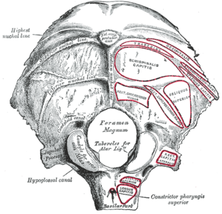
The foramen magnum is a large oval opening (foramen) in the occipital bone of the skull in humans and many other animals. It is one of the several oval or circular openings (foramina) in the base of the skull. The spinal cord, an extension of the medulla oblongata, passes through the foramen magnum as it exits the cranial cavity. Apart from the transmission of the medulla oblongata and its membranes, the foramen magnum transmits the vertebral arteries, the anterior and posterior spinal arteries, the tectorial membranes and alar ligaments. It also transmits the spinal component of the accessory nerve into the skull.
The occipital bone is a cranial dermal bone and the main bone of the occiput. It is trapezoidal in shape and curved on itself like a shallow dish. The occipital bone overlies the occipital lobes of the cerebrum. At the base of skull in the occipital bone, there is a large oval opening called the foramen magnum, which allows the passage of the spinal cord.

Dura mater is a thick membrane made of dense irregular connective tissue that surrounds the brain and spinal cord. It is the outermost of the three layers of membrane called the meninges that protect the central nervous system. The other two meningeal layers are the arachnoid mater and the pia mater. The dura surrounds the brain and the spinal cord and is responsible for keeping in the cerebrospinal fluid. It is derived from mesoderm.

The posterior cranial fossa is part of the cranial cavity, located between the foramen magnum and tentorium cerebelli. It contains the brainstem and cerebellum.
The emissary veins connect the extracranial venous system with the intracranial venous sinuses. They connect the veins outside the cranium to the venous sinuses inside the cranium. They drain from the scalp, through the skull, into the larger meningeal veins and dural venous sinuses.

The falx cerebelli is a small sickle shaped fold of dura mater, projecting forwards into the posterior cerebellar notch as well as projecting into the vallecula of the cerebellum between the two cerebellar hemispheres. The name comes from two Latin words: falx, meaning "curved blade or scythe", and cerebellum, meaning "brain". Its base is attached, above, to the under and back part of the tentorium cerebelli; its posterior margin, to the lower division of the vertical crest on the inner surface of the occipital bone. The falx cerebelli generally lies somewhere between 2.8 and 4.5 cm in length and is approximately 1–2 mm thick.

The condyloid process or condylar process is the process on the human mandible and some other species' mandibles that ends in a condyle, the mandibular condyle. It is thicker than the coronoid process of the mandible and consists of two portions: the condyle and the constricted portion which supports it, the neck.

The transverse sinuses, within the human head, are two areas beneath the brain which allow blood to drain from the back of the head. They run laterally in a groove along the interior surface of the occipital bone. They drain from the confluence of sinuses to the sigmoid sinuses, which ultimately connect to the internal jugular vein. See diagram : labeled under the brain as "SIN. TRANS.".

The occipital vein begins as a plexus at the posterior aspect of the scalp from the external occipital protuberance and superior nuchal line to the back part of the vertex of the skull.

The basilar part of the occipital bone extends forward and upward from the foramen magnum, and presents in front an area more or less quadrilateral in outline.

The mastoid foramen is a hole in the posterior border of the temporal bone. It transmits a Mastoid emissary vein to the sigmoid sinus and a small branch of the occipital artery, the posterior meningeal artery to the dura mater.

The occipital condyles are undersurface protuberances of the occipital bone in vertebrates, which function in articulation with the superior facets of the atlas vertebra.

The internal vertebral venous plexuses lie within the vertebral canal in the epidural space, and receive tributaries from the bones and from the spinal cord.

The base of skull, also known as the cranial base or the cranial floor, is the most inferior area of the skull. It is composed of the endocranium and the lower parts of the skull roof.

The following outline is provided as an overview of and topical guide to human anatomy:
The public domain consists of all the creative work to which no exclusive intellectual property rights apply. Those rights may have expired, been forfeited, expressly waived, or may be inapplicable.

Gray's Anatomy is an English written textbook of human anatomy originally written by Henry Gray and illustrated by Henry Vandyke Carter. Earlier editions were called Anatomy: Descriptive and Surgical, Anatomy of the Human Body and Gray's Anatomy: Descriptive and Applied, but the book's name is commonly shortened to, and later editions are titled, Gray's Anatomy. The book is widely regarded as an extremely influential work on the subject, and has continued to be revised and republished from its initial publication in 1858 to the present day. The latest edition of the book, the 41st, was published in September 2015.