
The skull is a bone protective cavity for the brain. The skull is composed of four types of bone i.e., cranial bones, facial bones, ear ossicles and hyoid bone, however two parts are more prominent: the cranium and the mandible. In humans, these two parts are the neurocranium (braincase) and the viscerocranium that includes the mandible as its largest bone. The skull forms the anterior-most portion of the skeleton and is a product of cephalisation—housing the brain, and several sensory structures such as the eyes, ears, nose, and mouth. In humans, these sensory structures are part of the facial skeleton.

A fontanelle is an anatomical feature of the infant human skull comprising soft membranous gaps (sutures) between the cranial bones that make up the calvaria of a fetus or an infant. Fontanelles allow for stretching and deformation of the neurocranium both during birth and later as the brain expands faster than the surrounding bone can grow. Premature complete ossification of the sutures is called craniosynostosis.

The humerus is a long bone in the arm that runs from the shoulder to the elbow. It connects the scapula and the two bones of the lower arm, the radius and ulna, and consists of three sections. The humeral upper extremity consists of a rounded head, a narrow neck, and two short processes. The body is cylindrical in its upper portion, and more prismatic below. The lower extremity consists of 2 epicondyles, 2 processes, and 3 fossae. As well as its true anatomical neck, the constriction below the greater and lesser tubercles of the humerus is referred to as its surgical neck due to its tendency to fracture, thus often becoming the focus of surgeons.
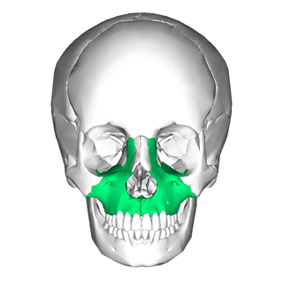
In vertebrates, the maxilla is the upper fixed bone of the jaw formed from the fusion of two maxillary bones. In humans, the upper jaw includes the hard palate in the front of the mouth. The two maxillary bones are fused at the intermaxillary suture, forming the anterior nasal spine. This is similar to the mandible, which is also a fusion of two mandibular bones at the mandibular symphysis. The mandible is the movable part of the jaw.

The occipital bone is a cranial dermal bone and the main bone of the occiput. It is trapezoidal in shape and curved on itself like a shallow dish. The occipital bone overlies the occipital lobes of the cerebrum. At the base of the skull in the occipital bone, there is a large oval opening called the foramen magnum, which allows the passage of the spinal cord.

In the human skull, the frontal bone or sincipital bone is a unpaired bone which consists of two portions. These are the vertically oriented squamous part, and the horizontally oriented orbital part, making up the bony part of the forehead, part of the bony orbital cavity holding the eye, and part of the bony part of the nose respectively. The name comes from the Latin word frons.
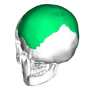
The parietal bones are two bones in the skull which, when joined at a fibrous joint, form the sides and roof of the neurocranium. In humans, each bone is roughly quadrilateral in form, and has two surfaces, four borders, and four angles. It is named from the Latin paries (-ietis), wall.

The crown is the top portion of the head behind the vertex. The anatomy of the crown varies between different organisms. The human crown is made of three layers of the scalp above the skull. The crown also covers a range of bone sutures, and contains blood vessels and branches of the trigeminal nerve.

In anatomy, a process is a projection or outgrowth of tissue from a larger body. For instance, in a vertebra, a process may serve for muscle attachment and leverage, or to fit, with another vertebra. The word is also used at the microanatomic level, where cells can have processes such as cilia or pedicels. Depending on the tissue, processes may also be called by other terms, such as apophysis, tubercle, or protuberance.

The brow ridge, or supraorbital ridge known as superciliary arch in medicine, is a bony ridge located above the eye sockets of all primates and some other animals. In humans, the eyebrows are located on their lower margin.

The frontal suture is a fibrous joint that divides the two halves of the frontal bone of the skull in infants and children. Typically, it completely fuses between three and nine months of age, with the two halves of the frontal bone being fused together. It is also called the metopic suture, although this term may also refer specifically to a persistent frontal suture.
The squamous part of the frontal bone is the superior portion when viewed in standard anatomical orientation. There are two surfaces of the squamous part of the frontal bone: the external surface, and the internal surface.
Haasiophis, consisting of the sole species Haasiophis terrasanctus, is an extinct genus of snakes with hind limbs. It is one of three genera of Cenomanian snakes known to have possessed hindlimbs.

The parietal eminence is a convex, smooth eminence on the external surface of the parietal bone of the skull. It is the site where intramembranous ossification of the parietal bone begins during embryological development. It tends to be slightly more prominent in men than in women, so may be used to help to identify the sex of a skull.

The zygomatic processes are three processes (protrusions) from other bones of the skull which each articulate with the zygomatic bone. The three processes are:

The clivus or Blumenbach clivus is a part of the occipital bone at the base of the skull. It is a shallow depression behind the dorsum sellae of the sphenoid bone. It slopes gradually to the anterior part of the basilar occipital bone at its junction with the sphenoid bone. It extends to the foramen magnum. It is related to the pons and the abducens nerve.

The calvaria is the top part of the skull. It is the superior part of the neurocranium and covers the cranial cavity containing the brain. It forms the main component of the skull roof.

In human anatomy, the neurocranium, also known as the braincase, brainpan, or brain-pan, is the upper and back part of the skull, which forms a protective case around the brain. In the human skull, the neurocranium includes the calvaria or skullcap. The remainder of the skull is the facial skeleton.

Metopism is the condition of having a persistent metopic suture, or persistence of the frontal metopic suture in the adult human skull. Metopism is the opposite of craniosynostosis. The main factor of the metopic suture is to increase the volume of the anterior cranial fossa. The frontal bone includes the forehead, and the roofs of the orbits of the eyes. The frontal bone has vertical portion (squama) and horizontal portion. Some adults have a metopic or frontal suture in the vertical portion. In uterine period in right and left half of frontal region of the fetus there is a membrane tissue. On each half a primary ossification center appears about the end of the second month of the fetus. Primary ossification center extends to form the corresponding half of the vertical part (squama) and horizontal part of the frontal bone.
In anatomy, eminence implies a protuberance, and may refer to a variety of structures:
