Body of sphenoid bone

Last updated
Body of sphenoid bone
Gray146.png
Figure 2: Sphenoid bone, anterior and inferior surfaces.
Gray147.png
Figure 3: Sphenoid bone, upper and posterior surfaces.
Details
Identifiers
Latin corpus ossis sphenoidalis
TA98 A02.1.05.002
TA2 585
FMA 52867
Anatomical terms of bone

The body of the sphenoid bone , more or less cubical in shape, is hollowed out in its interior to form two large cavities, the sphenoidal sinuses, which are separated from each other by a septum. [1]

Contents

Superior surface

The superior surface of the body [Fig. 1] presents in front a prominent spine, the ethmoidal spine, for articulation with the cribriform plate of the ethmoid bone; behind this is a smooth surface slightly raised in the middle line, and grooved on either side for the olfactory lobes of the brain.

This surface is bounded behind by a ridge, which forms the anterior border of a narrow, transverse groove, the prechiasmatic groove, above and behind which lies the optic chiasma; the groove ends on either side in the optic foramen, which transmits the optic nerve and ophthalmic artery into the orbital cavity.

Behind the chiasmatic groove is an elevation, the tuberculum sellae; and behind this is a deep depression, the saddle-shaped sella turcica (Turkish seat), the deepest part of which, the hypophyseal fossa, lodges the pituitary gland.

The anterior boundary of the sella turcica is completed by two small eminences, one on either side, called the middle clinoid processes, while the posterior boundary is formed by a square-shaped plate of bone, the dorsum sellae, ending at its superior angles in two tubercles, the posterior clinoid processes, the size and form of which vary considerably in different individuals.

The posterior clinoid processes deepen the sella turcica, and give attachment to the tentorium cerebelli.

On either side of the dorsum sellae is a notch for the passage of the abducent nerve, and below the notch a sharp process, the petrosal process, which articulates with the apex of the petrous portion of the temporal bone, and forms the medial boundary of the foramen lacerum.

Behind the dorsum sellae is a shallow depression, the clivus, which slopes obliquely backward, and is continuous with the groove on the basilar portion of the occipital bone; it supports the upper part of the pons.

Lateral surfaces

The lateral surfaces of the body are united with the greater wings of the sphenoid and the medial pterygoid plates.

Above the attachment of each greater wing is a broad groove, curved something like the italic letter f; it lodges the internal carotid artery and the cavernous sinus, and is named the carotid sulcus.

Along the posterior part of the lateral margin of this groove, in the angle between the body and greater wing, is a ridge of bone, called the sphenoidal lingula.

Posterior surfaces

The posterior surface, quadrilateral in form [Fig. 3], is joined, during infancy and adolescence, to the basilar part of the occipital bone by a plate of cartilage.

Between the eighteenth and twenty-fifth years this becomes ossified, ossification commencing above and extending downward.

Anterior surface

The anterior surface of the body [Fig. 2] presents, in the middle line, a vertical crest, the sphenoidal crest, which articulates with the perpendicular plate of the ethmoid, and forms part of the nasal septum.

On either side of the crest is an irregular opening leading into the corresponding sphenoidal air sinus.

These sinuses are two large, irregular cavities hollowed out of the interior of the body of the bone, and separated from one another by a bony septum, which is commonly bent to one or the other side.

They vary considerably in form and size, are seldom symmetrical, and are often partially subdivided by irregular bony laminae.

Occasionally, they extend into the basilar part of the occipital bone nearly as far as the foramen magnum. They begin to be developed before birth, and are of a considerable size by the age of six.

They are partially closed, in front and below, by two thin, curved plates of bone, the sphenoidal conchae, leaving in the articulated skull a round opening at the upper part of each sinus by which it communicates with the upper and back part of the nasal cavity and occasionally with the posterior ethmoidal air cells.

The lateral margin of the anterior surface is serrated, and articulates with the lamina papyracea of the ethmoid, completing the posterior ethmoidal cells; the lower margin articulates with the orbital process of the palatine bone, and the upper with the orbital plate of the frontal bone.

Inferior surface

The inferior surface presents, in the middle line, a triangular spine, the sphenoidal rostrum, which is continuous with the sphenoidal crest on the anterior surface, and is received in a deep fissure between the alæ of the vomer.

On either side of the rostrum is a projecting lamina, the vaginal process, directed medialward from the base of the medial pterygoid plate, with which it will be described.

Additional images

Related Research Articles

Articles related to anatomy include:

<span class="mw-page-title-main">Sphenoid bone</span> Bone of the neurocranium

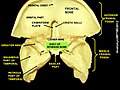
The sphenoid bone is an unpaired bone of the neurocranium. It is situated in the middle of the skull towards the front, in front of the basilar part of the occipital bone. The sphenoid bone is one of the seven bones that articulate to form the orbit. Its shape somewhat resembles that of a butterfly or bat with its wings extended.

<span class="mw-page-title-main">Occipital bone</span> Bone of the neurocranium

The occipital bone is a cranial dermal bone and the main bone of the occiput. It is trapezoidal in shape and curved on itself like a shallow dish. The occipital bone overlies the occipital lobes of the cerebrum. At the base of the skull in the occipital bone, there is a large oval opening called the foramen magnum, which allows the passage of the spinal cord.

<span class="mw-page-title-main">Orbit (anatomy)</span> Cavity or socket of the skull in which the eye and its appendages are situated

In anatomy, the orbit is the cavity or socket/hole of the skull in which the eye and its appendages are situated. "Orbit" can refer to the bony socket, or it can also be used to imply the contents. In the adult human, the volume of the orbit is 30 millilitres, of which the eye occupies 6.5 ml. The orbital contents comprise the eye, the orbital and retrobulbar fascia, extraocular muscles, cranial nerves II, III, IV, V, and VI, blood vessels, fat, the lacrimal gland with its sac and duct, the eyelids, medial and lateral palpebral ligaments, cheek ligaments, the suspensory ligament, septum, ciliary ganglion and short ciliary nerves.

<span class="mw-page-title-main">Sella turcica</span> Saddle-shaped depression in the sphenoid bone of the skull

The sella turcica is a saddle-shaped depression in the body of the sphenoid bone of the human skull and of the skulls of other hominids including chimpanzees, gorillas and orangutans. It serves as a cephalometric landmark. The pituitary gland or hypophysis is located within the most inferior aspect of the sella turcica, the hypophyseal fossa.

<span class="mw-page-title-main">Posterior cranial fossa</span> Area of the cranium containing the brainstem and cerebellum

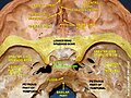
The posterior cranial fossa is the part of the cranial cavity located between the foramen magnum, and tentorium cerebelli. It is formed by the sphenoid bones, temporal bones, and occipital bone. It lodges the cerebellum, and parts of the brainstem.

<span class="mw-page-title-main">Orbital part of frontal bone</span>

The orbital or horizontal part of the frontal bone consists of two thin triangular plates, the orbital plates, which form the vaults of the orbits, and are separated from one another by a median gap, the ethmoidal notch.

<span class="mw-page-title-main">Ethmoid sinus</span> Air-filled space near the nasal cavity

The ethmoid sinuses or ethmoid air cells of the ethmoid bone are one of the four paired paranasal sinuses. Unlike the other three pairs of paranasal sinuses which consist of one or two large cavities, the ethmoidal sinuses entail a number of small air-filled cavities. The cells are located within the lateral mass (labyrinth) of each ethmoid bone and are variable in both size and number. The cells are grouped into anterior, middle, and posterior groups; the groups differ in their drainage modalities, though all ultimately drain into either the superior or the middle nasal meatus of the lateral wall of the nasal cavity.

<span class="mw-page-title-main">Sphenoid sinus</span> One of the four paired paranasal sinuses

The sphenoid sinus is a paired paranasal sinus occurring within the body of the sphenoid bone. It represents one pair of the four paired paranasal sinuses. The pair of sphenoid sinuses are separated in the middle by a septum of sphenoid sinuses. Each sphenoid sinus communicates with the nasal cavity via the opening of sphenoidal sinus. The two sphenoid sinuses vary in size and shape, and are usually asymmetrical.

<span class="mw-page-title-main">Sphenoidal conchae</span>

The sphenoidal conchae are two thin, curved plates, situated at the anterior and lower part of the body of the sphenoid. An aperture of variable size exists in the anterior wall of each, and through this the sphenoidal sinus opens into the nasal cavity.

<span class="mw-page-title-main">Greater wing of sphenoid bone</span> Large part of the skull front behind the eye socket

The greater wing of the sphenoid bone, or alisphenoid, is a bony process of the sphenoid bone, positioned in the skull behind each eye. There is one on each side, extending from the side of the body of the sphenoid and curving upward, laterally, and backward.

<span class="mw-page-title-main">Ethmoidal labyrinth</span>

The ethmoidal labyrinth or lateral mass of the ethmoid bone consists of a number of thin-walled cellular cavities, the ethmoid air cells, arranged in three groups, anterior, middle, and posterior, and interposed between two vertical plates of bone; the lateral plate forms part of the orbit, the medial plate forms part of the nasal cavity. In the disarticulated bone many of these cells are opened into, but when the bones are articulated, they are closed in at every part, except where they open into the nasal cavity.

<span class="mw-page-title-main">Anterior ethmoidal artery</span>

The anterior ethmoidal artery is a branch of the ophthalmic artery in the orbit. It exits the orbit through the anterior ethmoidal foramen alongside the anterior ethmoidal nerve. It contributes blood supply to the ethmoid sinuses, frontal sinuses, the dura mater, lateral nasal wall, and nasal septum. It issues a meningeal branch, and nasal branches.

<span class="mw-page-title-main">Petrous part of the temporal bone</span> Feature at the base of the human skull

The petrous part of the temporal bone is pyramid-shaped and is wedged in at the base of the skull between the sphenoid and occipital bones. Directed medially, forward, and a little upward, it presents a base, an apex, three surfaces, and three angles, and houses in its interior, the components of the inner ear. The petrous portion is among the most basal elements of the skull and forms part of the endocranium. Petrous comes from the Latin word petrosus, meaning "stone-like, hard". It is one of the densest bones in the body. In other mammals, it is a separate bone, the petrosal bone.

<span class="mw-page-title-main">Middle cranial fossa</span>

The middle cranial fossa is formed by the sphenoid bones, and the temporal bones. It lodges the temporal lobes, and the pituitary gland. It is deeper than the anterior cranial fossa, is narrow medially and widens laterally to the sides of the skull. It is separated from the posterior cranial fossa by the clivus and the petrous crest.

<span class="mw-page-title-main">Anterior cranial fossa</span>

The anterior cranial fossa is a depression in the floor of the cranial base which houses the projecting frontal lobes of the brain. It is formed by the orbital plates of the frontal, the cribriform plate of the ethmoid, and the small wings and front part of the body of the sphenoid; it is limited behind by the posterior borders of the small wings of the sphenoid and by the anterior margin of the chiasmatic groove. The lesser wings of the sphenoid separate the anterior and middle fossae.

<span class="mw-page-title-main">Dorsum sellae</span>

The dorsum sellae is part of the sphenoid bone in the skull. Together with the basilar part of the occipital bone it forms the clivus.

<span class="mw-page-title-main">Perpendicular plate of palatine bone</span>

The perpendicular plate of palatine bone is the vertical part of the palatine bone, and is thin, of an oblong form, and presents two surfaces and four borders.

<span class="mw-page-title-main">Base of skull</span> Inferior area of the skull, composed of the endocranium and lower parts of the skull roof

The base of skull, also known as the cranial base or the cranial floor, is the most inferior area of the skull. It is composed of the endocranium and the lower parts of the calvaria.

<span class="mw-page-title-main">Outline of human anatomy</span> Overview of and topical guide to human anatomy

The following outline is provided as an overview of and topical guide to human anatomy:

References

  1. Dutton, Jonathan J. (February 2, 2010). Radiology of the Orbit and Visual Pathways. Elsevier Health Sciences. p. 31. ISBN   9781455710676.