Middle nasal concha

Last updated
Middle nasal concha
Gray151.png
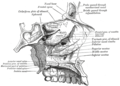
Ethmoid bone from behind.
Conchanasalismedius.PNG
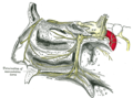
Lateral wall of nasal cavity, showing ethmoid bone in position. (Middle nasal concha is at bottom of pink region.)
Details
Identifiers
Latin concha media
TA98 A06.1.02.014
TA2 735
FMA 57459
Anatomical terms of bone

The medial surface of the labyrinth of ethmoid consists of a thin lamella, which descends from the under surface of the cribriform plate, and ends below in a free, convoluted margin, the middle nasal concha (middle nasal turbinate).

Contents

It is rough, and marked above by numerous grooves, directed nearly vertically downward from the cribriform plate; they lodge branches of the olfactory nerves, which are distributed to the mucous membrane covering the superior nasal concha.

The middle turbinates insert anteriorly into the frontal process of the maxilla and posteriorly into the perpendicular plate of the palatine bone. [1] There are three mutually perpendicular segments of the middle turbinate: from proximal to distal, there is the horizontal segment (axial plane), the basal lamella (coronal plane), and the vertical segment (sagittal plane).

Additional images

See also

Related Research Articles

<span class="mw-page-title-main">Maxilla</span> Upper jaw bone

The maxilla in vertebrates is the upper fixed bone of the jaw formed from the fusion of two maxillary bones. In humans, the upper jaw includes the hard palate in the front of the mouth. The two maxillary bones are fused at the intermaxillary suture, forming the anterior nasal spine. This is similar to the mandible, which is also a fusion of two mandibular bones at the mandibular symphysis. The mandible is the movable part of the jaw.

<span class="mw-page-title-main">Ethmoid bone</span> Bone in the human skull

The ethmoid bone is an unpaired bone in the skull that separates the nasal cavity from the brain. It is located at the roof of the nose, between the two orbits. The cubical bone is lightweight due to a spongy construction. The ethmoid bone is one of the bones that make up the orbit of the eye.

<span class="mw-page-title-main">Nasal bone</span> Two bones in the bridge of the human nose

The nasal bones are two small oblong bones, varying in size and form in different individuals; they are placed side by side at the middle and upper part of the face and by their junction, form the bridge of the upper one third of the nose.

<span class="mw-page-title-main">Inferior nasal concha</span>

The inferior nasal concha is one of the three paired nasal conchae in the nose. It extends horizontally along the lateral wall of the nasal cavity and consists of a lamina of spongy bone, curled upon itself like a scroll,. The inferior nasal conchae are considered a pair of facial bones. As the air passes through the turbinates, the air is churned against these mucosa-lined bones in order to receive warmth, moisture and cleansing. Superior to inferior nasal concha are the middle nasal concha and superior nasal concha which both arise from the ethmoid bone, of the cranial portion of the skull. Hence, these two are considered as a part of the cranial bones.

<span class="mw-page-title-main">Vomer</span> Unpaired facial bone of the skull

The vomer is one of the unpaired facial bones of the skull. It is located in the midsagittal line, and articulates with the sphenoid, the ethmoid, the left and right palatine bones, and the left and right maxillary bones. The vomer forms the inferior part of the nasal septum in humans, with the superior part formed by the perpendicular plate of the ethmoid bone. The name is derived from the Latin word for a ploughshare and the shape of the bone.

<span class="mw-page-title-main">Nasal cavity</span> Large, air-filled space above and behind the nose in the middle of the face

The nasal cavity is a large, air-filled space above and behind the nose in the middle of the face. The nasal septum divides the cavity into two cavities, also known as fossae. Each cavity is the continuation of one of the two nostrils. The nasal cavity is the uppermost part of the respiratory system and provides the nasal passage for inhaled air from the nostrils to the nasopharynx and rest of the respiratory tract.

<span class="mw-page-title-main">Nasal concha</span> Piece of bone in the breathing passage of humans and other animals

In anatomy, a nasal concha, also called a nasal turbinate or turbinal, is a long, narrow, curled shelf of bone that protrudes into the breathing passage of the nose in humans and various animals. The conchae are shaped like an elongated seashell, which gave them their name. A concha is any of the scrolled spongy bones of the nasal passages in vertebrates.

<span class="mw-page-title-main">Cribriform plate</span> Part of the ethmoid bone in the skull

In mammalian anatomy, the cribriform plate, horizontal lamina or lamina cribrosa is part of the ethmoid bone. It is received into the ethmoidal notch of the frontal bone and roofs in the nasal cavities. It supports the olfactory bulb, and is perforated by olfactory foramina for the passage of the olfactory nerves to the roof of the nasal cavity to convey smell to the brain. The foramina at the medial part of the groove allow the passage of the nerves to the upper part of the nasal septum while the foramina at the lateral part transmit the nerves to the superior nasal concha.

<span class="mw-page-title-main">Ethmoid sinus</span> Air-filled space near the nasal cavity

The ethmoid sinuses or ethmoid air cells of the ethmoid bone are one of the four paired paranasal sinuses. Unlike the other three pairs of paranasal sinuses which consist of one or two large cavities, the ethmoidal sinuses entail a number of small air-filled cavities. The cells are located within the lateral mass (labyrinth) of each ethmoid bone and are variable in both size and number. The cells are grouped into anterior, middle, and posterior groups; the groups differ in their drainage modalities, though all ultimately drain into either the superior or the middle nasal meatus of the lateral wall of the nasal cavity.

<span class="mw-page-title-main">Sphenoidal conchae</span>

The sphenoidal conchae are two thin, curved plates, situated at the anterior and lower part of the body of the sphenoid. An aperture of variable size exists in the anterior wall of each, and through this the sphenoidal sinus opens into the nasal cavity.

<span class="mw-page-title-main">Ethmoidal labyrinth</span>

The ethmoidal labyrinth or lateral mass of the ethmoid bone consists of a number of thin-walled cellular cavities, the ethmoid air cells, arranged in three groups, anterior, middle, and posterior, and interposed between two vertical plates of bone; the lateral plate forms part of the orbit, the medial plate forms part of the nasal cavity. In the disarticulated bone many of these cells are opened into, but when the bones are articulated, they are closed in at every part, except where they open into the nasal cavity.

<span class="mw-page-title-main">Perpendicular plate of ethmoid bone</span>

The perpendicular plate of the ethmoid bone is a thin, flattened lamina, polygonal in form, which descends from the under surface of the cribriform plate, and assists in forming the septum of the nose; it is generally deflected a little to one or other side. The anterior border articulates with the spine of the frontal bone and the crest of the nasal bones.

<span class="mw-page-title-main">Anterior ethmoidal artery</span>

The anterior ethmoidal artery is a branch of the ophthalmic artery in the orbit. It exits the orbit through the anterior ethmoidal foramen alongside the anterior ethmoidal nerve. It contributes blood supply to the ethmoid sinuses, frontal sinuses, the dura mater, lateral nasal wall, and nasal septum. It issues a meningeal branch, and nasal branches.

<span class="mw-page-title-main">Superior nasal concha</span>

The superior nasal concha is a small, curved plate of bone representing a medial bony process of the labyrinth of the ethmoid bone. The superior nasal concha forms the roof of the superior nasal meatus.

<span class="mw-page-title-main">Squamous part of the frontal bone</span> Upper two-thirds of the forward skull

The squamous part of the frontal bone is the superior portion when viewed in standard anatomical orientation. There are two surfaces of the squamous part of the frontal bone: the external surface, and the internal surface.

<span class="mw-page-title-main">Anterior cranial fossa</span>

The anterior cranial fossa is a depression in the floor of the cranial base which houses the projecting frontal lobes of the brain. It is formed by the orbital plates of the frontal, the cribriform plate of the ethmoid, and the small wings and front part of the body of the sphenoid; it is limited behind by the posterior borders of the small wings of the sphenoid and by the anterior margin of the chiasmatic groove. The lesser wings of the sphenoid separate the anterior and middle fossae.

<span class="mw-page-title-main">Frontal process of maxilla</span> Plate which forms part of the lateral boundary of the nose

The frontal process of maxilla is a strong plate, which projects upward, medialward, and backward from the maxilla, forming part of the lateral boundary of the nose.

<span class="mw-page-title-main">Human nose</span> Feature of the face

The human nose is the most protruding part of the face. It bears the nostrils and is the first organ of the respiratory system. It is also the principal organ in the olfactory system. The shape of the nose is determined by the nasal bones and the nasal cartilages, including the nasal septum which separates the nostrils and divides the nasal cavity into two. On average, the nose of a male is larger than that of a female.

<span class="mw-page-title-main">Nasal meatus</span> Nasal passage of the nasal cavity

In anatomy, the term nasal meatus can refer to any of the three meatuses (passages) through the skull's nasal cavity: the superior meatus, middle meatus, and inferior meatus.

<span class="mw-page-title-main">Plate (anatomy)</span> Index of articles associated with the same name

A plate in animal anatomy may refer to several things:

References

  1. Lee, Hye Yeon; Kim, Chang-Hoon; Kim, Jin Young; Kim, Jin Kook; Song, Mee Hyun; Yang, Hee Jun; Kim, Kyung-Su; Chung, In Hyunk; Lee, Jeung-Gweon; Yoon, Joo-Heon (September 2006). "Surgical anatomy of the middle turbinate". Clinical Anatomy (New York, N.Y.). 19 (6): 493–496. doi:10.1002/ca.20202. ISSN   0897-3806. PMID   16258979. S2CID   5720770.

PD-icon.svgThis article incorporates text in the public domain from page 156 of the 20th edition of Gray's Anatomy (1918)