
Temporomandibular joint dysfunction is an umbrella term covering pain and dysfunction of the muscles of mastication and the temporomandibular joints. The most important feature is pain, followed by restricted mandibular movement, and noises from the temporomandibular joints (TMJ) during jaw movement. Although TMD is not life-threatening, it can be detrimental to quality of life; this is because the symptoms can become chronic and difficult to manage.
In anatomy, the temporomandibular joints (TMJ) are the two joints connecting the jawbone to the skull. It is a bilateral synovial articulation between the temporal bone of the skull above and the mandible below; it is from these bones that its name is derived. This joint is unique in that it is a bilateral joint that functions as one unit. Since the TMJ is connected to the mandible, the right and left joints must function together and therefore are not independent of each other.

The humerus is a long bone in the arm that runs from the shoulder to the elbow. It connects the scapula and the two bones of the lower arm, the radius and ulna, and consists of three sections. The humeral upper extremity consists of a rounded head, a narrow neck, and two short processes. The body is cylindrical in its upper portion, and more prismatic below. The lower extremity consists of 2 epicondyles, 2 processes, and 3 fossae. As well as its true anatomical neck, the constriction below the greater and lesser tubercles of the humerus is referred to as its surgical neck due to its tendency to fracture, thus often becoming the focus of surgeons.

The jaws are a pair of opposable articulated structure at the entrance of the mouth, typically used for grasping and manipulating food. The term jaws is also broadly applied to the whole of the structures constituting the vault of the mouth and serving to open and close it and is part of the body plan of humans and most animals.

In anatomy, a process is a projection or outgrowth of tissue from a larger body. For instance, in a vertebra, a process may serve for muscle attachment and leverage, or to fit, with another vertebra. The word is also used at the microanatomic level, where cells can have processes such as cilia or pedicels. Depending on the tissue, processes may also be called by other terms, such as apophysis, tubercle, or protuberance.

The auriculotemporal nerve is a sensory branch of the mandibular nerve (CN V3) that runs with the superficial temporal artery and vein, and provides sensory innervation to parts of the external ear, scalp, and temporomandibular joint. The nerve also conveys post-ganglionic parasympathetic fibres from the otic ganglion to the parotid gland.

The lateral pterygoid muscle (or external pterygoid muscle) is a muscle of mastication. It has two heads. It lies superior to the medial pterygoid muscle. It is supplied by pterygoid branches of the maxillary artery, and the lateral pterygoid nerve (from the mandibular nerve, CN V3). It depresses and protrudes the mandible. When each muscle works independently, they can move the mandible side to side.
The temporomandibular ligament, also known as the external lateral ligament, is a ligament that connects the lower articular tubercle of the zygomatic arch to the lateral and posterior border of the neck of the mandible. It prevents posterior displacement of the mandible. It also prevents the condyloid process from being driven upward by a blow to the jaw, which would otherwise fracture the base of the skull.
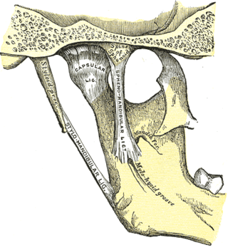
The sphenomandibular ligament is one of the three ligaments of the temporomandibular joint. It is situated medially to - and generally separate from - the articular capsule of the joint. Superiorly, it is attached to the spine of the sphenoid bone; inferiorly, it is attached to the lingula of mandible. The SML acts to limit inferior-ward movement of the mandible.

The condyloid process or condylar process is the process on the human and other mammalian species' mandibles that ends in a condyle, the mandibular condyle. It is thicker than the coronoid process of the mandible and consists of two portions: the condyle and the constricted portion which supports it, the neck.
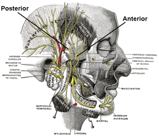
The deep temporal nerves are typically two nerves (one anterior and one posterior) which arise from the mandibular nerve (CN V3) and provide motor innervation to the temporalis muscle.

The squamous part of temporal bone, or temporal squama, forms the front and upper part of the temporal bone, and is scale-like, thin, and translucent.

The masseteric nerve is a nerve of the face. It is a branch of the mandibular nerve (CN V3). It passes through the mandibular notch to reach masseter muscle. It provides motor innervation the masseter muscle, and sensory innervation to the temporomandibular joint.

The mandibular fossa, also known as the glenoid fossa in some dental literature, is the depression in the temporal bone that articulates with the mandible.

The petrotympanic fissure is a fissure in the temporal bone that runs from the temporomandibular joint to the tympanic cavity.

The articular disk of the temporomandibular joint is a thin, oval plate made of non-vascular fibrous connective tissue located between the mandible's condyloid process and the cranium's mandibular fossa.
In dentistry, centric relation is the mandibular jaw position in which the head of the condyle is situated as far anterior and superior as it possibly can within the mandibular fossa/glenoid fossa.
In jawed vertebrates, the mandible, lower jaw, or jawbone is a bone that makes up the lower – and typically more mobile – component of the mouth.

The intercondylar area is the separation between the medial and lateral condyle on the upper extremity of the tibia. The anterior and posterior cruciate ligaments and the menisci attach to the intercondylar area.

Posselt's envelope of motion or Posselt's envelope of movement refers to the range of motion of the lower jaw bone, or mandible.

