Related Research Articles

The maxilla in vertebrates is the upper fixed bone of the jaw formed from the fusion of two maxillary bones. In humans, the upper jaw includes the hard palate in the front of the mouth. The two maxillary bones are fused at the intermaxillary suture, forming the anterior nasal spine. This is similar to the mandible, which is also a fusion of two mandibular bones at the mandibular symphysis. The mandible is the movable part of the jaw.

Rhinoplasty, commonly called nose job, medically called nasal reconstruction is a plastic surgery procedure for altering and reconstructing the nose. There are two types of plastic surgery used – reconstructive surgery that restores the form and functions of the nose and cosmetic surgery that changes the appearance of the nose. Reconstructive surgery seeks to resolve nasal injuries caused by various traumas including blunt, and penetrating trauma and trauma caused by blast injury. Reconstructive surgery can also treat birth defects, breathing problems, and failed primary rhinoplasties. Rhinoplasty may remove a bump, narrow nostril width, change the angle between the nose and the mouth, or address injuries, birth defects, or other problems that affect breathing, such as a deviated nasal septum or a sinus condition. Surgery only on the septum is called a septoplasty.

The ethmoid bone is an unpaired bone in the skull that separates the nasal cavity from the brain. It is located at the roof of the nose, between the two orbits. The cubical bone is lightweight due to a spongy construction. The ethmoid bone is one of the bones that make up the orbit of the eye.

The nasal bones are two small oblong bones, varying in size and form in different individuals; they are placed side by side at the middle and upper part of the face and by their junction, form the bridge of the upper one third of the nose.

The lacrimal bone is a small and fragile bone of the facial skeleton; it is roughly the size of the little fingernail. It is situated at the front part of the medial wall of the orbit. It has two surfaces and four borders. Several bony landmarks of the lacrimal bone function in the process of lacrimation or crying. Specifically, the lacrimal bone helps form the nasolacrimal canal necessary for tear translocation. A depression on the anterior inferior portion of the bone, the lacrimal fossa, houses the membranous lacrimal sac. Tears or lacrimal fluid, from the lacrimal glands, collect in this sac during excessive lacrimation. The fluid then flows through the nasolacrimal duct and into the nasopharynx. This drainage results in what is commonly referred to a runny nose during excessive crying or tear production. Injury or fracture of the lacrimal bone can result in posttraumatic obstruction of the lacrimal pathways.

The inferior nasal concha is one of the three paired nasal conchae in the nose. It extends horizontally along the lateral wall of the nasal cavity and consists of a lamina of spongy bone, curled upon itself like a scroll,. The inferior nasal conchae are considered a pair of facial bones. As the air passes through the turbinates, the air is churned against these mucosa-lined bones in order to receive warmth, moisture and cleansing. Superior to inferior nasal concha are the middle nasal concha and superior nasal concha which both arise from the ethmoid bone, of the cranial portion of the skull. Hence, these two are considered as a part of the cranial bones.
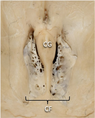
In mammalian anatomy, the cribriform plate, horizontal lamina or lamina cribrosa is part of the ethmoid bone. It is received into the ethmoidal notch of the frontal bone and roofs in the nasal cavities. It supports the olfactory bulb, and is perforated by olfactory foramina for the passage of the olfactory nerves to the roof of the nasal cavity to convey smell to the brain. The foramina at the medial part of the groove allow the passage of the nerves to the upper part of the nasal septum while the foramina at the lateral part transmit the nerves to the superior nasal concha.

In the ethmoid bone, a sickle shaped projection, the uncinate process, projects posteroinferiorly from the ethmoid labyrinth. Between the posterior edge of this process and the anterior surface of the ethmoid bulla, there is a two-dimensional space, resembling a crescent shape. This space continues laterally as a three-dimensional slit-like space - the ethmoidal infundibulum. This is bounded by the uncinate process, medially, the orbital lamina of ethmoid bone, laterally, and the ethmoidal bulla, posterosuperiorly. This concept is easier to understand if one imagine the infundibulum as a prism so that its medial face is the hiatus semilunaris. The "lateral face" of this infundibulum contains the ostium of the maxillary sinus, which, therefore, opens into the infundibulum.

The ethmoid sinuses or ethmoid air cells of the ethmoid bone are one of the four paired paranasal sinuses. Unlike the other three pairs of paranasal sinuses which consist of one or two large cavities, the ethmoidal sinuses entail a number of small air-filled cavities. The cells are located within the lateral mass (labyrinth) of each ethmoid bone and are variable in both size and number. The cells are grouped into anterior, middle, and posterior groups; the groups differ in their drainage modalities, though all ultimately drain into either the superior or the middle nasal meatus of the lateral wall of the nasal cavity.

The semilunar hiatus is a crescent-shaped/semicircular/ curved slit/groove upon the lateral wall of the nasal cavity at the middle nasal meatus just inferior to the ethmoidal bulla. It is the location of the openings for the frontal sinus, maxillary sinus, and anterior ethmoidal sinus. It is bounded inferiorly and anteriorly by the sharp concave margin of the uncinate process of the ethmoid bone, superiorly by the ethmoidal bulla, and posteriorly by the ethmoidal process of the inferior nasal concha. It leads into the ethmoidal infundibulum; it marks the medial limit of the ethmoidal infundibulum.

The sphenoidal conchae are two thin, curved plates, situated at the anterior and lower part of the body of the sphenoid. An aperture of variable size exists in the anterior wall of each, and through this the sphenoidal sinus opens into the nasal cavity.

The ethmoidal labyrinth or lateral mass of the ethmoid bone consists of a number of thin-walled cellular cavities, the ethmoid air cells, arranged in three groups, anterior, middle, and posterior, and interposed between two vertical plates of bone; the lateral plate forms part of the orbit, the medial plate forms part of the nasal cavity. In the disarticulated bone many of these cells are opened into, but when the bones are articulated, they are closed in at every part, except where they open into the nasal cavity.
The superior nasal concha is a small, curved plate of bone representing a medial bony process of the labyrinth of the ethmoid bone. The superior nasal concha forms the roof of the superior nasal meatus.
The medial surface of the labyrinth of ethmoid consists of a thin lamella, which descends from the under surface of the cribriform plate, and ends below in a free, convoluted margin, the middle nasal concha.
The ethmoid bulla is a rounded elevation upon the lateral wall of the middle nasal meatus produced by one or more of the underlying middle ethmoidal air cells. It varies significantly based on the size of the underlying air cells.
The maxillary hiatus is the opening of a maxillary sinus into the middle nasal meatus of the nasal cavity. It is situated superoposteriorly upon the lateral nasal wall, opening into the nasal cavity at the posterior portion of the ethmoidal infundibulum. Its opening in the maxillary sinus is present upon the superior part of the medial wall of the sinus near the roof of the sinus; because of the position, gravity cannot drain the maxillary sinus contents when the head is erect.

The frontal process of the maxilla is a strong plate, which projects upward, medialward, and backward from the maxilla, forming part of the lateral boundary of the nose.

Giovanni Domenico Santorini was an Italian anatomist. He was a native of Venice, earning his medical doctorate at Pisa in 1701. He is remembered for conducting anatomical dissections of the human body.

The human nose is the first organ of the respiratory system. It is also the principal organ in the olfactory system. The shape of the nose is determined by the nasal bones and the nasal cartilages, including the nasal septum which separates the nostrils and divides the nasal cavity into two.

In anatomy, the term nasal meatus can refer to any of the three meatuses (passages) through the skull's nasal cavity: the superior meatus, middle meatus, and inferior meatus.
References
| | This human musculoskeletal system article is a stub. You can help Wikipedia by expanding it. |