
The occipital bone is a cranial dermal bone and the main bone of the occiput. It is trapezoidal in shape and curved on itself like a shallow dish. The occipital bone overlies the occipital lobes of the cerebrum. At the base of the skull in the occipital bone, there is a large oval opening called the foramen magnum, which allows the passage of the spinal cord.
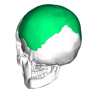
The parietal bones are two bones in the skull which, when joined at a fibrous joint known as a cranial suture, form the sides and roof of the neurocranium. In humans, each bone is roughly quadrilateral in form, and has two surfaces, four borders, and four angles. It is named from the Latin paries (-ietis), wall.

The temporal bones are situated at the sides and base of the skull, and lateral to the temporal lobes of the cerebral cortex.

The internal carotid artery is an artery in the neck which supplies the anterior and middle cerebral circulation.
The foramen lacerum is a triangular hole in the base of skull. It is located between the sphenoid bone, the apex of the petrous part of the temporal bone, and the basilar part of the occipital bone.

The posterior cranial fossa is the part of the cranial cavity located between the foramen magnum, and tentorium cerebelli. It is formed by the sphenoid bones, temporal bones, and occipital bone. It lodges the cerebellum, and parts of the brainstem.

The tympanic cavity is a small cavity surrounding the bones of the middle ear. Within it sit the ossicles, three small bones that transmit vibrations used in the detection of sound.

The cavernous sinus within the human head is one of the dural venous sinuses creating a cavity called the lateral sellar compartment bordered by the temporal bone of the skull and the sphenoid bone, lateral to the sella turcica.
The inferior petrosal sinuses are two small sinuses situated on the inferior border of the petrous part of the temporal bone, one on each side. Each inferior petrosal sinus drains the cavernous sinus into the internal jugular vein.

A jugular foramen is one of the two large foramina (openings) in the base of the skull, located behind the carotid canal. It is formed by the temporal bone and the occipital bone. It allows many structures to pass, including the inferior petrosal sinus, three cranial nerves, the sigmoid sinus, and meningeal arteries.

The greater wing of the sphenoid bone, or alisphenoid, is a bony process of the sphenoid bone, positioned in the skull behind each eye. There is one on each side, extending from the side of the body of the sphenoid and curving upward, laterally, and backward.
The transverse sinuses, within the human head, are two areas beneath the brain which allow blood to drain from the back of the head. They run laterally in a groove along the interior surface of the occipital bone. They drain from the confluence of sinuses to the sigmoid sinuses, which ultimately connect to the internal jugular vein. See diagram : labeled under the brain as "SIN. TRANS.".

The squamous part of temporal bone, or temporal squama, forms the front and upper part of the temporal bone, and is scale-like, thin, and translucent.

The mastoid part of the temporal bone is the posterior (back) part of the temporal bone, one of the bones of the skull. Its rough surface gives attachment to various muscles and it has openings for blood vessels. From its borders, the mastoid part articulates with two other bones.

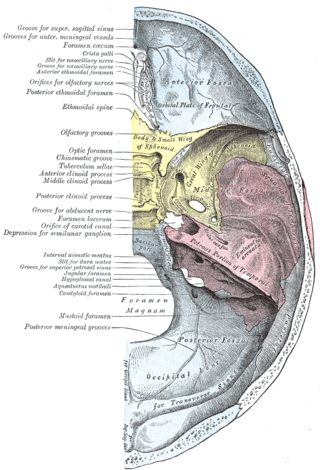
The petrous part of the temporal bone is pyramid-shaped and is wedged in at the base of the skull between the sphenoid and occipital bones. Directed medially, forward, and a little upward, it presents a base, an apex, three surfaces, and three angles, and houses in its interior the components of the inner ear. The petrous portion is among the most basal elements of the skull and forms part of the endocranium. Petrous comes from the Latin word petrosus, meaning "stone-like, hard". It is one of the densest bones in the body. In other mammals, it is a separate bone, the petrosal bone.
The tympanic part of the temporal bone is a curved plate of bone lying below the squamous part of the temporal bone, in front of the mastoid process, and surrounding the external part of the ear canal.

The middle cranial fossa is formed by the sphenoid bones, and the temporal bones. It lodges the temporal lobes, and the pituitary gland. It is deeper than the anterior cranial fossa, is narrow medially and widens laterally to the sides of the skull. It is separated from the posterior cranial fossa by the clivus and the petrous crest.
The petrosquamous sinus is a fetal vein that generally disappears by birth and, when present, runs backward along the junction of the squama and petrous portion of the temporal, and opens into the transverse sinus.

The calvaria is the top part of the skull. It is the superior part of the neurocranium and covers the cranial cavity containing the brain. It forms the main component of the skull roof.
Koerner's septum is an anatomic boundary in the temporal bone formed by the petrosquamous suture between the petrous and squamosal portions of the mastoid air cells, at the anatomic level of the mastoid antrum. Along with the middle ear ossicles, it is usually eroded in middle ear cholesteatomas. Superiorly, this continues as the petrosquamous suture, a normal anatomic structure that can be mistaken for fractures on temporal bone CT. It is surgically important as it may cause difficulty in locating the antrum and the deeper cells and thus may lead to incomplete removal of disease at mastoidectomy.

