| Crista galli | |
|---|---|
 Ethmoid bone from above. | |
 Ethmoid bone from behind. | |
| Details | |
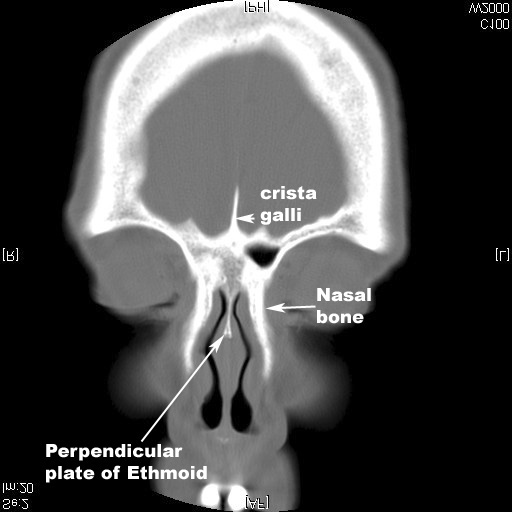
| Part of | Perpendicular plate of the ethmoid bone |
| System | Skeletal |
| Identifiers | |
| Latin | crista galli |
| TA98 | A02.1.07.004 |
| TA2 | 724 |
| FMA | 57442 |
| Anatomical terms of bone | |
The crista galli (Latin: "crest of the rooster") is a wedge-shaped, vertical, midline upward continuation of the perpendicular plate of the ethmoid bone of the skull, [1] projecting above the cribriform plate [2] into the cranial cavity. It serves as an attachment for the membranes surrounding the brain. [1]