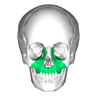
In vertebrates, the maxilla is the upper fixed bone of the jaw formed from the fusion of two maxillary bones. In humans, the upper jaw includes the hard palate in the front of the mouth. The two maxillary bones are fused at the intermaxillary suture, forming the anterior nasal spine. This is similar to the mandible, which is also a fusion of two mandibular bones at the mandibular symphysis. The mandible is the movable part of the jaw.

The occipital bone is a cranial dermal bone and the main bone of the occiput. It is trapezoidal in shape and curved on itself like a shallow dish. The occipital bone overlies the occipital lobes of the cerebrum. At the base of the skull in the occipital bone, there is a large oval opening called the foramen magnum, which allows the passage of the spinal cord.

In the human skull, the frontal bone or sincipital bone is a unpaired bone which consists of two portions. These are the vertically oriented squamous part, and the horizontally oriented orbital part, making up the bony part of the forehead, part of the bony orbital cavity holding the eye, and part of the bony part of the nose respectively. The name comes from the Latin word frons.

The lacrimal bones are two small and fragile bones of the facial skeleton; they are roughly the size of the little fingernail and situated at the front part of the medial wall of the orbit. They each have two surfaces and four borders. Several bony landmarks of the lacrimal bones function in the process of lacrimation. Specifically, the lacrimal bones help form the nasolacrimal canal necessary for tear translocation. A depression on the anterior inferior portion of one bone, the lacrimal fossa, houses the membranous lacrimal sac. Tears, from the lacrimal glands, collect in this sac during excessive lacrimation. The fluid then flows through the nasolacrimal duct and into the nasopharynx. This drainage results in what is commonly referred to a runny nose during excessive crying or tear production. Injury or fracture of the lacrimal bone can result in posttraumatic obstruction of the lacrimal pathways.

The inferior nasal concha is one of the three paired nasal conchae in the nose. It extends horizontally along the lateral wall of the nasal cavity and consists of a lamina of spongy bone, curled upon itself like a scroll,. The inferior nasal conchae are considered a pair of facial bones. As the air passes through the turbinates, the air is churned against these mucosa-lined bones in order to receive warmth, moisture and cleansing. Superior to inferior nasal concha are the middle nasal concha and superior nasal concha which both arise from the ethmoid bone, of the cranial portion of the skull. Hence, these two are considered as a part of the cranial bones.

The greater wing of the sphenoid bone, or alisphenoid, is a bony process of the sphenoid bone, positioned in the skull behind each eye. There is one on each side, extending from the side of the body of the sphenoid and curving upward, laterally, and backward.
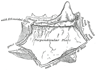
The perpendicular plate of the ethmoid bone is a thin, flattened lamina, polygonal in form, which descends from the under surface of the cribriform plate, and assists in forming the septum of the nose; it is generally deflected a little to one or other side. The anterior border articulates with the spine of the frontal bone and the crest of the nasal bones.
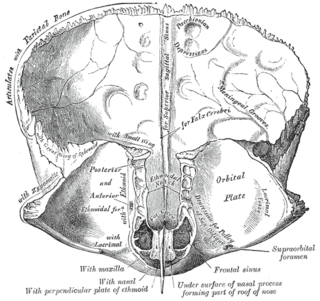
The frontal crest of the frontal bone ends below in a small notch which is converted into a foramen, the foramen cecum, by articulation with the ethmoid.

The squamous part of the frontal bone is the superior portion when viewed in standard anatomical orientation. There are two surfaces of the squamous part of the frontal bone: the external surface, and the internal surface.

The anterior cranial fossa is a depression in the floor of the cranial base which houses the projecting frontal lobes of the brain. It is formed by the orbital plates of the frontal, the cribriform plate of the ethmoid, and the small wings and front part of the body of the sphenoid; it is limited behind by the posterior borders of the small wings of the sphenoid and by the anterior margin of the chiasmatic groove. The lesser wings of the sphenoid separate the anterior and middle fossae.
The sagittal sulcus is a midline groove that runs across the internal surfaces of part of the squamous part of the frontal bone, the parietal bones, and part of the occipital bones. The sagittal sulcus accommodates the superior sagittal sinus. The falx cerebri attaches to the edge of the sagittal sulcus on either side.

The lacrimal sac or lachrymal sac is the upper dilated end of the nasolacrimal duct, and is lodged in a deep groove formed by the lacrimal bone and frontal process of the maxilla. It connects the lacrimal canaliculi, which drain tears from the eye's surface, and the nasolacrimal duct, which conveys this fluid into the nasal cavity. Lacrimal sac occlusion leads to dacryocystitis.
A frontal eminence is either of two rounded elevations on the frontal bone of the skull. They lie about 3 cm above the supraorbital margin on each side of the frontal suture. They are the site of ossification of the frontal bone during embryological development, although may not be the first site.

The posterior lacrimal crest is a vertical bony ridge on the orbital surface of the lacrimal bone. It divides the bone into two parts. It gives origin to the lacrimal part of the orbicularis oculi muscle.
The anterior lacrimal crest is a bony projection on the frontal process of the maxilla. It creates the lateral margin of the lacrimal sac fossa and is continuous with the orbital margin. The medial palpebral ligament is attached to anterior lacrimal crest. It is an important structure to avoid damaging during rhinoplasty.

The medial palpebral ligament is a ligament of the face. It attaches to the frontal process of the maxilla, the lacrimal groove, and the tarsus of each eyelid. It has a superficial (anterior) and a deep (posterior) layer, with many surrounding attachments. It connects the medial canthus of each eyelid to the medial part of the orbit. It is a useful point of fixation during eyelid reconstructive surgery.

The frontal process of the maxilla is a strong plate, which projects upward, medialward, and backward from the maxilla, forming part of the lateral boundary of the nose.

The supraorbital artery is a branch of the ophthalmic artery. It passes anteriorly within the orbit to exit the orbit through the supraorbital foramen or notch alongside the supraorbital nerve, splitting into two terminal branches which go on to form anastomoses with arteries of the head.

The lateral margin of the groove of the frontal process of the maxilla is named the anterior lacrimal crest, and is continuous below with the orbital margin; at its junction with the orbital surface is a small tubercle, the lacrimal tubercle, which serves as a guide to the position of the lacrimal sac.

The medial frontal gyrus is a continuation of the superior frontal gyrus from its most anterior border onto the medial surface of the hemisphere. The medial and superior frontal gyri are two of the frontal gyri of the frontal lobe. The portion on the lateral surface of the hemisphere is usually more or less completely subdivided into an upper and a lower part by an antero-posterior sulcus, the paramedial sulcus, which, however, is frequently interrupted by bridging gyri.

