
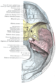
The facial nerve, also known as the seventh cranial nerve, cranial nerve VII, or simply CN VII, is a cranial nerve that emerges from the pons of the brainstem, controls the muscles of facial expression, and functions in the conveyance of taste sensations from the anterior two-thirds of the tongue. The nerve typically travels from the pons through the facial canal in the temporal bone and exits the skull at the stylomastoid foramen. It arises from the brainstem from an area posterior to the cranial nerve VI and anterior to cranial nerve VIII.
Articles related to anatomy include:

The vestibulocochlear nerve or auditory vestibular nerve, also known as the eighth cranial nerve, cranial nerve VIII, or simply CN VIII, is a cranial nerve that transmits sound and equilibrium (balance) information from the inner ear to the brain. Through olivocochlear fibers, it also transmits motor and modulatory information from the superior olivary complex in the brainstem to the cochlea.

The temporal bones are situated at the sides and base of the skull, and lateral to the temporal lobes of the cerebral cortex.

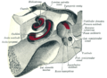
The saccule is a bed of sensory cells in the inner ear. It translates head movements into neural impulses for the brain to interpret. The saccule detects linear accelerations and head tilts in the vertical plane. When the head moves vertically, the sensory cells of the saccule are disturbed and the neurons connected to them begin transmitting impulses to the brain. These impulses travel along the vestibular portion of the eighth cranial nerve to the vestibular nuclei in the brainstem.

The posterior cranial fossa is the part of the cranial cavity located between the foramen magnum, and tentorium cerebelli. It is formed by the sphenoid bones, temporal bones, and occipital bone. It lodges the cerebellum, and parts of the brainstem.

The tympanic cavity is a small cavity surrounding the bones of the middle ear. Within it sit the ossicles, three small bones that transmit vibrations used in the detection of sound.

A jugular foramen is one of the two large foramina (openings) in the base of the skull, located behind the carotid canal. It is formed by the temporal bone and the occipital bone. It allows many structures to pass, including the inferior petrosal sinus, three cranial nerves, the sigmoid sinus, and meningeal arteries.

The greater wing of the sphenoid bone, or alisphenoid, is a bony process of the sphenoid bone, positioned in the skull behind each eye. There is one on each side, extending from the side of the body of the sphenoid and curving upward, laterally, and backward.

The labyrinthine artery is a branch of either the anterior inferior cerebellar artery or the basilar artery. It accompanies the vestibulocochlear nerve through the internal acoustic meatus. It supplies blood to the internal ear.

The carotid canal is a passage in the petrous part of the temporal bone of the skull through which the internal carotid artery and its internal carotid (nervous) plexus pass from the neck into the cranial cavity.

The squamous part of temporal bone, or temporal squama, forms the front and upper part of the temporal bone, and is scale-like, thin, and translucent.

The petrous part of the temporal bone is pyramid-shaped and is wedged in at the base of the skull between the sphenoid and occipital bones. Directed medially, forward, and a little upward, it presents a base, an apex, three surfaces, and three angles, and houses in its interior, the components of the inner ear. The petrous portion is among the most basal elements of the skull and forms part of the endocranium. Petrous comes from the Latin word petrosus, meaning "stone-like, hard". It is one of the densest bones in the body. In other mammals, it is a separate bone, the petrosal bone.

The facial canal is a Z-shaped canal in the temporal bone of the skull. It extends between the internal acoustic meatus and stylomastoid foramen. It transmits the facial nerve.

The middle cranial fossa is formed by the sphenoid bones, and the temporal bones. It lodges the temporal lobes, and the pituitary gland. It is deeper than the anterior cranial fossa, is narrow medially and widens laterally to the sides of the skull. It is separated from the posterior cranial fossa by the clivus and the petrous crest.

The subarcuate fossa is a shallow depression upon the internal surface of the petrous part of the temporal bone forming the wall of the posterior cranial fossa. The fossa accommodates the flocculus of the cerebellum. It is situated lateral/posterior to the internal auditory meatus.

The vestibule is the central part of the bony labyrinth in the inner ear, and is situated medial to the eardrum, behind the cochlea, and in front of the three semicircular canals.

The base of skull, also known as the cranial base or the cranial floor, is the most inferior area of the skull. It is composed of the endocranium and the lower parts of the calvaria.

The following outline is provided as an overview of and topical guide to human anatomy:


