Articles related to anatomy include:

The occipital bone is a cranial dermal bone and the main bone of the occiput. It is trapezoidal in shape and curved on itself like a shallow dish. The occipital bone overlies the occipital lobes of the cerebrum. At the base of the skull in the occipital bone, there is a large oval opening called the foramen magnum, which allows the passage of the spinal cord.

The frontal bone is a bone in the human skull. The bone consists of two portions. These are the vertically oriented squamous part, and the horizontally oriented orbital part, making up the bony part of the forehead, part of the bony orbital cavity holding the eye, and part of the bony part of the nose respectively. The name comes from the Latin word frons.

The parietal bones are two bones in the skull which, when joined at a fibrous joint, form the sides and roof of the cranium. In humans, each bone is roughly quadrilateral in form, and has two surfaces, four borders, and four angles. It is named from the Latin paries (-ietis), wall.

The great cerebral vein is one of the large blood vessels in the skull draining the cerebrum of the brain. It is also known as the vein of Galen, named for its discoverer, the Greek physician Galen.

In anatomy, the epidural space is the potential space between the dura mater and vertebrae (spine).

The falx cerebri is a large, crescent-shaped fold of dura mater that descends vertically into the longitudinal fissure between the cerebral hemispheres of the human brain, separating the two hemispheres and supporting dural sinuses that provide venous and CSF drainage to the brain. It is attached to the crista galli anteriorly, and blends with the tentorium cerebelli posteriorly.

The cerebellar tentorium or tentorium cerebelli is an extension of the dura mater between the inferior aspect of the occipital lobes and the superior aspect of the cerebellum. The free border of the tentorium gives passage to the midbrain.

The ophthalmic nerve (CN V1) is a sensory nerve of the head. It is one of three divisions of the trigeminal nerve (CN V), a cranial nerve. It has three major branches which provide sensory innervation to the eye, and the skin of the upper face and anterior scalp, as well as other structures of the head.

The frontal sinuses are one of the four pairs of paranasal sinuses that are situated behind the brow ridges. Sinuses are mucosa-lined airspaces within the bones of the face and skull. Each opens into the anterior part of the corresponding middle nasal meatus of the nose through the frontonasal duct which traverses the anterior part of the labyrinth of the ethmoid. These structures then open into the semilunar hiatus in the middle meatus.

The straight sinus, also known as tentorial sinus or the sinus rectus, is an area within the skull beneath the brain. It receives blood from the inferior sagittal sinus and the great cerebral vein, and drains into the confluence of sinuses.
The superior sagittal sinus, within the human head, is an unpaired area along the attached margin of the falx cerebri. It allows blood to drain from the lateral aspects of anterior cerebral hemispheres to the confluence of sinuses. Cerebrospinal fluid drains through arachnoid granulations into the superior sagittal sinus and is returned to venous circulation.
The inferior sagittal sinus, within the human head, is an area beneath the brain which allows blood to drain outwards posteriorly from the center of the head. It drains to the straight sinus, which connects to the transverse sinuses. See diagram : labeled in the brain as "SIN. SAGITTALIS INF.".
The inferior petrosal sinuses are two small sinuses situated on the inferior border of the petrous part of the temporal bone, one on each side. Each inferior petrosal sinus drains the cavernous sinus into the internal jugular vein.

The anterior ethmoidal artery is a branch of the ophthalmic artery in the orbit. It exits the orbit through the anterior ethmoidal foramen alongside the anterior ethmoidal nerve. It contributes blood supply to the ethmoid sinuses, frontal sinuses, the dura mater, lateral nasal wall, and nasal septum. It issues a meningeal branch, and nasal branches.

The squamous part of occipital bone is situated above and behind the foramen magnum, and is curved from above downward and from side to side.

The squamous part of the frontal bone is the superior portion when viewed in standard anatomical orientation. There are two surfaces of the squamous part of the frontal bone: the external surface, and the internal surface.

The anterior cranial fossa is a depression in the floor of the cranial base which houses the projecting frontal lobes of the brain. It is formed by the orbital plates of the frontal, the cribriform plate of the ethmoid, and the small wings and front part of the body of the sphenoid; it is limited behind by the posterior borders of the small wings of the sphenoid and by the anterior margin of the chiasmatic groove. The lesser wings of the sphenoid separate the anterior and middle fossae.
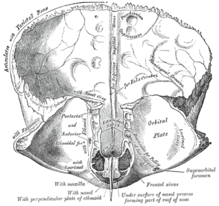
The frontal crest is a ridge on the internal surface of the squamous part of the frontal bone formed by the inferior convergence of the two edges of the sagittal sulcus. The frontal crest gives attachment to the falx cerebri.

The calvaria is the top part of the skull. It is the superior part of the neurocranium and covers the cranial cavity containing the brain. It forms the main component of the skull roof.
