
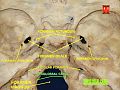
The hypoglossal nerve, also known as the twelfth cranial nerve, cranial nerve XII, or simply CN XII, is a cranial nerve that innervates all the extrinsic and intrinsic muscles of the tongue except for the palatoglossus, which is innervated by the vagus nerve. CN XII is a nerve with a sole motor function. The nerve arises from the hypoglossal nucleus in the medulla as a number of small rootlets, pass through the hypoglossal canal and down through the neck, and eventually passes up again over the tongue muscles it supplies into the tongue.

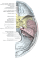
The foramen magnum is a large, oval-shaped opening in the occipital bone of the skull. It is one of the several oval or circular openings (foramina) in the base of the skull. The spinal cord, an extension of the medulla oblongata, passes through the foramen magnum as it exits the cranial cavity. Apart from the transmission of the medulla oblongata and its membranes, the foramen magnum transmits the vertebral arteries, the anterior and posterior spinal arteries, the tectorial membranes and alar ligaments. It also transmits the accessory nerve into the skull.

The occipital bone is a cranial dermal bone and the main bone of the occiput. It is trapezoidal in shape and curved on itself like a shallow dish. The occipital bone overlies the occipital lobes of the cerebrum. At the base of the skull in the occipital bone, there is a large oval opening called the foramen magnum, which allows the passage of the spinal cord.

The temporal bones are situated at the sides and base of the skull, and lateral to the temporal lobes of the cerebral cortex.
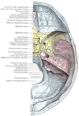
The foramen lacerum is a triangular hole in the base of skull. It is located between the sphenoid bone, the apex of the petrous part of the temporal bone, and the basilar part of the occipital bone.

The posterior cranial fossa is the part of the cranial cavity located between the foramen magnum, and tentorium cerebelli. It is formed by the sphenoid bones, temporal bones, and occipital bone. It lodges the cerebellum, and parts of the brainstem.
The jugular fossa is a deep depression in the inferior part of the temporal bone at the base of the skull. It lodges the bulb of the internal jugular vein.

A jugular foramen is one of the two large foramina (openings) in the base of the skull, located behind the carotid canal. It is formed by the temporal bone and the occipital bone. It allows many structures to pass, including the inferior petrosal sinus, three cranial nerves, the sigmoid sinus, and meningeal arteries.

The greater wing of the sphenoid bone, or alisphenoid, is a bony process of the sphenoid bone, positioned in the skull behind each eye. There is one on each side, extending from the side of the body of the sphenoid and curving upward, laterally, and backward.

The carotid canal is a passage in the petrous part of the temporal bone of the skull through which the internal carotid artery and its internal carotid (nervous) plexus pass from the neck into the cranial cavity.

The mastoid part of the temporal bone is the posterior (back) part of the temporal bone, one of the bones of the skull. Its rough surface gives attachment to various muscles and it has openings for blood vessels. From its borders, the mastoid part articulates with two other bones.

The petrous part of the temporal bone is pyramid-shaped and is wedged in at the base of the skull between the sphenoid and occipital bones. Directed medially, forward, and a little upward, it presents a base, an apex, three surfaces, and three angles, and houses in its interior, the components of the inner ear. The petrous portion is among the most basal elements of the skull and forms part of the endocranium. Petrous comes from the Latin word petrosus, meaning "stone-like, hard". It is one of the densest bones in the body. In other mammals, it is a separate bone, the petrosal bone.

The temporal styloid process is a slender bony process of the temporal bone extending downward and forward from the undersurface of the temporal bone just below the ear. The styloid process gives attachments to several muscles, and ligaments.

The lateral parts of the occipital bone are situated at the sides of the foramen magnum; on their under surfaces are the condyles for articulation with the superior facets of the atlas.

The occipitomastoid suture or occipitotemporal suture is the cranial suture between the occipital bone and the mastoid portion of the temporal bone.

The mastoid foramen is a hole in the posterior border of the temporal bone. It transmits an emissary vein between the sigmoid sinus and the suboccipital venous plexus, and a small branch of the occipital artery, the posterior meningeal artery to the dura mater.
The jugular process is a quadrilateral or triangular bony plate projecting lateralward from the posterior half of the occipital condyle; it is a part of the lateral part of the occipital bone.
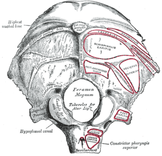
The condylar canal is a canal in the condyloid fossa of the lateral parts of occipital bone behind the occipital condyle. Resection of the rectus capitis posterior major and minor muscles reveals the bony recess leading to the condylar canal, which is situated posterior and lateral to the occipital condyle. It is immediately superior to the extradural vertebral artery, which makes a loop above the posterior C1 ring to enter the foramen magnum. The anteriomedial wall of the condylar canal thickens to join the foramen magnum rim and connect to the occipital condyle.
The jugular tubercle is a rounded prominence/oval elevation upon the superior surface of the occipital condyle at the junction of the basilar part and lateral part of the occipital bone, just medial to the jugular foramen on either side of the foramen magnum.

The occipital condyles are undersurface protuberances of the occipital bone in vertebrates, which function in articulation with the superior facets of the atlas vertebra.

