Middle cranial fossa

Last updated
Middle cranial fossa
Middle cranial fossa boundaries.svg
Superior view of the skull base. Middle cranial fossa shown in green.

1: Sphenoidal limbus (anterior margin of the chiasmatic groove)
2: Posterior borders of the lesser wings of the sphenoid
3: Dorsum sellae of the sphenoid bone

4: Superior borders of the

Contents

petrous part of the temporal bone
Gray193 - Cranial fossae.png
Base of the skull. Upper surface. (Middle cranial fossa is the centermost of the three indentations, in pink and yellow.)
Details
Identifiers
Latin fossa cranii media
MeSH D035301
TA98 A02.1.00.049
TA2 452
FMA 54369
Anatomical terminology

The middle cranial fossa is formed by the sphenoid bones, and the temporal bones. It lodges the temporal lobes, and the pituitary gland. [1] [2] It is deeper than the anterior cranial fossa, is narrow medially and widens laterally to the sides of the skull. It is separated from the posterior cranial fossa by the clivus and the petrous crest.

It is bounded in front by the posterior margins of the lesser wings of the sphenoid bone, the anterior clinoid processes, and the ridge forming the anterior margin of the chiasmatic groove; behind, by the superior angles of the petrous portions of the temporal bones and the dorsum sellae; laterally by the temporal squamae, sphenoidal angles of the parietals, and greater wings of the sphenoid. It is traversed by the squamosal, sphenoparietal, sphenosquamosal, and sphenopetrosal sutures.

Anatomy

Features

Middle cranial fossa surgical anatomy as demonstrated in a right cadaver temporal bone by Dr Jack M Kartush - view from above. Middle cranial fossa surgical anatomy - J M Kartush.png
Middle cranial fossa surgical anatomy as demonstrated in a right cadaver temporal bone by Dr Jack M Kartush - view from above.

Middle part

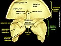
The middle part of the fossa presents, in front, the chiasmatic groove and tuberculum sellae; the chiasmatic groove ends on either side at the optic foramen, which transmits the optic nerve and ophthalmic artery to the orbital cavity.

Behind the optic foramen the anterior clinoid process is directed backward and medialward and gives attachment to the cerebellar tentorium .

Behind the tuberculum sellae is a deep depression, the sella turcica, containing the fossa hypophyseos, which lodges the pituitary gland, and presents on its anterior wall the middle clinoid processes.

The sella turcica is bounded posteriorly by a quadrilateral plate of bone, the dorsum sellae, the upper angles of which are surmounted by the posterior clinoid processes: these afford attachment to the cerebellar tentorium, and below each is a notch for the abducent nerve.

On either side of the sella turcica is the carotid groove, which is broad, shallow, and curved somewhat like the italic letter f.

It begins behind at the foramen lacerum, and ends on the medial side of the anterior clinoid process, where it is sometimes converted into a foramen (carotico-clinoid) by the union of the anterior with the middle clinoid process; posteriorly, it is bounded laterally by the lingula.

This groove lodges the cavernous sinus and the internal carotid artery, the latter being surrounded by a plexus of sympathetic nerves.

Lateral parts

The lateral parts of the middle fossa are of considerable depth, and support the temporal lobes of the brain.

They are marked by depressions for the brain convolutions and traversed by furrows for the anterior and posterior branches of the middle meningeal vessels.

These furrows begin near the foramen spinosum, and the anterior runs forward and upward to the sphenoidal angle of the parietal, where it is sometimes converted into a bony canal; the posterior runs lateralward and backward across the temporal squama and passes on to the parietal near the middle of its lower border.

The following apertures are also to be seen.

In front is the superior orbital fissure, bounded above by the small wing, below, by the great wing, and medially, by the body of the sphenoid; it is usually completed laterally by the orbital plate of the frontal bone.

It transmits to the orbital cavity the oculomotor, the trochlear, the ophthalmic division of the trigeminal, and the abducent nerves, some filaments from the cavernous plexus of the sympathetic, and the orbital branch of the middle meningeal artery; and from the orbital cavity a recurrent branch from the lacrimal artery to the dura mater, and the ophthalmic veins.

Behind the medial end of the superior orbital fissure is the foramen rotundum, for the passage of the maxillary nerve.

Behind and lateral to the foramen rotundum is the foramen ovale, which transmits the mandibular nerve, the accessory meningeal artery, and the lesser superficial petrosal nerve.

Medial to the foramen ovale is the foramen Vesalii, which varies in size in different individuals, and is often absent; when present, it opens below at the lateral side of the scaphoid fossa, and transmits a small vein.

Lateral to the foramen ovale is the foramen spinosum, for the passage of the middle meningeal vessels, and a recurrent branch from the mandibular nerve.

Medial to the foramen ovale is the foramen lacerum; in the fresh state the lower part of this aperture is filled up by a layer of fibrocartilage, while its upper and inner parts transmit the internal carotid artery surrounded by a plexus of sympathetic nerves.

The nerve of the pterygoid canal and a meningeal branch from the ascending pharyngeal artery pierce the layer of fibrocartilage.

On the anterior surface of the petrous portion of the temporal bone are seen the eminence caused by the projection of the superior semicircular canal; in front of and a little lateral to this a depression corresponding to the roof of the tympanic cavity; the groove leading to the hiatus of the facial canal, for the transmission of the greater superficial petrosal nerve and the petrosal branch of the middle meningeal artery; beneath it, the smaller groove, for the passage of the lesser superficial petrosal nerve; and, near the apex of the bone, the depression for the semilunar ganglion and the orifice of the carotid canal.

Clinical significance

A middle fossa craniotomy is one means to surgically remove acoustic neuromas (vestibular schwannoma) growing within the internal auditory canal of the temporal bone.

See also

Related Research Articles

Articles related to anatomy include:

<span class="mw-page-title-main">Sphenoid bone</span> Bone at the front of the skull

The sphenoid bone is an unpaired bone of the neurocranium. It is situated in the middle of the skull towards the front, in front of the basilar part of the occipital bone. The sphenoid bone is one of the seven bones that articulate to form the orbit. Its shape somewhat resembles that of a butterfly or bat with its wings extended.

<span class="mw-page-title-main">Internal carotid artery</span> Artery of the human brain

The internal carotid artery is an artery in the neck which supplies the anterior circulation of the brain.

<span class="mw-page-title-main">Orbit (anatomy)</span> Cavity or socket of the skull in which the eye and its appendages are situated

In anatomy, the orbit is the cavity or socket/hole of the skull in which the eye and its appendages are situated. "Orbit" can refer to the bony socket, or it can also be used to imply the contents. In the adult human, the volume of the orbit is 30 millilitres, of which the eye occupies 6.5 ml. The orbital contents comprise the eye, the orbital and retrobulbar fascia, extraocular muscles, cranial nerves II, III, IV, V, and VI, blood vessels, fat, the lacrimal gland with its sac and duct, the eyelids, medial and lateral palpebral ligaments, cheek ligaments, the suspensory ligament, septum, ciliary ganglion and short ciliary nerves.

<span class="mw-page-title-main">Middle meningeal artery</span> Artery supplying the meninges

The middle meningeal artery is typically the third branch of the first portion of the maxillary artery. After branching off the maxillary artery in the infratemporal fossa, it runs through the foramen spinosum to supply the dura mater and the calvaria. The middle meningeal artery is the largest of the three (paired) arteries that supply the meninges, the others being the anterior meningeal artery and the posterior meningeal artery.

<span class="mw-page-title-main">Posterior cranial fossa</span> Area of the cranium containing the brainstem and cerebellum

The posterior cranial fossa is the part of the cranial cavity located between the foramen magnum, and tentorium cerebelli. It is formed by the sphenoid bones, temporal bones, and occipital bone. It lodges the cerebellum, and parts of the brainstem.

<span class="mw-page-title-main">Tympanic cavity</span> Small cavity surrounding the bones of the middle ear

The tympanic cavity is a small cavity surrounding the bones of the middle ear. Within it sit the ossicles, three small bones that transmit vibrations used in the detection of sound.

<span class="mw-page-title-main">Greater petrosal nerve</span>

The greater petrosal nerve is a nerve of the head mainly containing pre-ganglionic parasympathetic fibres which ultimately synapse in the pterygopalatine ganglion. It branches from the facial nerve and is derived from the parasympathetic part of the nervus intermedius component of CN VII, with its cell bodies located in the superior salivary nucleus. In the connective tissue substance of the foramen lacerum, the greater petrosal nerve unites with the (sympathetic) deep petrosal nerve to form the nerve of the pterygoid canal which proceeds to the pterygopalatine ganglion.

<span class="mw-page-title-main">Cavernous sinus</span> Sinus in the human head

The cavernous sinus within the human head is one of the dural venous sinuses creating a cavity called the lateral sellar compartment bordered by the temporal bone of the skull and the sphenoid bone, lateral to the sella turcica.

<span class="mw-page-title-main">Foramen spinosum</span> Hole in the sphenoid bone of the skull

The foramen spinosum is a small open hole in the greater wing of the sphenoid bone that gives passage to the middle meningeal artery and vein, and the meningeal branch of the mandibular nerve.

<span class="mw-page-title-main">Greater wing of sphenoid bone</span> Large part of the skull front behind the eye socket

The greater wing of the sphenoid bone, or alisphenoid, is a bony process of the sphenoid bone; there is one on each side, extending from the side of the body of the sphenoid and curving upward, laterally, and backward.

<span class="mw-page-title-main">Lesser wing of sphenoid bone</span>

The lesser wings of the sphenoid or orbito-sphenoids are two thin triangular plates, which arise from the upper and anterior parts of the body, and, projecting lateralward, end in sharp points [Fig. 1].

<span class="mw-page-title-main">Optic canal</span>

The optic foramen is the opening to the optic canal. The canal is located in the sphenoid bone; it is bounded medially by the body of the sphenoid and laterally by the lesser wing of the sphenoid.

<span class="mw-page-title-main">Carotid canal</span> Hole in the temporal bone in the skull for the internal carotid artery to enter the skull

The carotid canal is a passage in the petrous part of the temporal bone of the skull through which the internal carotid artery and its internal carotid (nervous) plexus pass from the neck into the cranial cavity.

<span class="mw-page-title-main">Petrous part of the temporal bone</span> Feature at the base of the human skull

The petrous part of the temporal bone is pyramid-shaped and is wedged in at the base of the skull between the sphenoid and occipital bones. Directed medially, forward, and a little upward, it presents a base, an apex, three surfaces, and three angles, and houses in its interior, the components of the inner ear. The petrous portion is among the most basal elements of the skull and forms part of the endocranium. Petrous comes from the Latin word petrosus, meaning "stone-like, hard". It is one of the densest bones in the body. In other mammals, it is a separate bone, the petrosal bone.

<span class="mw-page-title-main">Anterior cranial fossa</span>

The anterior cranial fossa is a depression in the floor of the cranial base which houses the projecting frontal lobes of the brain. It is formed by the orbital plates of the frontal, the cribriform plate of the ethmoid, and the small wings and front part of the body of the sphenoid; it is limited behind by the posterior borders of the small wings of the sphenoid and by the anterior margin of the chiasmatic groove. The lesser wings of the sphenoid separate the anterior and middle fossae.

<span class="mw-page-title-main">Infratemporal fossa</span> Cavity that is part of the skull

The infratemporal fossa is an irregularly shaped cavity that is a part of the skull. It is situated below and medial to the zygomatic arch. It is not fully enclosed by bone in all directions. It contains superficial muscles, including the lower part of the temporalis muscle, the lateral pterygoid muscle, and the medial pterygoid muscle. It also contains important blood vessels such as the middle meningeal artery, the pterygoid plexus, and the retromandibular vein, and nerves such as the mandibular nerve (CN V3) and its branches.

<span class="mw-page-title-main">Body of sphenoid bone</span>

The body of the sphenoid bone, more or less cubical in shape, is hollowed out in its interior to form two large cavities, the sphenoidal sinuses, which are separated from each other by a septum.

<span class="mw-page-title-main">Base of skull</span> Inferior area of the skull, composed of the endocranium and lower parts of the skull roof

The base of skull, also known as the cranial base or the cranial floor, is the most inferior area of the skull. It is composed of the endocranium and the lower parts of the calvaria.

<span class="mw-page-title-main">Outline of human anatomy</span> Overview of and topical guide to human anatomy

The following outline is provided as an overview of and topical guide to human anatomy:

References

PD-icon.svgThis article incorporates text in the public domain from page 190 of the 20th edition of Gray's Anatomy (1918)

  1. Standring, Susan (2020). Gray's Anatomy: The Anatomical Basis of Clinical Practice (42nd ed.). [New York]. ISBN   978-0-7020-7707-4. OCLC   1201341621.{{cite book}}: CS1 maint: location missing publisher (link)
  2. Mancall, Elliott L.; Brock, David G., eds. (2011). "Cranial Fossae". Gray's Clinical Anatomy. Elsevier Health Sciences. p. 154. ISBN   9781437735802.

Additional images