Fontanelle

Last updated

Fontanelle
Gray197.png
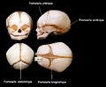
The skull at birth, showing the anterior and posterior fontanelles
Gray198.png
The skull at birth, showing the lateral fontanelles
Details
Identifiers
Latin fonticuli cranii
MeSH D055762
TA98 A02.1.00.027
TA2 431
FMA 75437
Anatomical terminology

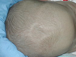
A fontanelle (or fontanel) (colloquially, soft spot) is an anatomical feature of the infant human skull comprising soft membranous gaps (sutures) between the cranial bones that make up the calvaria of a fetus or an infant. [1] Fontanelles allow for stretching and deformation of the neurocranium both during birth and later as the brain expands faster than the surrounding bone can grow. [2] Premature complete ossification of the sutures is called craniosynostosis.

Contents

After infancy, the anterior fontanelle is known as the bregma.

Structure

An infant's skull consists of five main bones: two frontal bones, two parietal bones, and one occipital bone. These are joined by fibrous sutures, which allow movement that facilitates childbirth and brain growth.

During birth, fontanelles enable the bony plates of the skull to flex, allowing the child's head to pass through the birth canal. The ossification of the bones of the skull causes the anterior fontanelle to close over by 9 to 18 months. [3] The sphenoidal and posterior fontanelles close during the first few months of life. The closures eventually form the sutures of the neurocranium. Other than the anterior and posterior fontanelles, the mastoid fontanelle and the sphenoidal fontanelle are also significant.

Closure

In humans, the sequence of fontanelle closure is as follows: [2] [4]

  1. The posterior fontanelle generally closes 2 to 3 months after birth;
  2. The sphenoidal fontanelle is the next to close around 6 months after birth;
  3. The mastoid fontanelle closes next from 6 to 18 months after birth; and
  4. The anterior fontanelle is generally the last to close between 12 and 18 months.

Clinical significance

The fontanelle may pulsate, and although the precise cause of this is not known, it is normal and seems to echo the heartbeat, perhaps via the arterial pulse within the brain vasculature, or in the meninges. This pulsating action is how the soft spot got its name – fontanelle is borrowed from the old French word fontenele, which is a diminutive of fontaine, meaning "spring". It is assumed that the term spring is used because of the analogy of the dent in a rock or earth where a spring arises. [5]

Parents may worry that their infant may be more prone to injury at the fontanelles. In fact, although they may colloquially be called "soft-spots", the membrane covering the fontanelles is extremely tough and difficult to penetrate. [6]

Fontanelles allow the infant brain to be imaged using ultrasonography. Once they are closed, most of the brain is inaccessible to ultrasound imaging, because the bony skull presents an acoustic barrier. [6]

Disorders

Bulging

A very tense or bulging anterior fontanelle indicates raised intracranial pressure. Increased cranial pressure in infants may cause the fontanelles to bulge or the head to begin to enlarge abnormally. [7] It can occur due to: [4]

Sunken

A sunken (also called "depressed") fontanelle indicates dehydration or malnutrition. [9]

Anterior fontanelle of a 1-month-old infant Human anterior fontanelle 1 month dscn1449.jpg
Anterior fontanelle of a 1-month-old infant

Enlarged

The fontanelles may be enlarged, may be slow to close, or may never close, most commonly due to causes like: [10]

Rarer causes include: [10]

Third

Sometimes there is a third bigger fontanelle other than posterior and anterior ones in a newborn. In one study, the frequency of third fontanelles in an unselected population of newborn infants was 6.3%. It is very common in Down syndrome and some congenital infections. If present, the physician should rule out serious conditions associated with the third fontanelle. [11]

Other animals

Primates

In apes the fontanelles fuse soon after birth. In chimpanzees the anterior fontanelle is fully closed by 3 months of age. [2]

Dogs

One of the more serious problems that can affect canines is known as an "open fontanelle", which occurs when the skull bones at the top of the head fail to close. The problem is often found in conjunction with hydrocephalus, which is a condition in which too much fluid is found within and around the brain, placing pressure on the brain and surrounding tissues. Often the head will appear dome-shaped, and the open fontanelle is noticeable as a "soft spot" on the top of the dog's head. The fluid-filled spaces within the brain, known as ventricles, also become swollen. The increased pressure damages or prevents the development of brain tissue. [12]

Not all open fontanelles are connected with hydrocephalus. In many young dogs the skull bones are not fused at birth, but instead will close slowly over a three- to six-month period. Occasionally these bones fail to close, but the dog is still healthy. In these cases, however, the dog's owners need to be very careful, since any injury or bumps to the animal's head could cause significant brain damage, as well as conditions like epilepsy.

An open fontanelle, known as a "molera", is a recognized feature of the Chihuahua breed. The American Kennel Club breed standard states that the skull of the Chihuahua should be domed, with or without the molera being present. [13] However, the Fédération Cynologique Internationale (FCI) standard for the Chihuahua lists an open fontanelle as a disqualification. [14]

Additional images

Related Research Articles

<span class="mw-page-title-main">Skull</span> Bony structure that forms the head in vertebrates

The skull is a bone protective cavity for the brain. The skull is composed of four types of bone i.e., cranial bones, facial bones, ear ossicles and hyoid bone, however two parts are more prominent: the cranium and the mandible. In humans, these two parts are the neurocranium (braincase) and the viscerocranium that includes the mandible as its largest bone. The skull forms the anterior-most portion of the skeleton and is a product of cephalisation—housing the brain, and several sensory structures such as the eyes, ears, nose, and mouth. In humans, these sensory structures are part of the facial skeleton.

<span class="mw-page-title-main">Sphenoid bone</span> Bone at the front of the skull

The sphenoid bone is an unpaired bone of the neurocranium. It is situated in the middle of the skull towards the front, in front of the basilar part of the occipital bone. The sphenoid bone is one of the seven bones that articulate to form the orbit. Its shape somewhat resembles that of a butterfly or bat with its wings extended.

<span class="mw-page-title-main">Occipital bone</span> Saucer-shaped membrane bone situated at the back and lower part of the cranium

The occipital bone is a cranial dermal bone and the main bone of the occiput. It is trapezoidal in shape and curved on itself like a shallow dish. The occipital bone overlies the occipital lobes of the cerebrum. At the base of the skull in the occipital bone, there is a large oval opening called the foramen magnum, which allows the passage of the spinal cord.

<span class="mw-page-title-main">Parietal bone</span> Bone in the human skull which, when joined together, forms the sides and roof of the cranium

The parietal bones are two bones in the skull which, when joined at a fibrous joint, form the sides and roof of the cranium. In humans, each bone is roughly quadrilateral in form, and has two surfaces, four borders, and four angles. It is named from the Latin paries (-ietis), wall.

<span class="mw-page-title-main">Temporal bone</span> Sides and base of skull, connecting to the jaw and occipital bone

The temporal bones are situated at the sides and base of the skull, and lateral to the temporal lobes of the cerebral cortex.

A molera is a "soft spot" on the top of a Chihuahua's skull; it is the equivalent to the bregmatic or anterior fontanelle in human babies, but unlike most mammals Chihuahua's fontanelle persist into maturity. Historically it has been very common amongst Chihuahuas and was regarded as a mark of purity for this miniature dog breed. It is still mentioned in many Chihuahua breed standards, however, it is considered a fault in European countries because of concern that this might reflect underlying malformations such as hydrocephalus and ventriculomegaly, Chiari-like malformation and syringomyelia. Fontanelles are fibrous, membrane-covered gaps that lie between the skull bones and at the intersection of the cranial sutures. The cranial sutures are the junctions between cranial bones. The fontanelles serve as the major sites of bone expansion during post-natal skull growth which accommodates the enlarging brain. The Chihuahua likely has a molera because of neuroparenchymal disproportion i.e. a proportionally big brain for the skull. This is likely because there is premature closure of the skull base cranial sutures. To accommodate the developing brain there is increased growth of the skull bone in a parallel plane giving the dog a characterised domed or "apple-headed" appearance.

<span class="mw-page-title-main">Crouzon syndrome</span> Genetic disorder of the skull and face

Crouzon syndrome is an autosomal dominant genetic disorder known as a branchial arch syndrome. Specifically, this syndrome affects the first branchial arch, which is the precursor of the maxilla and mandible. Since the branchial arches are important developmental features in a growing embryo, disturbances in their development create lasting and widespread effects.

<span class="mw-page-title-main">Craniosynostosis</span> Premature fusion of bones in the skull

Craniosynostosis is a condition in which one or more of the fibrous sutures in a young infant's skull prematurely fuses by turning into bone (ossification), thereby changing the growth pattern of the skull. Because the skull cannot expand perpendicular to the fused suture, it compensates by growing more in the direction parallel to the closed sutures. Sometimes the resulting growth pattern provides the necessary space for the growing brain, but results in an abnormal head shape and abnormal facial features. In cases in which the compensation does not effectively provide enough space for the growing brain, craniosynostosis results in increased intracranial pressure leading possibly to visual impairment, sleeping impairment, eating difficulties, or an impairment of mental development combined with a significant reduction in IQ.

<span class="mw-page-title-main">Frontal suture</span> Midline joint in the frontal bone of the forehead

The frontal suture is a fibrous joint that divides the two halves of the frontal bone of the skull in infants and children. Typically, it completely fuses between three and nine months of age, with the two halves of the frontal bone being fused together. It is also called the metopic suture, although this term may also refer specifically to a persistent frontal suture.

<span class="mw-page-title-main">Lambdoid suture</span> Connective tissue between the parietal bones and the occipital bone of the skull

The lambdoid suture is a dense, fibrous connective tissue joint on the posterior aspect of the skull that connects the parietal bones with the occipital bone. It is continuous with the occipitomastoid suture.

<span class="mw-page-title-main">Cranial cavity</span> Space inside the skull formed by eight cranial bones known as the neurocranium

The cranial cavity, also known as intracranial space, is the space within the skull that accommodates the brain. The skull minus the mandible is called the cranium. The cavity is formed by eight cranial bones known as the neurocranium that in humans includes the skull cap and forms the protective case around the brain. The remainder of the skull is called the facial skeleton. Meninges are protective membranes that surround the brain to minimize damage to the brain in the case of head trauma. Meningitis is the inflammation of meninges caused by bacterial or viral infections.

<span class="mw-page-title-main">Bregma</span> Meeting point of the coronal suture and the sagittal suture of the skull

The bregma is the anatomical point on the skull at which the coronal suture is intersected perpendicularly by the sagittal suture.

<span class="mw-page-title-main">Mastoid part of the temporal bone</span> Back part of the sides of the skull base

The mastoid part of the temporal bone is the posterior (back) part of the temporal bone, one of the bones of the skull. Its rough surface gives attachment to various muscles and it has openings for blood vessels. From its borders, the mastoid part articulates with two other bones.

<span class="mw-page-title-main">Fibrous joint</span> Fixed joints between bones held together by dense, fibrous tissue

In anatomy, fibrous joints are joints connected by fibrous tissue, consisting mainly of collagen. These are fixed joints where bones are united by a layer of white fibrous tissue of varying thickness. In the skull the joints between the bones are called sutures. Such immovable joints are also referred to as synarthroses.

<span class="mw-page-title-main">Calvaria (skull)</span> Top part of the skull

The calvaria is the top part of the skull. It is the superior part of the neurocranium and covers the cranial cavity containing the brain. It forms the main component of the skull roof.

<span class="mw-page-title-main">Base of skull</span> Inferior area of the skull, composed of the endocranium and lower parts of the skull roof

The base of skull, also known as the cranial base or the cranial floor, is the most inferior area of the skull. It is composed of the endocranium and the lower parts of the calvaria.

<span class="mw-page-title-main">Outline of human anatomy</span> Overview of and topical guide to human anatomy

The following outline is provided as an overview of and topical guide to human anatomy:

The fetal head, from an obstetrical viewpoint, and in particular its size, is important because an essential feature of labor is the adaptation between the fetal head and the maternal bony pelvis. Only a comparatively small part of the head at term is represented by the face. The rest of the head is composed of the firm skull, which is made up of two frontal, two parietal, and two temporal bones, along with the upper portion of the occipital bone and the wings of the sphenoid.

<span class="mw-page-title-main">McGillivray syndrome</span> Medical condition

McGillivray syndrome is a rare syndrome characterized mainly by heart defects, skull and facial abnormalities and ambiguous genitalia. The symptoms of this syndrome are ventricular septal defect, patent ductus arteriosus, small jaw, undescended testes, and webbed fingers. Beside to these symptoms there are more symptoms which is related with bone structure and misshape.

Cranial ultrasound is a technique for scanning the brain using high-frequency sound waves. It is used almost exclusively in babies because their fontanelle provides an "acoustic window". A different form of ultrasound-based brain scanning, transcranial Doppler, can be used in any age group. This uses Doppler ultrasound to assess blood flow through the major arteries in the brain, and can scan through bone. It is not usual for this technique to be referred to simply as "cranial ultrasound". Additionally, cranial ultrasound can be used for intra-operative imaging in adults undergoing neurosurgery once the skull has been opened, for example to help identify the margins of a tumour.

References

  1. "fontanelle". TheFreeDictionary. Retrieved 24 April 2013.
  2. 1 2 3 Beasley, Melanie. "Age of Closure of Fontanelles / Sutures". The Center for Academic Research and Training in Anthropogeny (CARTA). Retrieved 24 April 2013.
  3. "USMLE Step 2: Secrets".editor1=Theodore X. O'Connell.editor2=Adam Brochert.book=USMLE Step 2: Secrets.ed=3rd.page=271
  4. 1 2 MedlinePlus Encyclopedia : Fontanelles – bulging
  5. "Online Etymology Dictionary". www.etymonline.com. Retrieved 2 April 2016.
  6. 1 2 "Fontanels". Boundless. Retrieved 3 October 2016.
  7. Waxman, Stephen G. Clinical Neuroanatomy. 25th ed. New York: Lange Medical /McGraw-Hill, Medical Pub. Division, 2003.
  8. "Craniosynostosis". www.hopkinsmedicine.org. Retrieved 4 October 2022.
  9. MedlinePlus Encyclopedia : Fontanelles – sunken
  10. 1 2 MedlinePlus Encyclopedia : Fontanelles – enlarged
  11. Chemke, Juan; Robinson, Arthur (1969). "The third fontanelle". The Journal of Pediatrics. 75 (4): 617–622. doi:10.1016/S0022-3476(69)80457-9. PMID   4241462.
  12. "Open Skull Bones May, May Not Be Sign of Deadly Disorder". Terrificpets.com. Retrieved 24 October 2012.
  13. "American Kennel Club – Chihuahua breed standard" (PDF).
  14. "FCI Chihuahua breed standard" (PDF).