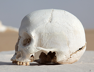
The skull is a bony structure that forms the head in vertebrates. It supports the structures of the face and provides a protective cavity for the brain. The skull is composed of two parts: the cranium and the mandible. In humans, these two parts are the neurocranium and the viscerocranium that includes the mandible as its largest bone. The skull forms the anterior-most portion of the skeleton and is a product of cephalisation—housing the brain, and several sensory structures such as the eyes, ears, nose, and mouth. In humans these sensory structures are part of the facial skeleton.

A fontanelle is an anatomical feature of the infant human skull comprising any of the soft membranous gaps (sutures) between the cranial bones that make up the calvaria of a fetus or an infant. Fontanelles allow for rapid stretching and deformation of the neurocranium as the brain expands faster than the surrounding bone can grow. Premature complete ossification of the sutures is called craniosynostosis.
The occipital bone is a cranial dermal bone and the main bone of the occiput. It is trapezoidal in shape and curved on itself like a shallow dish. The occipital bone overlies the occipital lobes of the cerebrum. At the base of skull in the occipital bone, there is a large oval opening called the foramen magnum, which allows the passage of the spinal cord.

The parietal bones are two bones in the skull which, when joined together at a fibrous joint, form the sides and roof of the cranium. In humans, each bone is roughly quadrilateral in form, and has two surfaces, four borders, and four angles. It is named from the Latin paries (-ietis), wall.

The temporal bones are situated at the sides and base of the skull, and lateral to the temporal lobes of the cerebral cortex.
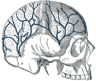
The diploic veins are large, thin-walled valveless veins that channel in the diploë between the inner and outer layers of the cortical bone in the skull. They are lined by a single layer of endothelium supported by elastic tissue. They develop fully by the age of two years. The diploic veins drain this area into the dural venous sinuses. The four major trunks of the diploic veins found on each side of the head are frontal, anterior temporal, posterior temporal, and occipital diploic veins.

A skull fracture is a break in one or more of the eight bones that form the cranial portion of the skull, usually occurring as a result of blunt force trauma. If the force of the impact is excessive, the bone may fracture at or near the site of the impact and cause damage to the underlying structures within the skull such as the membranes, blood vessels, and brain.

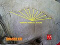
The coronal suture is a dense, fibrous connective tissue joint that separates the two parietal bones from the frontal bone of the skull.

The asterion is the point on the skull corresponding to the posterior end of the parietomastoid suture.

The transverse sinuses, within the human head, are two areas beneath the brain which allow blood to drain from the back of the head. They run laterally in a groove along the interior surface of the occipital bone. They drain from the confluence of sinuses to the sigmoid sinuses, which ultimately connect to the internal jugular vein. See diagram : labeled under the brain as "SIN. TRANS.".

The squamous part of temporal bone, or temporal squama, forms the front and upper part of the temporal bone, and is scale-like, thin, and translucent.

The mastoid part of the temporal bone is the posterior (back) part of the temporal bone, one of the bones of the skull. Its rough surface gives attachment to various muscles and it has openings for blood vessels. From its borders, the mastoid part articulates with two other bones.

The petrous part of the temporal bone is pyramid-shaped and is wedged in at the base of the skull between the sphenoid and occipital bones. Directed medially, forward, and a little upward, it presents a base, an apex, three surfaces, and three angles, and houses in its interior, the components of the inner ear. The petrous portion is among the most basal elements of the skull and forms part of the endocranium. Petrous comes from the Latin word petrosus, meaning "stone-like, hard". It is one of the densest bones in the body.

The Sphenoparietal suture is the cranial suture between the sphenoid bone and the parietal bone. It is one of the sutures that comprises the pterion.

The pterion is the region where the frontal, parietal, temporal, and sphenoid bones join together. It is located on the side of the skull, just behind the temple.
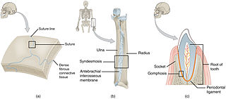
In anatomy, fibrous joints are joints connected by fibrous tissue, consisting mainly of collagen. These are fixed joints where bones are united by a layer of white fibrous tissue of varying thickness. In the skull the joints between the bones are called sutures. Such immovable joints are also referred to as synarthroses.
Angonisaurus is an extinct genus of kannemeyeriiform dicynodont from the Middle Triassic of Africa between 247 and 242 million years ago. Only one species, Angonisaurus cruickshanki has been assigned to this genus. This genus is thought to have been widely spread but rare in southern Gondwana. Though few in number, the fossil record of Angonisaurus cruickshanki contains multiple specimens giving it a measurable stratigraphic range. Sexually dimorphic features are found in Angonisaurus which include presence or absence of tusks and difference is size and robustness of the temporal arch and the rostral.
Dianopachysaurus is an extinct genus of pachypleurosaur known from the lower Middle Triassic of Yunnan Province, southwestern China. It was found in the Middle Triassic Lagerstatte of the Guanling Formation. It was first named by Jun Liu, Olivier Rieppel, Da-Yong Jiang, Jonathan C. Aitchison, Ryosuke Motani, Qi-Yue Zhang, Chang-Yong Zhou and Yuan-Yuan Sun in 2011 and the type species is Dianopachysaurus dingi, thanking a Professor Ding for his help.
Koerner's septum is an anatomic boundary in the temporal bone formed by the petrosquamous suture between the petrous and squamosal portions of the mastoid air cells, at the anatomic level of the antrum. Along with the middle ear ossicles, it is usually eroded in middle ear cholesteatomas. Superiorly, this continues as the petrosquamous suture, a normal anatomic structure that can be mistaken for fractures on temporal bone CT. It is surgically important as it may cause difficulty in locating the antrum and the deeper cells and thus may lead to incomplete removal of disease at mastoidectomy.
Glaurung is an extinct genus of weigeltisaurid reptile from the Upper Permian of Germany. The only known species is Glaurung schneideri. Originally considered a specimen of Coelurosauravus, a later study named it as a new genus after noting that it had several unique characteristics relative to other weigeltisaurids. These characteristics included a low skull, small eyes, smooth parietal and squamosal bones, and spiny jugal bones.