
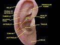
The outer ear, external ear, or auris externa is the external part of the ear, which consists of the auricle and the ear canal. It gathers sound energy and focuses it on the eardrum.

In vertebrates, an ear is the organ that enables hearing and body balance using the vestibular system. In humans, the ear is described as having three parts: the outer ear, the middle ear and the inner ear. The outer ear consists of the auricle and the ear canal. Since the outer ear is the only visible portion of the ear, the word "ear" often refers to the external part (auricle) alone. The middle ear includes the tympanic cavity and the three ossicles. The inner ear sits in the bony labyrinth, and contains structures which are key to several senses: the semicircular canals, which enable balance and eye tracking when moving; the utricle and saccule, which enable balance when stationary; and the cochlea, which enables hearing. The ear canal is cleaned via earwax, which naturally migrates to the auricle.

The auricle or auricula is the visible part of the ear that is outside the head. It is also called the pinna, a term that is used more in zoology.
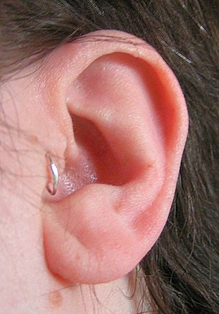
A tragus piercing is the perforation of the tragus, which projects immediately in front of the ear canal, for the purpose of inserting and wearing a piece of jewelry. The piercing itself is usually made with a small gauge hollow piercing needle, and typical jewelry would be a small diameter captive bead ring or small gauge post style piercing jewelry. A related piercing is known as the antitragus piercing.
Otoplasty is a procedure for correcting the deformities and defects of the auricle, whether these defects are congenital conditions or caused by trauma. Otoplastic surgeons may reshape, move, or augment the cartilaginous support framework of the auricle to correct these defects.
The helix is the prominent rim of the auricle. Where the helix turns downwards posteriorly, a small tubercle is sometimes seen, namely the auricular tubercle of Darwin.
The tragus is a small pointed eminence of the external ear, situated in front of the concha, and projecting backward over the meatus. It also is the name of hair growing at the entrance of the ear. Its name comes from the Ancient Greek tragos, meaning 'goat', and is descriptive of its general covering on its under surface with a tuft of hair, resembling a goat's beard. The nearby antitragus projects forwards and upwards.

In human anatomy, the superficial temporal artery is a major artery of the head. It arises from the external carotid artery when it splits into the superficial temporal artery and maxillary artery.

The antitragicus is an intrinsic muscle of the outer ear.

The tragicus, also called the tragus muscle or Valsalva muscle, is an intrinsic muscle of the outer ear.
The oblique muscle of auricle is an intrinsic muscle of the outer ear.
The transverse muscle of auricle is an intrinsic muscle of the outer ear.
The Helicis minor is a small skeletal muscle. The helicis minor is an intrinsic muscle of the outer ear. The muscle runs obliques and covers the helical crus, part of the helix located just above the tragus.
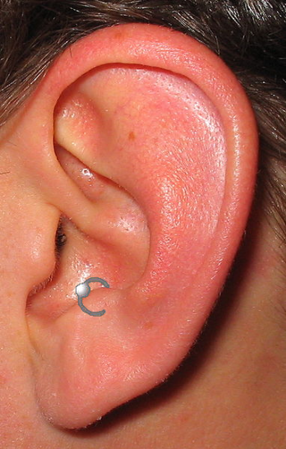
An antitragus piercing is a perforation of the outer ear cartilage for the purpose of inserting and wearing a piece of jewelry. It is placed in the antitragus, a piece of cartilage opposite the ear canal. Overall, the piercing has characteristics similar to the tragus piercing; the piercings are performed and cared for in much the same way.

The anterior auricular branches of the superficial temporal artery are distributed to the anterior portion of the auricula, the lobule, and part of the external meatus, anastomosing with the posterior auricular. They supply the external acoustic meatus and the visible part of the ear.
A cartilage piercing can refer to any area of cartilage on the body with a perforation created for the purpose of wearing jewelry. The two most common areas with cartilage piercings are the ear and the nose. Outside of the body modification community, many people commonly refer to a helix piercing as a "cartilage piercing." The cartilage ear piercing is known to be more sore than the lobe as in the cartilage there is less blood so it takes longer to heal.
The posterior auricular muscle is a muscle behind the auricle of the outer ear. It arises from the mastoid part of the temporal bone, and inserts into the lower part of the cranial surface of the auricle of the outer ear. It draws the auricle backwards, usually a very slight effect.

The superior auricular muscle is a muscle above the auricle of the outer ear. It originates from the epicranial aponeurosis, and inserts into the upper part of the medial surface of the auricle. It draws the auricle upwards.

The anterior auricular muscle, the smallest of the three auricular muscles, is thin and fan-shaped, and its fibers are pale and indistinct. It arises from the lateral edge of the epicranial aponeurosis, and its fibers converge to be inserted into a projection on the front of the helix.
The intertragic notch is an anatomical feature of the ears of mammals. In humans, it is the space that separates the tragus from the antitragus in the outer ear.


