Vestibule of the ear

Last updated
Vestibule of the ear
Sobo 1911 772.png
Schematic overview of the vestibulocochlear organ, vestibule centre left.
Details
Part of Bony labyrinth of the inner ear
Identifiers
Latin vestibulum labyrinthi, vestibulum auris
MeSH D014722
TA98 A15.3.03.004
TA2 6942
FMA 60183
Anatomical terminology

The vestibule is the central part of the bony labyrinth in the inner ear, and is situated medial to the eardrum, behind the cochlea, and in front of the three semicircular canals. [1]

Contents

The name comes from the Latin vestibulum, literally an entrance hall.

Structure

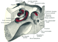
The vestibule is somewhat oval in shape, but flattened transversely; it measures about 5 mm from front to back, the same from top to bottom, and about 3 mm across.

In its lateral or tympanic wall is the oval window, closed, in the fresh state, by the base of the stapes and annular ligament.

On its medial wall, at the forepart, is a small circular depression, the recessus sphæricus, which is perforated, at its anterior and inferior part, by several minute holes (macula cribrosa media) for the passage of filaments of the acoustic nerve to the saccule; and behind this depression is an oblique ridge, the crista vestibuli, the anterior end of which is named the pyramid of the vestibule.

This ridge bifurcates below to enclose a small depression, the fossa cochlearis, which is perforated by a number of holes for the passage of filaments of the acoustic nerve which supply the vestibular end of the cochlear duct.

The orifice of the vestibular aqueduct is the hind part of the medial wall; it extends to the posterior surface of the petrous portion of the temporal bone.

It transmits a small vein and contains a tubular prolongation of the membranous labyrinth, the endolymphatic duct, which ends in a cul-de-sac between the layers of the dura mater within the cranial cavity.

On the upper wall or roof, there is a transversely oval depression, the recessus ellipticus, separated from the recessus sphæricus by the crista vestibuli already mentioned.

The pyramid and adjoining part of the recessus ellipticus are perforated by a number of holes (macula cribrosa superior).

The apertures in the pyramid transmit the nerves to the utricle; those in the recessus ellipticus are the nerves to the ampullæ of the superior and lateral semicircular ducts.

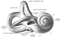
Behind, the five orifices of the semicircular canals can be found.

In the frontal view, there is an elliptical opening which communicates with the vestibular duct of the cochlea.

Additional images

Related Research Articles

<span class="mw-page-title-main">Inner ear</span> Innermost part of the vertebrate ear

The inner ear is the innermost part of the vertebrate ear. In vertebrates, the inner ear is mainly responsible for sound detection and balance. In mammals, it consists of the bony labyrinth, a hollow cavity in the temporal bone of the skull with a system of passages comprising two main functional parts:

<span class="mw-page-title-main">Cochlea</span> Snail-shaped part of inner ear involved in hearing

The cochlea is the part of the inner ear involved in hearing. It is a spiral-shaped cavity in the bony labyrinth, in humans making 2.75 turns around its axis, the modiolus. A core component of the cochlea is the organ of Corti, the sensory organ of hearing, which is distributed along the partition separating the fluid chambers in the coiled tapered tube of the cochlea.

Articles related to anatomy include:

<span class="mw-page-title-main">Semicircular canals</span> Organ located in innermost part of ear

The semicircular canals are three semicircular interconnected tubes located in the innermost part of each ear, the inner ear. The three canals are the lateral, anterior and posterior semicircular canals. They are the part of the bony labyrinth, a periosteum-lined cavity on the petrous part of the temporal bone filled with perilymph.

<span class="mw-page-title-main">Utricle (ear)</span> Membranous labyrinth in the vestibule of ear

The utricle and saccule are the two otolith organs in the vertebrate inner ear. They are part of the balancing system in the vestibule of the bony labyrinth. They use small stones and a viscous fluid to stimulate hair cells to detect motion and orientation. The utricle detects linear accelerations and head-tilts in the horizontal plane. The word utricle comes from Latin uter 'leather bag'.

<span class="mw-page-title-main">Saccule</span> Bed of sensory cells in the inner ear

The saccule is a bed of sensory cells in the inner ear. It translates head movements into neural impulses for the brain to interpret. The saccule detects linear accelerations and head tilts in the vertical plane. When the head moves vertically, the sensory cells of the saccule are disturbed and the neurons connected to them begin transmitting impulses to the brain. These impulses travel along the vestibular portion of the eighth cranial nerve to the vestibular nuclei in the brainstem.

<span class="mw-page-title-main">Tympanic cavity</span> Small cavity surrounding the bones of the middle ear

The tympanic cavity is a small cavity surrounding the bones of the middle ear. Within it sit the ossicles, three small bones that transmit vibrations used in the detection of sound.

<span class="mw-page-title-main">Perilymph</span> Extracellular fluid located within the inner ear

Perilymph is an extracellular fluid located within the inner ear. It is found within the scala tympani and scala vestibuli of the cochlea. The ionic composition of perilymph is comparable to that of plasma and cerebrospinal fluid. The major cation in perilymph is sodium, with the values of sodium and potassium concentration in the perilymph being 138 mM and 6.9 mM, respectively. It is also named Cotunnius' liquid and liquor cotunnii for Domenico Cotugno.

<span class="mw-page-title-main">Tympanic duct</span>

The tympanic duct or scala tympani is one of the perilymph-filled cavities in the inner ear of humans. It is separated from the cochlear duct by the basilar membrane, and it extends from the round window to the helicotrema, where it continues as vestibular duct.

<span class="mw-page-title-main">Cochlear duct</span> Cavity in the cochlea of the inner ear

The cochlear duct is an endolymph filled cavity inside the cochlea, located between the tympanic duct and the vestibular duct, separated by the basilar membrane and the vestibular membrane respectively. The cochlear duct houses the organ of Corti.

<span class="mw-page-title-main">Petrous part of the temporal bone</span> Feature at the base of the human skull

The petrous part of the temporal bone is pyramid-shaped and is wedged in at the base of the skull between the sphenoid and occipital bones. Directed medially, forward, and a little upward, it presents a base, an apex, three surfaces, and three angles, and houses in its interior, the components of the inner ear. The petrous portion is among the most basal elements of the skull and forms part of the endocranium. Petrous comes from the Latin word petrosus, meaning "stone-like, hard". It is one of the densest bones in the body. In other mammals, it is a separate bone, the petrosal bone.

<span class="mw-page-title-main">Lateral parts of occipital bone</span>

The lateral parts of the occipital bone are situated at the sides of the foramen magnum; on their under surfaces are the condyles for articulation with the superior facets of the atlas.

<span class="mw-page-title-main">Ampullary cupula</span>

The ampullary cupula, or cupula, is a structure in the vestibular system, providing the sense of spatial orientation.

<span class="mw-page-title-main">Endolymphatic duct</span> Canal

From the posterior wall of the saccule a canal, the endolymphatic duct, is given off; this duct is joined by the ductus utriculosaccularis, and then passes along the aquaeductus vestibuli and ends in a blind pouch on the posterior surface of the petrous portion of the temporal bone, where it is in contact with the dura mater.

<span class="mw-page-title-main">Vestibular aqueduct</span> Orifice

At the hinder part of the medial wall of the vestibule is the orifice of the vestibular aqueduct, which extends to the posterior surface of the petrous portion of the temporal bone. The vestibular aqueduct parallels the petrous apex, in contrast to the cochlear aqueduct, which lies perpendicular to the petrous apex.

<span class="mw-page-title-main">Crista ampullaris</span> Sensory organ in the inner ear

The crista ampullaris is the sensory organ of rotation. They are found in the ampullae of each of the semicircular canals of the inner ear, meaning that there are three pairs in total. The function of the crista ampullaris is to sense angular acceleration and deceleration.

The saccule is the smaller sized vestibular sac ; it is globular in form, and lies in the recessus sphæricus near the opening of the scala vestibuli of the cochlea. Its anterior part exhibits an oval thickening, the macula of saccule, to which are distributed the saccular filaments of the acoustic nerve.

<span class="mw-page-title-main">Outline of human anatomy</span> Overview of and topical guide to human anatomy

The following outline is provided as an overview of and topical guide to human anatomy:

<span class="mw-page-title-main">Vestibulocochlear dysfunction progressive familial</span> Medical condition

Vestibulocochlear dysfunction progressive familial, known also as familial progressive vestibulocochlear dysfunction is an autosomal dominant disease that results in sensorineural hearing loss and vestibular areflexia. Patients report feelings of vague dizziness, blurred vision, dysequilibrium in the dark, and progressive hearing impairment.

<span class="mw-page-title-main">Vestibular duct</span>

The vestibular duct or scala vestibuli is a perilymph-filled cavity inside the cochlea of the inner ear that conducts sound vibrations to the cochlear duct.

References

PD-icon.svgThis article incorporates text in the public domain from page 1047 of the 20th edition of Gray's Anatomy (1918)

  1. Treuting, Piper M.; Dintzis, Suzanne M.; Sellers, Rani (2018). "Special Senses". Comparative Anatomy and Histology. Elsevier. pp. 471–485. doi:10.1016/b978-0-12-802900-8.00022-1. ISBN   978-0-12-802900-8. The vestibule sits between and connects the cochlea and semicircular canals and helps to maintain equilibrium.

See also