The inner ear is the innermost part of the vertebrate ear. In vertebrates, the inner ear is mainly responsible for sound detection and balance. In mammals, it consists of the bony labyrinth, a hollow cavity in the temporal bone of the skull with a system of passages comprising two main functional parts:
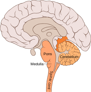
The pons is part of the brainstem that in humans and other bipeds lies inferior to the midbrain, superior to the medulla oblongata and anterior to the cerebellum.

The sphenoid bone is an unpaired bone of the neurocranium. It is situated in the middle of the skull towards the front, in front of the basilar part of the occipital bone. The sphenoid bone is one of the seven bones that articulate to form the orbit. Its shape somewhat resembles that of a butterfly or bat with its wings extended.
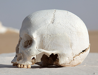
The occipital bone is a cranial dermal bone and the main bone of the occiput. It is trapezoidal in shape and curved on itself like a shallow dish. The occipital bone overlies the occipital lobes of the cerebrum. At the base of skull in the occipital bone, there is a large oval opening called the foramen magnum, which allows the passage of the spinal cord.

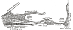
The basilar membrane is a stiff structural element within the cochlea of the inner ear which separates two liquid-filled tubes that run along the coil of the cochlea, the scala media and the scala tympani. The basilar membrane moves up and down in response to incoming sound waves, which are converted to traveling waves on the basilar membrane.

The basilar artery is one of the arteries that supplies the brain with oxygen-rich blood.

The posterior cerebral artery (PCA) is one of a pair of arteries that supply oxygenated blood to the occipital lobe, part of the back of the human brain. The two arteries originate from the distal end of the basilar artery, where it bifurcates into the left and right posterior cerebral arteries. These anastomose with the middle cerebral arteries and internal carotid arteries via the posterior communicating arteries.

In human anatomy, the left and right posterior communicating arteries are arteries at the base of the brain that form part of the circle of Willis. Each posterior communicating artery connects the three cerebral arteries of the same side. Anteriorly, it connects to the internal carotid artery (ICA) prior to the terminal bifurcation of the ICA into the anterior cerebral artery and middle cerebral artery. Posteriorly, it communicates with the posterior cerebral artery.

The superior cerebellar artery (SCA) arises near the termination of the basilar artery.

The labyrinthine artery is a branch of the anterior inferior cerebellar artery or basilar artery. It accompanies the vestibulocochlear nerve through the internal acoustic meatus, and supplies blood to the internal ear.

The petrous part of the temporal bone is pyramid-shaped and is wedged in at the base of the skull between the sphenoid and occipital bones. Directed medially, forward, and a little upward, it presents a base, an apex, three surfaces, and three angles, and houses in its interior, the components of the inner ear. The petrous portion is among the most basal elements of the skull and forms part of the endocranium. Petrous comes from the Latin word petrosus, meaning "stone-like, hard". It is one of the densest bones in the body.

The basilar part of the occipital bone extends forward and upward from the foramen magnum, and presents in front an area more or less quadrilateral in outline.
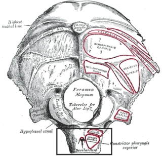
The pharyngeal tubercle is a part of the occipital bone of the head and neck. It is located on the lower surface of the basilar part of occipital bone, about 1 cm. anterior to the foramen magnum. The pharyngeal tubercle gives attachment to the fibrous raphe of the pharynx, also known as the pharyngeal raphe.

The basilar plexus consists of several interlacing venous channels between the layers of the dura mater over the basilar part of the occipital bone, and serves to connect the two inferior petrosal sinuses.
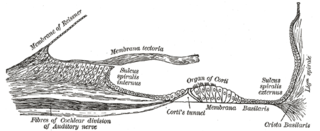
The basilar crest lies within the cochlear duct in the inner ear. It gives attachment to the outer edge of the basilar membrane and is a spiral ligament that projects inward below as a triangular prominence.
The pharyngeal aponeurosis, is situated between the mucous and muscular layers.

The basilar part of pons, also known as basis pontis, is the ventral part of the pons; the dorsal part is known as the pontine tegmentum.

The basilar membrane stretches from the tympanic lip of the osseous spiral lamina to the basilar crest and consists of two parts, an inner and an outer. The inner is thin, and is named the inner tunnel : it supports the spiral organ of Corti.
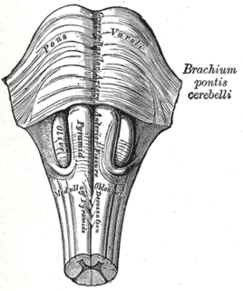
The basilar sulcus is a groove in the pons, part of the brainstem.