Roots of the ciliary ganglion

Last updated
Ciliary ganglion
Ciliary ganglion pathways.png
Pathways in the ciliary ganglion. Green = parasympathetic; Red = sympathetic; Blue = sensory
Anatomical terms of neuroanatomy

The ciliary ganglion is a parasympathetic ganglion located just behind the eye in the posterior orbit. Three types of axons enter the ciliary ganglion but only the preganglionic parasympathetic axons synapse there. The entering axons are arranged into three roots of the ciliary ganglion, which join enter the posterior surface of the ganglion.

Contents

Sympathetic root

The sympathetic root of ciliary ganglion is one of three roots of the ciliary ganglion. It contains postganglionic sympathetic fibers whose cell bodies are located in the superior cervical ganglion. Their axons ascend with the internal carotid artery as a plexus of nerves, the internal carotid plexus. Sympathetic fibers supplying the eye separate from the carotid plexus within the cavernous sinus. They run forward through the superior orbital fissure and merge with the long ciliary nerves (branches of the nasociliary nerve) and the short ciliary nerves (from the ciliary ganglion). Sympathetic fibers in the short ciliary nerves pass through the ciliary ganglion without forming synapses.

Preganglionic sympathetic fibers originate from neurons in the intermediolateral column of the thoracic spinal cord, at the level of thoracic spinal nerve 1 (T1) and thoracic spinal nerve 2 (T2). They form synapses in the superior cervical ganglion. The ratio of incoming to outgoing fibers (the “convergence”) in this ganglion is approximately 100:1. Sympathetic motor neurons in the spinal cord are controlled by supranuclear pathways that descend through the brainstem and spinal cord. Interruption of the sympathetic chain at any level (from the brainstem to the ciliary ganglion) will produce pupillary constriction (miosis) and eyelid droop (ptosis) – the classic signs of Horner's syndrome.

Sympathetic fibers from the superior cervical ganglion innervate blood vessels (vasoconstriction), sweat glands, and 4 eye muscles: the dilator pupillae, the superior tarsal muscle, the inferior tarsal muscle and the orbitalis.

The dilator pupillae dilates the pupil; its action is antagonistic to the sphincter pupillae. Pupil size is therefore under the dual control of sympathetic and parasympathetic nerves.

Postsynaptic sympathetic signals that originate in the superior cervical ganglion are carried by the nasociliary nerve or directly extend from the internal carotid plexus and pass through the ciliary ganglion.

The superior tarsal muscle elevates the upper eyelid. The levator palpebrae superioris, which is supplied by a branch of the oculomotor nerve, also elevates the upper eyelid. Eyelid elevation is therefore under both voluntary and involuntary control. Interruption of either pathway will result in eyelid droop (ptosis).

The other two eye muscles with sympathetic supply (the inferior tarsal muscle and the orbitalis) are vestigial in humans. They are variable and often incompletely developed.

Sensory root

Sensory fibers from the eyeball (the cornea, iris, and ciliary body) run posteriorly through the short ciliary nerves and pass through the ciliary ganglion without forming synapses. They leave the ciliary ganglion in the sensory root of ciliary ganglion, which joins the nasociliary nerve—a branch of the ophthalmic nerve. From there, the signal travels back through the ophthalmic nerve to the trigeminal nerve and back into specific nuclei in the thalamus where they are relayed to areas in the cerebral cortex.

The exact distribution of sensory fibers, like the distribution of sympathetic fibers, is anatomically variable. There are other pathways to the eye for both sympathetic and sensory fibers, and the precise anatomy varies from person to person. Since the result is the same regardless of how the fibers reach the eye, the presence of sympathetic and sensory fibers in the ciliary ganglion (the contributions of the “sensory” and “sympathetic” roots) is of no functional significance.

Parasympathetic root

The parasympathetic root of ciliary ganglion provides parasympathetic supply to the ciliary ganglion.

The ciliary ganglion is a parasympathetic ganglion. Incoming parasympathetic nerve fibers form synapses with the dendrites of nerve cells within the ganglion. However, the ciliary ganglion is not simply a relay station connecting preganglionic to postganglionic nerve fibers. There are roughly twice as many incoming parasympathetic fibers as outgoing parasympathetic fibers. Neural processing occurs as incoming signals converge onto target neurons.

Presynaptic parasympathetic fibers originate in the Edinger-Westphal nucleus, the parasympathetic motor nucleus associated with the oculomotor nucleus in the brainstem. Axons from the Edinger-Westphal nucleus and the oculomotor nucleus run together in the brainstem and exit together as the oculomotor nerve. The oculomotor nerve passes through the lateral wall of the cavernous sinus and enters the orbit through the superior orbital fissure. It divides into branches that innervate the levator palpebrae superioris and four of the six extraocular muscles. Parasympathetic fibers initially run in the inferior division of the oculomotor nerve. They exit as one or two short “motor roots” that synapse in the ciliary ganglion.

Postsynaptic parasympathetic fibers leave the ciliary ganglion in multiple (six to ten) short ciliary nerves. These nerves enter the posterior aspect of the eyeball to supply the sphincter pupillae and ciliaris muscles. The sphincter pupillae constricts the iris. The ciliaris muscle changes the shape of the lens, allowing the eye to focus on nearby objects (accommodation). Neither of these muscles is under voluntary control.

Related Research Articles

<span class="mw-page-title-main">Autonomic nervous system</span> Division of the nervous system supplying internal organs, smooth muscle and glands

The autonomic nervous system (ANS), formerly referred to as the vegetative nervous system, is a division of the nervous system that operates internal organs, smooth muscle and glands. The autonomic nervous system is a control system that acts largely unconsciously and regulates bodily functions, such as the heart rate, its force of contraction, digestion, respiratory rate, pupillary response, urination, and sexual arousal. This system is the primary mechanism in control of the fight-or-flight response.

<span class="mw-page-title-main">Parasympathetic nervous system</span> Division of the autonomic nervous system

The parasympathetic nervous system (PSNS) is one of the three divisions of the autonomic nervous system, the others being the sympathetic nervous system and the enteric nervous system. The enteric nervous system is sometimes considered part of the autonomic nervous system, and sometimes considered an independent system.

<span class="mw-page-title-main">Facial nerve</span> Cranial nerve VII, for the face and tasting

The facial nerve, also known as the seventh cranial nerve, cranial nerve VII, or simply CN VII, is a cranial nerve that emerges from the pons of the brainstem, controls the muscles of facial expression, and functions in the conveyance of taste sensations from the anterior two-thirds of the tongue. The nerve typically travels from the pons through the facial canal in the temporal bone and exits the skull at the stylomastoid foramen. It arises from the brainstem from an area posterior to the cranial nerve VI and anterior to cranial nerve VIII.

Articles related to anatomy include:

<span class="mw-page-title-main">Oculomotor nerve</span> Cranial nerve III, for eye movements

The oculomotor nerve, also known as the third cranial nerve, cranial nerve III, or simply CN III, is a cranial nerve that enters the orbit through the superior orbital fissure and innervates extraocular muscles that enable most movements of the eye and that raise the eyelid. The nerve also contains fibers that innervate the intrinsic eye muscles that enable pupillary constriction and accommodation. The oculomotor nerve is derived from the basal plate of the embryonic midbrain. Cranial nerves IV and VI also participate in control of eye movement.

<span class="mw-page-title-main">Glossopharyngeal nerve</span> Cranial nerve IX, for the tongue and pharynx

The glossopharyngeal nerve, also known as the ninth cranial nerve, cranial nerve IX, or simply CN IX, is a cranial nerve that exits the brainstem from the sides of the upper medulla, just anterior to the vagus nerve. Being a mixed nerve (sensorimotor), it carries afferent sensory and efferent motor information. The motor division of the glossopharyngeal nerve is derived from the basal plate of the embryonic medulla oblongata, whereas the sensory division originates from the cranial neural crest.
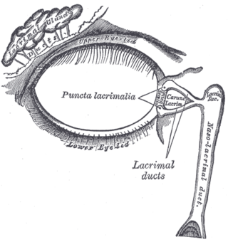
<span class="mw-page-title-main">Lacrimal gland</span> Exocrine gland, one for each eye, that secrete tears

The lacrimal glands are paired exocrine glands, one for each eye, found in most terrestrial vertebrates and some marine mammals, that secrete the aqueous layer of the tear film. In humans, they are situated in the upper lateral region of each orbit, in the lacrimal fossa of the orbit formed by the frontal bone. Inflammation of the lacrimal glands is called dacryoadenitis. The lacrimal gland produces tears which are secreted by the lacrimal ducts, and flow over the ocular surface, and then into canals that connect to the lacrimal sac. From that sac, the tears drain through the lacrimal duct into the nose.

<span class="mw-page-title-main">Horner's syndrome</span> Facial disorder due to damage of the sympathetic nerves

Horner's syndrome, also known as oculosympathetic paresis, is a combination of symptoms that arises when a group of nerves known as the sympathetic trunk is damaged. The signs and symptoms occur on the same side (ipsilateral) as it is a lesion of the sympathetic trunk. It is characterized by miosis, partial ptosis, apparent anhidrosis, with apparent enophthalmos.

<span class="mw-page-title-main">Otic ganglion</span> Parasympathetic ganglion of the head and neck

The otic ganglion is a small parasympathetic ganglion located immediately below the foramen ovale in the infratemporal fossa and on the medial surface of the mandibular nerve. It is functionally associated with the glossopharyngeal nerve and innervates the parotid gland for salivation.

<span class="mw-page-title-main">Pterygopalatine ganglion</span> Parasympathetic ganglion in the pterygopalatine fossa

The pterygopalatine ganglion is a parasympathetic ganglion in the pterygopalatine fossa. It is one of four parasympathetic ganglia of the head and neck,.

<span class="mw-page-title-main">Ciliary ganglion</span> Bundle of nerves, parasympathetic ganglion

The ciliary ganglion is a bundle of nerves, parasympathetic ganglion located just behind the eye in the posterior orbit. It is 1–2 mm in diameter and in humans contains approximately 2,500 neurons. The ganglion contains postganglionic parasympathetic neurons. These neurons supply the pupillary sphincter muscle, which constricts the pupil, and the ciliary muscle which contracts to make the lens more convex. Both of these muscles are involuntary since they are controlled by the parasympathetic division of the autonomic nervous system.
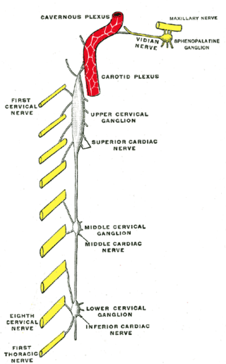
<span class="mw-page-title-main">Superior cervical ganglion</span> Largest of the cervical ganglia

The superior cervical ganglion (SCG) is the upper-most and largest of the cervical sympathetic ganglia of the sympathetic trunk. It probably formed by the union of four sympathetic ganglia of the cervical spinal nerves C1–C4. It is the only ganglion of the sympathetic nervous system that innervates the head and neck. The SCG innervates numerous structures of the head and neck.

<span class="mw-page-title-main">Long ciliary nerves</span>

The long ciliary nerves are 2-3 nerves that arise from the nasociliary nerve (itself a branch of the ophthalmic branch (CN V1) of the trigeminal nerve (CN V)). They enter the eyeball to provide sensory innervation to parts of the eye, and sympathetic visceral motor innervation to the dilator pupillae muscle.

<span class="mw-page-title-main">Submandibular ganglion</span>

The submandibular ganglion is part of the human autonomic nervous system. It is one of four parasympathetic ganglia of the head and neck..

<span class="mw-page-title-main">Parasympathetic ganglia</span> Autonomic ganglia of the parasympathetic nervous system

Parasympathetic ganglia are the autonomic ganglia of the parasympathetic nervous system. Most are small terminal ganglia or intramural ganglia, so named because they lie near or within (respectively) the organs they innervate. The exceptions are the four paired parasympathetic ganglia of the head and neck.

<span class="mw-page-title-main">Short ciliary nerves</span> Nerves of the orbit around the eye

The short ciliary nerves are nerves of the orbit around the eye. They are branches of the ciliary ganglion. They supply parasympathetic and sympathetic nerve fibers to the ciliary muscle, iris, and cornea. Damage to the short ciliary nerve may result in loss of the pupillary light reflex, or mydriasis.

<span class="mw-page-title-main">Lateral grey column</span>

The lateral grey column is one of the three grey columns of the spinal cord ; the others being the anterior and posterior grey columns. The lateral grey column is primarily involved with activity in the sympathetic division of the autonomic motor system. It projects to the side as a triangular field in the thoracic and upper lumbar regions of the postero-lateral part of the anterior grey column.

<span class="mw-page-title-main">Ciliospinal center</span>

The ciliospinal center is a cluster of pre-ganglionic sympathetic neuron cell bodies located in the intermediolateral cell column of the spinal cord at the (C8) T1-T2 spinal levels.

<span class="mw-page-title-main">Outline of the human nervous system</span> Overview of and topical guide to the human nervous system

The following diagram is provided as an overview of and topical guide to the human nervous system:

References