Peripheral vision

Last updated
Double system e.jpg
Peripheral vision of the human eye Peripheral vision.svg
Peripheral vision of the human eye
Field of view of the human eye Field of view.svg
Field of view of the human eye

Peripheral vision, or indirect vision, is vision as it occurs outside the point of fixation, i.e. away from the center of gaze or, when viewed at large angles, in (or out of) the "corner of one's eye". The vast majority of the area in the visual field is included in the notion of peripheral vision. "Far peripheral" vision refers to the area at the edges of the visual field, "mid-peripheral" vision refers to medium eccentricities, and "near-peripheral", sometimes referred to as "para-central" vision, exists adjacent to the center of gaze. [1]

Contents

Boundaries

Inner boundaries

The inner boundaries of peripheral vision can be defined in any of several ways depending on the context. In everyday language the term "peripheral vision" is often used to refer to what in technical usage would be called "far peripheral vision." This is vision outside of the range of stereoscopic vision. It can be conceived as bounded at the center by a circle 60° in radius or 120° in diameter, centered around the fixation point, i.e., the point at which one's gaze is directed. [2] However, in common usage, peripheral vision may also refer to the area outside a circle 30° in radius or 60° in diameter. [3] [4] In vision-related fields such as physiology, ophthalmology, optometry, or vision science in general, the inner boundaries of peripheral vision are defined more narrowly in terms of one of several anatomical regions of the central retina, in particular the fovea and the macula. [1]

The fovea is a cone-shaped depression in the central retina measuring 1.5 mm in diameter, corresponding [5] to 5° of the visual field. [6] The outer boundaries of the fovea are visible under a microscope, or with microscopic imaging technology such as OCT or microscopic MRI. When viewed through the pupil, as in an eye exam (using an ophthalmoscope or retinal photography), only the central portion of the fovea may be visible. Anatomists refer to this as the clinical fovea, and say that it corresponds to the anatomical foveola, a structure with a diameter of 0.35 mm corresponding to 1 degree of the visual field. In clinical usage the central part of the fovea is typically referred to simply as the fovea. [7] [8] [9]

In terms of visual acuity, "foveal vision" may be defined as vision using the part of the retina in which a visual acuity of at least 20/20 (6/6 metric or 0.0 LogMAR; internationally 1.0) is attained. This corresponds to using the foveal avascular zone (FAZ) with a diameter of 0.5 mm representing 1.5° of the visual field (although often idealized as perfect circles, the central structures of the retina tend to be irregular ovals). Thus, foveal vision may also be defined as the central 1.5–2° of the visual field. Vision within the fovea is generally called central vision, while vision outside of the fovea, or even outside the foveola, is called peripheral, or indirect vision. [1]

A ring-shaped region surrounding the fovea, known as the parafovea, is sometimes taken to represent an intermediate form of vision called paracentral vision. [10] The parafovea has an outer diameter of 2.5 mm representing 8° of the visual field. [11] [12]

The macula, the next larger region of the retina, is defined as having at least two layers of ganglia (bundles of nerves and neurons) and is sometimes taken as defining the boundaries of central vs. peripheral vision [13] [14] [15] (but this is controversial [16] ). Estimates of the macula’s size differ, [17] its diameter estimated at 6° – 10° [18] (corresponding to 1.7 – 2.9 mm), up to 17° of the visual field (5.5 mm [5] ). [19] [12] The term is familiar in the general public through the widespread macular degeneration (AMD) at older age, where central vision is lost. When viewed from the pupil, as in an eye exam, only the central portion of the macula may be visible. Known to anatomists as the clinical macula (and in clinical setting as simply the macula) this inner region is thought to correspond to the anatomical fovea. [20]

A dividing line between near and mid peripheral vision at 30° radius can be based on several features of visual performance. Visual acuity declines systematically up to 30° eccentricity: At 2°, acuity is half the foveal value, at 4° one-third, at 6° one-fourth etc. At 30°, it is one-sixteenth the foveal value. [21] [1] From thereon the decline is steeper. [22] [23] (Note that it would be wrong to say, the value were halved every 2°, as said in some textbooks or in previous versions of this article.) [16] Color perception is strong at 20° but weak at 40°. [24] In dark-adapted vision, light sensitivity corresponds to rod density,[ citation needed ] which peaks just at 18°. From 18° towards the center, rod density declines rapidly. From 18° away from the center, rod density declines more gradually, in a curve with distinct inflection points resulting in two humps. The outer edge of the second hump is at about 30°, and corresponds to the outer edge of good night vision. [25] [26] [27]

Outer boundaries

Classical image of the shape and size of the visual field Traquair 1938 Fig 1 modified.png
Classical image of the shape and size of the visual field

The outer boundaries of peripheral vision correspond to the boundaries of the visual field as a whole. For a single eye, the extent of the visual field can be (roughly) defined in terms of four angles, each measured from the fixation point, i.e., the point at which one's gaze is directed. These angles, representing four cardinal directions, are 60° upwards, 60° nasally (towards the nose), 70–75° downwards, and 100–110° temporally (away from the nose and towards the temple). [29] [28] [30] [31] [32] [33] For both eyes the combined visual field is 130–135° vertically [34] [35] and 200–220° horizontally. [28] [36] [33]

Characteristics

The loss of peripheral vision while retaining central vision is known as tunnel vision, and the loss of central vision while retaining peripheral vision is known as central scotoma [ citation needed ].

Peripheral vision is weak in humans, especially at distinguishing detail, color, and shape. This is because the density of receptor and ganglion cells in the retina is greater at the center and lowest at the edges, and, moreover, the representation in the visual cortex is much smaller than that of the fovea [1] (see visual system for an explanation of these concepts). The distribution of receptor cells across the retina is different between the two main types, rod cells and cone cells. Rod cells are unable to distinguish color and peak in density in the near periphery (at 18° eccentricity), while cone cell density is highest in the very center, the fovea. Note that this does not mean that there is a lack of cones representing in the periphery; colors can be distinguished in peripheral vision. [37]

Flicker fusion thresholds decline towards the periphery, but do that at a lower rate than other visual functions; so the periphery has a relative advantage at noticing flicker. [1] Peripheral vision is also relatively good at detecting motion (a feature of Magno cells).

Central vision is relatively weak in the dark (scotopic vision) since cone cells lack sensitivity at low light levels. Rod cells, which are concentrated further away from the fovea, operate better than cone cells in low light. This makes peripheral vision useful for detecting faint light sources at night (like faint stars). Because of this, pilots are taught to use peripheral vision to scan for aircraft at night. [ citation needed ]

Ovals A, B and C show which portions of the chess situation chess masters can reproduce correctly with their peripheral vision. Lines show path of foveal fixation during 5 seconds when the task is to memorize the situation as correctly as possible. Image from based on data by Eye movements of a chess champion nc.jpg
Ovals A, B and C show which portions of the chess situation chess masters can reproduce correctly with their peripheral vision. Lines show path of foveal fixation during 5 seconds when the task is to memorize the situation as correctly as possible. Image from based on data by

The distinctions between foveal (sometimes also called central) and peripheral vision are reflected in subtle physiological and anatomical differences in the visual cortex. Different visual areas contribute to the processing of visual information coming from different parts of the visual field, and a complex of visual areas located along the banks of the interhemispheric fissure (a deep groove that separates the two brain hemispheres) has been linked to peripheral vision. It has been suggested that these areas are important for fast reactions to visual stimuli in the periphery, and monitoring body position relative to gravity. [40]

Functions

The main functions of peripheral vision are: [38]

Extreme peripheral vision

Side-view of the human eye, viewed approximately 90deg temporal, illustrating how the iris and pupil appear rotated towards the viewer due to the optical properties of the cornea and the aqueous humor. Mairead cropped.png
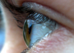
Side-view of the human eye, viewed approximately 90° temporal, illustrating how the iris and pupil appear rotated towards the viewer due to the optical properties of the cornea and the aqueous humor.

When viewed at large angles, the iris and pupil appear to be rotated toward the viewer due to the optical refraction in the cornea. As a result, the pupil may still be visible at angles greater than 90°. [41] [42] [43]

Cone-rich rim of the retina

The rim of the retina contains a large concentration of cone cells. The retina extends farthest in the superior-nasal 45° quadrant (in the direction from the pupil to the bridge of the nose) with the greatest extent of the visual field in the opposite direction, the inferior temporal 45° quadrant (from the pupil of either eye towards the bottom of the nearest ear). Vision at this extreme part of the visual field is thought to be possibly concerned with threat detection, measuring optical flow, color constancy, or circadian rhythm. [44] [45] [46]

See also

Related Research Articles

<span class="mw-page-title-main">Retina</span> Part of the eye

The retina is the innermost, light-sensitive layer of tissue of the eye of most vertebrates and some molluscs. The optics of the eye create a focused two-dimensional image of the visual world on the retina, which then processes that image within the retina and sends nerve impulses along the optic nerve to the visual cortex to create visual perception. The retina serves a function which is in many ways analogous to that of the film or image sensor in a camera.

<span class="mw-page-title-main">Eye</span> Organ that detects light and converts it into electro-chemical impulses in neurons

An eye is a sensory organ that allows an organism to perceive visual information. It detects light and converts it into electro-chemical impulses in neurons (neurones). It is part of an organism's visual system.

<span class="mw-page-title-main">Macula</span> Oval-shaped pigmented area near the center of the retina

The macula (/ˈmakjʊlə/) or macula lutea is an oval-shaped pigmented area in the center of the retina of the human eye and in other animals. The macula in humans has a diameter of around 5.5 mm (0.22 in) and is subdivided into the umbo, foveola, foveal avascular zone, fovea, parafovea, and perifovea areas.

<span class="mw-page-title-main">Field of view</span> Extent of the observable world seen at any given moment

The field of view (FOV) is the angular extent of the observable world that is seen at any given moment. In the case of optical instruments or sensors, it is a solid angle through which a detector is sensitive to electromagnetic radiation. It is further relevant in photography.

<span class="mw-page-title-main">Retinoschisis</span> Eye disease involving splitting of the retina

Retinoschisis is an eye disease characterized by the abnormal splitting of the retina's neurosensory layers, usually in the outer plexiform layer. Retinoschisis can be divided into degenerative forms which are very common and almost exclusively involve the peripheral retina and hereditary forms which are rare and involve the central retina and sometimes the peripheral retina. The degenerative forms are asymptomatic and involve the peripheral retina only and do not affect the visual acuity. Some rarer forms result in a loss of vision in the corresponding visual field.

<span class="mw-page-title-main">Cone cell</span> Photoreceptor cells responsible for color vision made to function in bright light

Cone cells or cones are photoreceptor cells in the retinas of vertebrates' eyes. They respond differently to light of different wavelengths, and the combination of their responses is responsible for color vision. Cones function best in relatively bright light, called the photopic region, as opposed to rod cells, which work better in dim light, or the scotopic region. Cone cells are densely packed in the fovea centralis, a 0.3 mm diameter rod-free area with very thin, densely packed cones which quickly reduce in number towards the periphery of the retina. Conversely, they are absent from the optic disc, contributing to the blind spot. There are about six to seven million cones in a human eye, with the highest concentration being towards the macula.

<span class="mw-page-title-main">Visual acuity</span> Clarity of vision

Visual acuity (VA) commonly refers to the clarity of vision, but technically rates an animal's ability to recognize small details with precision. Visual acuity depends on optical and neural factors. Optical factors of the eye influence the sharpness of an image on its retina. Neural factors include the health and functioning of the retina, of the neural pathways to the brain, and of the interpretative faculty of the brain.

<span class="mw-page-title-main">Fovea centralis</span> Small pit in the retina of the eye responsible for all central vision

The fovea centralis is a small, central pit composed of closely packed cones in the eye. It is located in the center of the macula lutea of the retina.

The visual field is "that portion of space in which objects are visible at the same moment during steady fixation of the gaze in one direction"; in ophthalmology and neurology the emphasis is on the structure inside the visual field and it is then considered “the field of functional capacity obtained and recorded by means of perimetry”.

<span class="mw-page-title-main">Central retinal artery</span>

The central retinal artery branches off the ophthalmic artery, running inferior to the optic nerve within its dural sheath to the eyeball.

In neuroscience, cortical magnification describes how many neurons in an area of the visual cortex are 'responsible' for processing a stimulus of a given size, as a function of visual field location. In the center of the visual field, corresponding to the center of the fovea of the retina, a very large number of neurons process information from a small region of the visual field. If the same stimulus is seen in the periphery of the visual field, it would be processed by a much smaller number of neurons. The reduction of the number of neurons per visual field area from foveal to peripheral representations is achieved in several steps along the visual pathway, starting already in the retina.

<span class="mw-page-title-main">Mammalian eye</span>

Mammals normally have a pair of eyes. Although mammalian vision is not so excellent as bird vision, it is at least dichromatic for most of mammalian species, with certain families possessing a trichromatic color perception.

<span class="mw-page-title-main">Foveola</span>

The foveola is located within a region called the macula, a yellowish, cone photoreceptor filled portion of the human retina. Approximately 0.35 mm in diameter, the foveola lies in the center of the fovea and contains only cone cells and a cone-shaped zone of Müller cells. In this region the cone receptors are found to be longer, slimmer, and more densely packed than anywhere else in the retina, thus allowing that region to have the potential to have the highest visual acuity in the eye.

<span class="mw-page-title-main">Crowding</span>

Crowding is a perceptual phenomenon where the recognition of objects presented away from the fovea is impaired by the presence of other neighbouring objects. It has been suggested that crowding occurs due to mandatory integration of the crowded objects by a texture-processing neural mechanism, but there are several competing theories about the underlying mechanisms. It is considered a kind of grouping since it is "a form of integration over space as target features are spuriously combined with flanker features."

<span class="mw-page-title-main">Eagle eye</span>

The eagle eye is among the sharpest in the animal kingdom, with an eyesight estimated at 4 to 8 times stronger than that of the average human. Although an eagle may only weigh 10 pounds (4.5 kg), its eyes are roughly the same size as those of a human. Eagle weight varies: a small eagle could weigh 700 grams (1.5 lb), while a larger one could weigh 6.5 kilograms (14 lb); an eagle of about 10 kilograms (22 lb) weight could have eyes as big as that of a human who weighs 200 pounds (91 kg). Although the size of the eagle eye is about the same as that of a human being, the back side shape of the eagle eye is flatter. Their eyes are stated to be larger in size than their brain, by weight. Color vision with resolution and clarity are the most prominent features of eagles' eyes, hence sharp-sighted people are sometimes referred to as "eagle-eyed". Eagles can identify five distinctly colored squirrels and locate their prey even if hidden.

<span class="mw-page-title-main">Vernier acuity</span>

Vernier acuity is a type of visual acuity – more precisely of hyperacuity – that measures the ability to discern a disalignment among two line segments or gratings. A subject's vernier acuity is the smallest visible offset between the stimuli that can be detected. Because the disalignments are often much smaller than the diameter and spacing of retinal receptors, vernier acuity requires neural processing and "pooling" to detect it. Because vernier acuity exceeds acuity by far, the phenomenon has been termed hyperacuity. Vernier acuity develops rapidly during infancy and continues to slowly develop throughout childhood. At approximately three to twelve months old, it surpasses grating acuity in foveal vision in humans. However, vernier acuity decreases more quickly than grating acuity in peripheral vision. Vernier acuity was first explained by Ewald Hering in 1899, based on earlier data by Alfred Volkmann in 1863 and results by Ernst Anton Wülfing in 1892.

<span class="mw-page-title-main">Parafovea</span>

Parafovea or the parafoveal belt is a region in the retina that circumscribes the fovea and is part of the macula lutea. It is circumscribed by the perifovea.

Microperimetry, sometimes called fundus-controlled perimetry, is a type of visual field test which uses one of several technologies to create a "retinal sensitivity map" of the quantity of light perceived in specific parts of the retina in people who have lost the ability to fixate on an object or light source. The main difference with traditional perimetry instruments is that, microperimetry includes a system to image the retina and an eye tracker to compensate eye movements during visual field testing.

Occult macular dystrophy (OMD) is a rare inherited degradation of the retina, characterized by progressive loss of function in the most sensitive part of the central retina (macula), the location of the highest concentration of light-sensitive cells (photoreceptors) but presenting no visible abnormality. "Occult" refers to the degradation in the fundus being difficult to discern. The disorder is called "dystrophy" instead of "degradation" to distinguish its genetic origin from other causes, such as age. OMD was first reported by Y. Miyake et al. in 1989.

Sickle cell retinopathy can be defined as retinal changes due to blood vessel damage in the eye of a person with a background of sickle cell disease. It can likely progress to loss of vision in late stages due to vitreous hemorrhage or retinal detachment. Sickle cell disease is a structural red blood cell disorder leading to consequences in multiple systems. It is characterized by chronic red blood cell destruction, vascular injury, and tissue ischemia causing damage to the brain, eyes, heart, lungs, kidneys, spleen, and musculoskeletal system.

References

  1. 1 2 3 4 5 6 Strasburger, Hans; Rentschler, Ingo; Jüttner, Martin (2011). "Peripheral vision and pattern recognition: A review". Journal of Vision. 11 (5): 13. doi: 10.1167/11.5.13 . ISSN   1534-7362. PMID   22207654.
  2. Sardegna, Jill; Shelly, Susan; Rutzen, Allan Richard; Scott M Steidl (2002). The Encyclopedia of Blindness and Vision Impairment. Infobase Publishing. p. 253. ISBN   978-0-8160-6623-0 . Retrieved 30 November 2014.
  3. Grosvenor, Theodore; Grosvenor, Theodore P. (2007). Primary Care Optometry. Elsevier Health Sciences. p. 129. ISBN   978-0-7506-7575-8 . Retrieved 29 November 2014.
  4. Bhise, Vivek D. (15 September 2011). Ergonomics in the Automotive Design Process. CRC Press. p. 68. ISBN   978-1-4398-4210-2 . Retrieved 30 November 2014.
  5. 1 2 1 mm = 3.436°
  6. Millodot, Michel (30 July 2014). Dictionary of Optometry and Visual Science. Elsevier Health Sciences UK. p. 250. ISBN   978-0-7020-5188-3 . Retrieved 30 November 2014.
  7. Small, Robert G. (15 August 1994). The Clinical Handbook of Ophthalmology. CRC Press. p. 134. ISBN   978-1-85070-584-0 . Retrieved 29 November 2014.
  8. Peyman, Gholam A.; Meffert, Stephen A.; Chou, Famin; Mandi D. Conway (27 November 2000). Vitreoretinal Surgical Techniques. CRC Press. pp. 6–7. ISBN   978-1-85317-585-5 . Retrieved 29 November 2014.
  9. Alfaro, D. Virgil (2006). Age-related Macular Degeneration: A Comprehensive Textbook. Lippincott Williams & Wilkins. p. 3. ISBN   978-0-7817-3899-6 . Retrieved 29 November 2014.
  10. Colman, Andrew M. (2009). A Dictionary of Psychology. Oxford University Press. p. 546. ISBN   978-0-19-953406-7 . Retrieved 30 November 2014.
  11. Swanson, William H.; Fish, Gary E. (1995). "Color matches in diseased eyes with good acuity: detection of deficits in cone optical density and in chromatic discrimination". Journal of the Optical Society of America A. 12 (10): 2230–2236. Bibcode:1995JOSAA..12.2230S. doi:10.1364/JOSAA.12.002230. ISSN   1084-7529. PMID   7500203.
  12. 1 2 Polyak, S. L. (1941). The Retina. Chicago: The University of Chicago Press.
  13. Morris, Christopher G. (1992). Academic Press Dictionary of Science and Technology. Gulf Professional Publishing. p. 1610. ISBN   978-0-12-200400-1 . Retrieved 29 November 2014.
  14. Landolt, Edmund (1879). Swan M.Burnett (ed.). A Manual of Examination of the Eyes. D.G. Brinton. p.  201 . Retrieved 29 November 2014.
  15. Johnston, J. Milton (1892). Eye Studies; a Series of Lessons on Vision and Visual Tests. Johnston. p.  56 . Retrieved 29 November 2014.
  16. 1 2 Strasburger, Hans (2019). "Seven myths on crowding and peripheral vision". PeerJ Preprints. 6:e27353.
  17. since there are no precise borders
  18. Oyster, Clyde W. (1999). The Human Eye, Structure and Function. Sunderland, Mass.: Sinauer Associates. ISBN   0-87893-645-9.; sizes based on data of Polyak, Oesterberg, and Curcio.
  19. Gupta, AK.; Mazumdar, Shahana; Choudhry, Saurabh (2010). Practical Approach to Ophthalmoscopic Retinal Diagnosis. Jaypee Brothers Publishers. p. 4. ISBN   978-81-8448-877-7 . Retrieved 30 November 2014.
  20. Alfaro, D. Virgil; Kerrison, John B. (4 September 2014). Age-Related Macular Degeneration. Wolters Kluwer Health. pp. 36–7. ISBN   978-1-4698-8964-1 . Retrieved 30 November 2014.
  21. The decline is according to E2/(E2+E), where E is eccentricity in degrees visual angle, and E2 is a constant of approximately 2°. An E2 value of 2° results from Anstis’s (1974) Figure 1, with the foveal value assumed to be standard 20/20 acuity.
  22. Anstis, S. M. (1974). "A chart demonstrating variations in acuity with retinal position". Vision Research. 14 (7): 589–592. doi:10.1016/0042-6989(74)90049-2. PMID   4419807.
  23. Besharse, Joseph C.; Bok, Dean (2011). The Retina and Its Disorders. Academic Press. p. 4. ISBN   978-0-12-382198-0.
  24. Abramov, Israel; Gordon, James; Chan, Hoover (1991). "Color appearance in the peripheral retina: effects of stimulus size". Journal of the Optical Society of America A. 8 (2): 404–414. Bibcode:1991JOSAA...8..404A. doi:10.1364/JOSAA.8.000404. ISSN   1084-7529. PMID   2007915.
  25. Sebag, J. (October 2014). Vitreous. Springer. p. 484. ISBN   978-1-4939-1086-1 . Retrieved 2 December 2014.
  26. Li Zhaoping (8 May 2014). Understanding Vision: Theory, Models, and Data. OUP Oxford. p. 37. ISBN   978-0-19-100830-6 . Retrieved 2 December 2014.
  27. McIlwain, James T. (28 November 1996). An Introduction to the Biology of Vision . Cambridge University Press. p.  92. ISBN   978-0-521-49890-6 . Retrieved 2 December 2014.
  28. 1 2 3 Traquair, Harry Moss (1938). An Introduction to Clinical Perimetry, Chpt. 1. London: Henry Kimpton. pp. 4–5.
  29. Rönne, Henning (1915). "Zur Theorie und Technik der Bjerrrumschen Gesichtsfelduntersuchung". Archiv für Augenheilkunde. 78 (4): 284–301.
  30. Savino, Peter J.; Danesh-Meyer, Helen V. (1 May 2012). Color Atlas and Synopsis of Clinical Ophthalmology -- Wills Eye Institute -- Neuro-Ophthalmology. Lippincott Williams & Wilkins. p. 12. ISBN   978-1-60913-266-8 . Retrieved 9 November 2014.
  31. Ryan, Stephen J.; Schachat, Andrew P.; Wilkinson, Charles P.; David R. Hinton; SriniVas R. Sadda; Peter Wiedemann (1 November 2012). Retina. Elsevier Health Sciences. p. 342. ISBN   978-1-4557-3780-2 . Retrieved 9 November 2014.
  32. Trattler, William B.; Kaiser, Peter K.; Friedman, Neil J. (5 January 2012). Review of Ophthalmology: Expert Consult - Online and Print. Elsevier Health Sciences. p. 255. ISBN   978-1-4557-3773-4 . Retrieved 9 November 2014.
  33. 1 2 Strasburger H, Jüttner M (April 2024), [JOV "Erratum. Corrections to: Strasburger, Rentschler & Jüttner (2011), Peripheral Vision and Pattern Recognition"], Journal of Vision, vol. 24, no. 15{{citation}}: Check |url= value (help)
  34. Dagnelie, Gislin (21 February 2011). Visual Prosthetics: Physiology, Bioengineering, Rehabilitation. Springer Science & Business Media. p.  398. ISBN   978-1-4419-0754-7 . Retrieved 9 November 2014.
  35. Dohse, K.C. (2007). Effects of Field of View and Stereo Graphics on Memory in Immersive Command and Control. p. 6. ISBN   978-0-549-33503-0 . Retrieved 9 November 2014.
  36. Szinte, Martin; Cavanagh, Patrick (15 October 2012), "Apparent Motion from Outside the Visual Field, Retinotopic Cortices May Register Extra-Retinal Positions", PLOS ONE, 7 (10): e47386, Bibcode:2012PLoSO...747386S, doi: 10.1371/journal.pone.0047386 , PMC   3471811 , PMID   23077606, With our head and eyes steady, our normal binocular vision covers a visual field of about 200 to 220 degrees of visual angle.
  37. Tyler, Christopher (2015). "Peripheral Color Demo". i-Perception. 6 (6): 5. doi: 10.1177/2041669515613671 . PMC   4975120 . PMID   27551354.
  38. 1 2 Hans-Werner Hunziker, (2006) Im Auge des Lesers: foveale und periphere Wahrnehmung – vom Buchstabieren zur Lesefreude [In the eye of the reader: foveal and peripheral perception - from letter recognition to the joy of reading] Transmedia Stäubli Verlag Zürich 2006 ISBN   978-3-7266-0068-6
  39. DE GROOT, A. : Perception and memory in chess; an experimental study of the heuristics of the professional eye. Mimeograph; Psychologisch Laboratorium Universiteit van Amsterdam, Seminarium September 1969
  40. Palmer SM, Rosa MG (2006). "A distinct anatomical network of cortical areas for analysis of motion in far peripheral vision". Eur J Neurosci. 24 (8): 2389–405. doi:10.1111/j.1460-9568.2006.05113.x. PMID   17042793. S2CID   21562682.
  41. Spring, K. H.; Stiles, W. S. (1948). "Apparent Shape and Size of the Pupil Viewed Obliquely". British Journal of Ophthalmology. 32 (6): 347–354. doi:10.1136/bjo.32.6.347. ISSN   0007-1161. PMC   510837 . PMID   18170457.
  42. Fedtke, Cathleen; Manns, Fabrice; Ho, Arthur (2010). "The entrance pupil of the human eye: a three-dimensional model as a function of viewing angle". Optics Express. 18 (21): 22364–76. Bibcode:2010OExpr..1822364F. doi:10.1364/OE.18.022364. ISSN   1094-4087. PMC   3408927 . PMID   20941137.
  43. Mathur, A.; Gehrmann, J.; Atchison, D. A. (2013). "Pupil shape as viewed along the horizontal visual field". Journal of Vision. 13 (6): 3. doi: 10.1167/13.6.3 . ISSN   1534-7362. PMID   23648308.
  44. Mollon, J D; Regan, B C; Bowmaker, J K (1998). "What is the function of the cone-rich rim of the retina?" (PDF). Eye. 12 (3b): 548–552. doi: 10.1038/eye.1998.144 . ISSN   0950-222X. PMID   9775216.
  45. Williams, Robert W. (1991). "The human retina has a cone-enriched rim". Visual Neuroscience. 6 (4): 403–6. doi:10.1017/S0952523800006647. ISSN   0952-5238. PMID   1829378. S2CID   10369867.
  46. To, M.P.S.; Regan, B.C.; Wood, Dora; Mollon, J.D. (2011). "Vision out of the corner of the eye". Vision Research. 51 (1): 203–214. doi: 10.1016/j.visres.2010.11.008 . ISSN   0042-6989. PMID   21093472.