| Ventral posterolateral nucleus | |
|---|---|
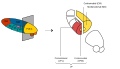
 Thalamic nuclei: MNG = Midline nuclear group AN = Anterior nuclear group MD = Medial dorsal nucleus VNG = Ventral nuclear group VA = Ventral anterior nucleus VL = Ventral lateral nucleus VPL = Ventral posterolateral nucleus VPM = Ventral posteromedial nucleus LNG = Lateral nuclear group PUL = Pulvinar MTh = Metathalamus LG = Lateral geniculate nucleus MG = Medial geniculate nucleus | |
 Thalamic nuclei | |
| Details | |
| Part of | Ventral posterior nucleus |
| Identifiers | |
| Latin | nucleus ventralis posterolateralis |
| NeuroNames | 344 |
| NeuroLex ID | birnlex_737 |
| TA98 | A14.1.08.641 A14.1.08.656 |
| TA2 | 5692 |
| FMA | 62200 |
| Anatomical terms of neuroanatomy | |
The ventral posterolateral nucleus (VPL) is one of the subdivisions of the ventral posterior nucleus in the ventral nuclear group of the thalamus. [1] It relays sensory information from second-order neurons of the neospinothalamic tract and the medial lemniscus (of the dorsal column-medial lemniscus pathway), which synapse with third-order neurons in the nucleus. These then project to the primary somatosensory cortex in the postcentral gyrus. [2] [ citation needed ]
Contents
There is uncertainty regarding the location of VMpo (posterior part of ventral medial nucleus), as determined by spinothalamic tract (STT) terminations and calcium-binding protein staining, and several authorities do not consider its existence as being proved. [1] [3]
The term "ventral posterolateral nucleus" was introduced by Le Gros Clark in 1930. [4] [5]