Primary motor cortex

Last updated
Primary motor cortex
Ba4.png
Brodmann area 4 of the human brain.
Blausen 0103 Brain Sensory&Motor.png
Primary motor cortex shown in green.
Details
Part of Precentral gyrus
Artery Anterior cerebral
Middle cerebral
Identifiers
Latin cortex motorius primus
NeuroNames 1910
NeuroLex ID nlx_143555
FMA 224854
Anatomical terms of neuroanatomy
Animation. Primary motor cortex (Brodmann area 4) of the left cerebral hemisphere shown in red. Brodmann area 4 animation.gif
Animation. Primary motor cortex (Brodmann area 4) of the left cerebral hemisphere shown in red.

The primary motor cortex (Brodmann area 4) is a brain region that in humans is located in the dorsal portion of the frontal lobe. It is the primary region of the motor system and works in association with other motor areas including premotor cortex, the supplementary motor area, posterior parietal cortex, and several subcortical brain regions, to plan and execute voluntary movements. Primary motor cortex is defined anatomically as the region of cortex that contains large neurons known as Betz cells, which, along with other cortical neurons, send long axons down the spinal cord to synapse onto the interneuron circuitry of the spinal cord and also directly onto the alpha motor neurons in the spinal cord which connect to the muscles.

Contents

At the primary motor cortex, motor representation is orderly arranged (in an inverted fashion) from the toe (at the top of the cerebral hemisphere) to mouth (at the bottom) along a fold in the cortex called the central sulcus. However, some body parts may be controlled by partially overlapping regions of cortex. Each cerebral hemisphere of the primary motor cortex only contains a motor representation of the opposite (contralateral) side of the body. The amount of primary motor cortex devoted to a body part is not proportional to the absolute size of the body surface, but, instead, to the relative density of cutaneous motor receptors on said body part. The density of cutaneous motor receptors on the body part is generally indicative of the necessary degree of precision of movement required at that body part. For this reason, the human hands and face have a much larger representation than the legs.

For the discovery of the primary motor cortex and its relationship to other motor cortical areas, see the main article on the motor cortex.

Structure

The human primary motor cortex is located on the anterior wall of the central sulcus. It also extends anteriorly out of the sulcus partly onto the precentral gyrus. Anteriorly, the primary motor cortex is bordered by a set of areas that lie on the precentral gyrus and that are generally considered to compose the lateral premotor cortex. Posteriorly, the primary motor cortex is bordered by the primary somatosensory cortex, which lies on the posterior wall of the central sulcus. Ventrally the primary motor cortex is bordered by the insular cortex in the lateral sulcus. The primary motor cortex extends dorsally to the top of the hemisphere and then continues onto the medial wall of the hemisphere.

The location of the primary motor cortex is most obvious on histological examination due to the presence of the distinctive Betz cells. Layer V of the primary motor cortex contains giant (70-100 μm) pyramidal neurons which are the Betz cells. These neurons send long axons to the contralateral motor nuclei of the cranial nerves and to the lower motor neurons in the ventral horn of the spinal cord. These axons form a part of the corticospinal tract. The Betz cells account for only a small percentage of the corticospinal tract. By some measures, they account for about 10% of the primary motor cortex neurons projecting to the spinal cord [1] or about 2-3% of the total cortical projection to the spinal cord. [2] Though the Betz cells do not compose the entire motor output of the cortex, they nonetheless provide a clear marker for the primary motor cortex. This region of cortex, characterized by the presence of Betz cells, was termed area 4 by Brodmann.

Cellular components

The primary motor cortex alone has been shown to have as many as 116 different types of cells differentiated in their morphology, electrophysiological properties (including firing patterns) and gene expression profile (for example, by type of neurotransmitter released (GABA, glutamate etc.). [3]

Pathway

As the primary motor axons travel down through the cerebral white matter, they move closer together and form part of the posterior limb of the internal capsule.

They continue down into the brainstem, where some of them, after crossing over to the contralateral side, distribute to the cranial nerve motor nuclei. (Note: a few motor fibers synapse with lower motor neurons on the same side of the brainstem).

After crossing over to the contralateral side in the medulla oblongata (pyramidal decussation), the axons travel down the spinal cord as the lateral corticospinal tract .

Fibers that do not cross over in the brainstem travel down the separate ventral corticospinal tract, and most of them cross over to the contralateral side in the spinal cord, shortly before reaching the lower motor neurons. In addition to the main corticospinal tract, Motor cortex projects to other cortical and subcortical areas, including the striatum, hypothalamus, midbrain and hindbrain, as well as the thalamus, basal ganglia, midbrain and medulla [4]

Corticomotorneurons

Corticomotorneurons are neurons in the primary cortex which project directly to motor neurons in the ventral horn of the spinal cord. [5] [6] Axons of corticomotorneurons terminate on the spinal motor neurons of multiple muscles as well as on spinal interneurons. [5] [6] They are unique to primates and it has been suggested that their function is the adaptive control of the distal extremities (e.g. the hands) including the relatively independent control of individual fingers. [6] Corticomotorneurons have so far only been found in the primary motor cortex and not in secondary motor areas. [6]

Blood supply

Branches of the middle cerebral artery provide most of the arterial blood supply for the primary motor cortex.

The medial aspect (leg areas) is supplied by branches of the anterior cerebral artery.

Function

Homunculus

There is a broad representation of the different body parts in the primary motor cortex in an arrangement called a motor homunculus (Latin: little person). [7] The leg area is located close to the midline, in interior sections of the motor area folding into the medial longitudinal fissure. The lateral, convex side of the primary motor cortex is arranged from top to bottom in areas that correspond to the buttocks, torso, shoulder, elbow, wrist, fingers, thumb, eyelids, lips, and jaw. The arm and hand motor area is the largest, and occupies the part of precentral gyrus between the leg and face area.

These areas are not proportional to their size in the body with the lips, face parts, and hands represented by particularly large areas due to the comparative enrichment and density of motor receptor in these regions. Following amputation or paralysis, motor areas can shift to adopt new parts of the body.

Neural input from the thalamus

The primary motor cortex receives thalamic inputs from different thalamic nuclei. Among others:

- Ventral lateral nucleus for cerebellar afferents

- Ventral anterior nucleus for basal ganglia afferents

Alternative maps

Map of the body in the human brain Human motor map.jpg
Map of the body in the human brain

At least two modifications to the classical somatotopic ordering of body parts have been reported in the primary motor cortex of primates.

First, the arm representation may be organized in a core and surround manner. In the monkey cortex, the digits of the hand are represented in a core area at the posterior edge of the primary motor cortex. This core area is surrounded on three sides (on the dorsal, anterior, and ventral sides) by a representation of the more proximal parts of the arm including the elbow and shoulder. [8] [9] In humans, the digit representation is surrounded dorsally, anteriorly, and ventrally, by a representation of the wrist. [10]

A second modification of the classical somatotopic ordering of body parts is a double representation of the digits and wrist studied mainly in the human motor cortex. One representation lies in a posterior region called area 4p, and the other lies in an anterior region called area 4a. The posterior area can be activated by attention without any sensory feedback and has been suggested to be important for initiation of movements, while the anterior area is dependent on sensory feedback. [11] It can also be activated by imaginary finger movements [12] and listening to speech while making no actual movements. This anterior representation area has been suggested to be important in executing movements involving complex sensoriomotor interactions. [13] It is possible that area 4a in humans corresponds to some parts of the caudal premotor cortex as described in the monkey cortex.

In 2009, it was reported, that there are two evolutionary distinct regions, an older one on the outer surface, and a new one found in the cleft. The older one connects to the spinal motorneurons through interneurons in the spinal cord. The newer one, found only in monkeys and apes, connects directly to the spinal motorneurons. [14] The direct connections form after birth, are dominant over the indirect connections, and are more flexible in the circuits they can develop which allows the post-natal learning of complex fine motor skills. "The emergence of the 'new' M1 region during evolution of the primate lineage is therefore likely to have been important for the enhanced manual dexterity of the human hand." [15]

Common misconceptions

Certain misconceptions about the primary motor cortex are common in secondary reviews, textbooks, and popular material. Three of the more common misconceptions are listed here.

Segregated map of the body

One of the most common misconceptions about the primary motor cortex is that the map of the body is cleanly segregated. Yet it is not a map of individuated muscles or even individuated body parts. The map contains considerable overlap. This overlap increases in more anterior regions of the primary motor cortex. One of the main goals in the history of work on the motor cortex was to determine just how much the different body parts are overlapped or segregated in the motor cortex. Researchers who addressed this issue found that the map of the hand, arm, and shoulder contained extensive overlap. [7] [9] [10] [16] [17] [18] [19] [20] Studies that map the precise functional connectivity from cortical neurons to muscles show that even a single neuron in the primary motor cortex can influence the activity of many muscles related to many joints. [16] In experiments on cats and monkeys, as animals learn complex, coordinated movements, the map in the primary motor cortex becomes more overlapping, evidently learning to integrate the control of many muscles. [21] [22] In monkeys, when electrical stimulation is applied to the motor cortex on a behavioral timescale, it evokes complex, highly integrated movements such as reaching with the hand shaped to grasp, or bringing the hand to the mouth and opening the mouth. [23] [24] This type of evidence suggests that the primary motor cortex, while containing a rough map of the body, may participate in integrating muscles in meaningful ways rather than in segregating the control of individual muscle groups. It has been suggested that a deeper principle of organization may be a map of the statistical correlations in the behavioral repertoire, rather than a map of body parts. [24] [25] To the extent that the movement repertoire breaks down partly into the actions of separate body parts, the map contains a rough and overlapping body arrangement.

M1 and primary motor cortex

The term "M1" and the term "primary motor cortex" are often used interchangeably. However, they come from different historical traditions and refer to different divisions of cortex. Some scientists suggested that the motor cortex could be divided into a primary motor strip that was more posterior and a lateral premotor strip that was more anterior. Early researchers who originally proposed this view included Campbell, [26] Vogt and Vogt, [27] Foerster, [28] and Fulton. [29] Others suggested that the motor cortex could not be divided in that manner. Instead, in this second view, the so-called primary motor and lateral premotor strips together composed a single cortical area termed M1. A second motor area on the medial wall of the hemisphere was termed M2 or the supplementary motor area. Proponents of this view included Penfield [7] and Woolsey. [30] Today the distinction between the primary motor cortex and the lateral premotor cortex is generally accepted. However, the term M1 is sometimes mistakenly used to refer to the primary motor cortex. Strictly speaking M1 refers to the single map that, according to some previous researchers, encompassed both the primary motor and the lateral premotor cortex.

Betz cells as the final common pathway

The Betz cells, or giant pyramidal cells in the primary motor cortex, are sometimes mistaken to be the only or main output from the cortex to the spinal cord. This mistake is old, dating back at least to Campbell in 1905. [26] Yet the Betz cells compose only about 2-3% of the neurons that project from the cortex to the spinal cord, [2] and only about 10% of the neurons that project specifically from the primary motor cortex to the spinal cord. [1] A range of cortical areas including the premotor cortex, the supplementary motor area, and even the primary somatosensory cortex, project to the spinal cord. Even when the Betz cells are damaged, the cortex can still communicate to subcortical motor structures and control movement. If the primary motor cortex with its Betz cells is damaged, a temporary paralysis results and other cortical areas can evidently take over some of the lost function.

Clinical significance

Lesions of the precentral gyrus result in paralysis of the contralateral side of the body (facial palsy, arm-/leg monoparesis, hemiparesis) - see upper motor neuron.

Movement coding

Evarts [31] suggested that each neuron in the motor cortex contributes to the force in a muscle. As the neuron becomes active, it sends a signal to the spinal cord, the signal is relayed to a motorneuron, the motorneuron sends a signal to a muscle, and the muscle contracts. The more activity in the motor cortex neuron, the more muscle force.

Georgopoulos and colleagues [32] [33] [34] suggested that muscle force alone was too simple a description. They trained monkeys to reach in various directions and monitored the activity of neurons in the motor cortex. They found that each neuron in the motor cortex was maximally active during a specific direction of reach, and responded less well to neighboring directions of reach. On this basis they suggested that neurons in motor cortex, by "voting" or pooling their influences into a "population code", could precisely specify a direction of reach.

The proposal that motor cortex neurons encode the direction of a reach became controversial. Scott and Kalaska [35] showed that each motor cortex neuron was better correlated with the details of joint movement and muscle force than with the direction of the reach. Schwartz and colleagues [36] showed that motor cortex neurons were well correlated with the speed of the hand. Strick and colleagues [37] found that some neurons in motor cortex were active in association with muscle force and some with the spatial direction of movement. Todorov [38] proposed that the many different correlations are the result of a muscle controller in which many movement parameters happen to be correlated with muscle force.

The code by which neurons in the primate motor cortex control the spinal cord, and thus movement, remains debated.

Some specific progress in understanding how motor cortex causes movement has also been made in the rodent model. The rodent motor cortex, like the monkey motor cortex, may contain subregions that emphasize different common types of actions. [39] [40] For example, one region appears to emphasize the rhythmic control of whisking. [39] [41] [42] Neurons in this region project to a specific subcortical nucleus in which a pattern generator coordinates the cyclic rhythm of the whiskers. This nucleus then projects to the muscles that control the whiskers.

Additional images

See also

Related Research Articles

<span class="mw-page-title-main">Central nervous system</span> Brain and spinal cord

The central nervous system (CNS) is the part of the nervous system consisting of the brain and spinal cord, the retina and optic nerve, and the olfactory nerve and epithelia. The CNS is so named because the brain integrates the received information and coordinates and influences the activity of all parts of the bodies of bilaterally symmetric and triploblastic animals—that is, all multicellular animals except sponges and diploblasts. It is a structure composed of nervous tissue positioned along the rostral to caudal axis of the body and may have an enlarged section at the rostral end which is a brain. Only arthropods, cephalopods and vertebrates have a true brain, though precursor structures exist in onychophorans, gastropods and lancelets.

<span class="mw-page-title-main">Motor neuron</span> Nerve cell sending impulse to muscle

A motor neuron is a neuron whose cell body is located in the motor cortex, brainstem or the spinal cord, and whose axon (fiber) projects to the spinal cord or outside of the spinal cord to directly or indirectly control effector organs, mainly muscles and glands. There are two types of motor neuron – upper motor neurons and lower motor neurons. Axons from upper motor neurons synapse onto interneurons in the spinal cord and occasionally directly onto lower motor neurons. The axons from the lower motor neurons are efferent nerve fibers that carry signals from the spinal cord to the effectors. Types of lower motor neurons are alpha motor neurons, beta motor neurons, and gamma motor neurons.

The motor system is the set of central and peripheral structures in the nervous system that support motor functions, i.e. movement. Peripheral structures may include skeletal muscles and neural connections with muscle tissues. Central structures include cerebral cortex, brainstem, spinal cord, pyramidal system including the upper motor neurons, extrapyramidal system, cerebellum, and the lower motor neurons in the brainstem and the spinal cord.

<span class="mw-page-title-main">Trigeminal nerve</span> Cranial nerve responsible for the faces senses and motor functions

In neuroanatomy, the trigeminal nerve (lit. triplet nerve), also known as the fifth cranial nerve, cranial nerve V, or simply CN V, is a cranial nerve responsible for sensation in the face and motor functions such as biting and chewing; it is the most complex of the cranial nerves. Its name (trigeminal, from Latin tri- 'three', and -geminus 'twin') derives from each of the two nerves (one on each side of the pons) having three major branches: the ophthalmic nerve (V1), the maxillary nerve (V2), and the mandibular nerve (V3). The ophthalmic and maxillary nerves are purely sensory, whereas the mandibular nerve supplies motor as well as sensory (or "cutaneous") functions. Adding to the complexity of this nerve is that autonomic nerve fibers as well as special sensory fibers (taste) are contained within it.

<span class="mw-page-title-main">Somatic nervous system</span> Part of the peripheral nervous system

The somatic nervous system (SNS) is made up of nerves that link the brain and spinal cord to voluntary or skeletal muscles that are under conscious control as well as to skin sensory receptors. Specialized nerve fiber ends called sensory receptors are responsible for detecting information within and outside of the body.

<span class="mw-page-title-main">Pyramidal tracts</span> The corticobulbar tract and the corticospinal tract

The pyramidal tracts include both the corticobulbar tract and the corticospinal tract. These are aggregations of efferent nerve fibers from the upper motor neurons that travel from the cerebral cortex and terminate either in the brainstem (corticobulbar) or spinal cord (corticospinal) and are involved in the control of motor functions of the body.

<span class="mw-page-title-main">Motor cortex</span> Region of the cerebral cortex

The motor cortex is the region of the cerebral cortex involved in the planning, control, and execution of voluntary movements. The motor cortex is an area of the frontal lobe located in the posterior precentral gyrus immediately anterior to the central sulcus.

<span class="mw-page-title-main">Upper motor neuron</span> Neurons in the brain that carry signals to lower motor neurons

Upper motor neurons (UMNs) is a term introduced by William Gowers in 1886. They are found in the cerebral cortex and brainstem and carry information down to activate interneurons and lower motor neurons, which in turn directly signal muscles to contract or relax. UMNs represent the major origin point for voluntary somatic movement.

<span class="mw-page-title-main">Precentral gyrus</span> Motor gyrus of the posterior frontal lobe of the brain

The precentral gyrus is a prominent gyrus on the surface of the posterior frontal lobe of the brain. It is the site of the primary motor cortex that in humans is cytoarchitecturally defined as Brodmann area 4.

<span class="mw-page-title-main">Betz cell</span> Giant pyramidal neurons of the primary motor cortex

Betz cells are giant pyramidal cells (neurons) located within the fifth layer of the grey matter in the primary motor cortex. These neurons are the largest in the central nervous system, sometimes reaching 100 μm in diameter.

<span class="mw-page-title-main">Premotor cortex</span>

The premotor cortex is an area of the motor cortex lying within the frontal lobe of the brain just anterior to the primary motor cortex. It occupies part of Brodmann's area 6. It has been studied mainly in primates, including monkeys and humans. The functions of the premotor cortex are diverse and not fully understood. It projects directly to the spinal cord and therefore may play a role in the direct control of behavior, with a relative emphasis on the trunk muscles of the body. It may also play a role in planning movement, in the spatial guidance of movement, in the sensory guidance of movement, in understanding the actions of others, and in using abstract rules to perform specific tasks. Different subregions of the premotor cortex have different properties and presumably emphasize different functions. Nerve signals generated in the premotor cortex cause much more complex patterns of movement than the discrete patterns generated in the primary motor cortex.

<span class="mw-page-title-main">Supplementary motor area</span> Midline region in front of the motor cortex of the brain

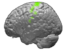
The supplementary motor area (SMA) is a part of the motor cortex of primates that contributes to the control of movement. It is located on the midline surface of the hemisphere just in front of the primary motor cortex leg representation. In monkeys the SMA contains a rough map of the body. In humans the body map is not apparent. Neurons in the SMA project directly to the spinal cord and may play a role in the direct control of movement. Possible functions attributed to the SMA include the postural stabilization of the body, the coordination of both sides of the body such as during bimanual action, the control of movements that are internally generated rather than triggered by sensory events, and the control of sequences of movements. All of these proposed functions remain hypotheses. The precise role or roles of the SMA is not yet known.

A topographic map is the ordered projection of a sensory surface, like the retina or the skin, or an effector system, like the musculature, to one or more structures of the central nervous system. Topographic maps can be found in all sensory systems and in many motor systems.

Premovement neuronal activity in neurophysiological literature refers to neuronal modulations that alter the rate at which neurons fire before a subject produces movement. Through experimentation with multiple animals, predominantly monkeys, it has been shown that several regions of the brain are particularly active and involved in initiation and preparation of movement. Two specific membrane potentials, the bereitschaftspotential, or the BP, and contingent negative variation, or the CNV, play a pivotal role in premovement neuronal activity. Both have been shown to be directly involved in planning and initiating movement. Multiple factors are involved with premovement neuronal activity including motor preparation, inhibition of motor response, programming of the target of movement, closed-looped and open-looped tasks, instructed delay periods, short-lead and long-lead changes, and mirror motor neurons.

<span class="mw-page-title-main">Spinal cord</span> Long, tubular central nervous system structure in the vertebral column

The spinal cord is a long, thin, tubular structure made up of nervous tissue that extends from the medulla oblongata in the brainstem to the lumbar region of the vertebral column (backbone) of vertebrate animals. The center of the spinal cord is hollow and contains a structure called central canal, which contains cerebrospinal fluid. The spinal cord is also covered by meninges and enclosed by the neural arches. Together, the brain and spinal cord make up the central nervous system.

<span class="mw-page-title-main">Anatomy of the cerebellum</span> Structures in the cerebellum, a part of the brain

The anatomy of the cerebellum can be viewed at three levels. At the level of gross anatomy, the cerebellum consists of a tightly folded and crumpled layer of cortex, with white matter underneath, several deep nuclei embedded in the white matter, and a fluid-filled ventricle in the middle. At the intermediate level, the cerebellum and its auxiliary structures can be broken down into several hundred or thousand independently functioning modules or compartments known as microzones. At the microscopic level, each module consists of the same small set of neuronal elements, laid out with a highly stereotyped geometry.

Michael Steven Anthony Graziano is an American scientist and novelist who is currently a professor of Psychology and Neuroscience at Princeton University. His scientific research focuses on the brain basis of awareness. He has proposed the "attention schema" theory, an explanation of how, and for what adaptive advantage, brains attribute the property of awareness to themselves. His previous work focused on how the cerebral cortex monitors the space around the body and controls movement within that space. Notably he has suggested that the classical map of the body in motor cortex, the homunculus, is not correct and is better described as a map of complex actions that make up the behavioral repertoire. His publications on this topic have had a widespread impact among neuroscientists but have also generated controversy. His novels rely partly on his background in psychology and are known for surrealism or magic realism. Graziano also composes music including symphonies and string quartets.

Nonprimary motor cortex is a functionally defined portion of the frontal lobe. It includes two subdivisions, the premotor cortex and the supplementary motor cortex. Largely coincident with the cytoarchitecturally defined area 6 of Brodmann (human), it is located primarily in the rostral portion of the precentral gyrus and caudal portions of the superior frontal gyrus and the middle frontal gyrus, It aids in cerebral control of movement. Anatomically speaking, several nonmprimary areas exist, and make direct connections with the spinal cord.

<span class="mw-page-title-main">Corticospinal tract</span> Pyramidal white matter motor pathway

The corticospinal tract is a white matter motor pathway starting at the cerebral cortex that terminates on lower motor neurons and interneurons in the spinal cord, controlling movements of the limbs and trunk. There are more than one million neurons in the corticospinal tract, and they become myelinated usually in the first two years of life.

<span class="mw-page-title-main">Eberhard Fetz</span> American neuroscientist, academic and researcher

Eberhard Erich Fetz is an American neuroscientist, academic and researcher. He is a Professor of Physiology and Biophysics and DXARTS at the University of Washington.

References

  1. 1 2 Rivara CB, Sherwood CC, Bouras C, Hof PR (2003). "Stereologic characterization and spatial distribution patterns of Betz cells in the human primary motor cortex". The Anatomical Record Part A: Discoveries in Molecular, Cellular, and Evolutionary Biology. 270 (2): 137–151. doi: 10.1002/ar.a.10015 . PMID   12524689.
  2. 1 2 Lassek, A.M. (1941). "The pyramidal tract of the monkey". J. Comp. Neurol. 74 (2): 193–202. doi:10.1002/cne.900740202. S2CID   83536088.
  3. Callaway, Edward M.; Dong, Hong-Wei; Ecker, Joseph R.; Hawrylycz, Michael J.; Huang, Z. Josh; Lein, Ed S.; Ngai, John; Osten, Pavel; Ren, Bing; Tolias, Andreas Savas; White, Owen (October 2021). "A multimodal cell census and atlas of the mammalian primary motor cortex". Nature. 598 (7879): 86–102. doi: 10.1038/s41586-021-03950-0 . ISSN   1476-4687. PMC   8494634 . PMID   34616075.
  4. "Section viewer". brainarchitecture.org. Retrieved 2020-11-20.
  5. 1 2 Siegelbaum, Steven A.; Hudspeth, A.J (2013). Principles of neural science. Kandel, Eric R. (5th ed.). New York. ISBN   9780071390118. OCLC   795553723.{{cite book}}: CS1 maint: location missing publisher (link)
  6. 1 2 3 4 Lemon, Roger N. (April 4, 2008). "Descending Pathways in Motor Control". Annual Review of Neuroscience. 31 (1): 195–218. doi:10.1146/annurev.neuro.31.060407.125547. ISSN   0147-006X. PMID   18558853.
  7. 1 2 3 Penfield, W. and Boldrey, E. (1937). "Somatic motor and sensory representation in the cerebral cortex of man as studied by electrical stimulation". Brain. 60 (4): 389–443. doi:10.1093/brain/60.4.389.{{cite journal}}: CS1 maint: multiple names: authors list (link)
  8. Kwan HC, MacKay WA, Murphy JT, Wong YC (1978). "Spatial organization of precentral cortex in awake primates. II. Motor outputs". J. Neurophysiol. 41 (5): 1120–1131. doi:10.1152/jn.1978.41.5.1120. PMID   100584.
  9. 1 2 Park, M.C., Belhaj-Saif, A., Gordon, M. and Cheney, P.D. (2001). "Consistent features in the forelimb representation of primary motor cortex in rhesus macaques". J. Neurosci. 21 (8): 2784–2792. doi: 10.1523/JNEUROSCI.21-08-02784.2001 . PMC   6762507 . PMID   11306630.{{cite journal}}: CS1 maint: multiple names: authors list (link)
  10. 1 2 Meier, J.D., Aflalo, T.N., Kastner, S. and Graziano, M.S.A. (2008). "Complex organization of human primary motor cortex: A high-resolution fMRI study". J. Neurophysiol. 100 (4): 1800–1812. doi:10.1152/jn.90531.2008. PMC   2576195 . PMID   18684903.{{cite journal}}: CS1 maint: multiple names: authors list (link)
  11. Binkofski F, Fink GR, Geyer S, Buccino G, Gruber O, Shah NJ, Taylor JG, Seitz RJ, Zilles K, Freund HJ (2002). "Neural activity in human primary motor cortex areas 4a and 4b is modulated differentially by attention to action". J. Neurophysiol. 88 (1): 514–519. doi:10.1152/jn.2002.88.1.514. hdl: 11858/00-001M-0000-0010-CA2B-E . PMID   12091573. S2CID   6459190.
  12. Nikhil Sharma; P.S. Jones; T.A. Carpenter; Jean-Claude Baron (2008). "Mapping the involvement of BA 4a and 4p during Motor Imagery". NeuroImage. 41 (1): 92–99. doi:10.1016/j.neuroimage.2008.02.009. PMID   18358742. S2CID   8673179.
  13. Terumitsu M, Ikeda K, Kwee IL, and Nakada, T (2009). "Participation of primary motor cortex area 4a in complex sensory processing: 3.0T fMRI study". NeuroReport. 20 (7): 679–683. doi:10.1097/WNR.0b013e32832a1820. PMID   19339906. S2CID   23674509.{{cite journal}}: CS1 maint: multiple names: authors list (link)
  14. Rathelot, J.-A.; strick, P. L. (Jan 20, 2009). "Subdivisions of primary motor cortex based on cortico-motoneuronal cells". Proc. Natl. Acad. Sci. 106 (3): 918–923. Bibcode:2009PNAS..106..918R. doi: 10.1073/pnas.0808362106 . PMC   2621250 . PMID   19139417.
  15. Costandi, Mo (2009). "The evolution of manual dexterity". Proceedings of the National Academy of Sciences. 106 (3): 918–923. doi: 10.1073/pnas.0808362106 . PMC   2621250 . PMID   19139417 . Retrieved 29 November 2015.
  16. 1 2 Cheney, P.D. & Fetz, E.E. (1985). "Comparable patterns of muscle facilitation evoked by individual corticomotoneuronal (CM) cells and by single intracortical microstimuli in primates: evidence for functional groups of CM cells". J. Neurophysiol. 53 (3): 786–804. doi:10.1152/jn.1985.53.3.786. PMID   2984354.
  17. Schieber, M.H. & Hibbard, L.S. (1993). "How somatotopic is the motor cortex hand area?". Science. 261 (5120): 489–492. Bibcode:1993Sci...261..489S. doi:10.1126/science.8332915. PMID   8332915.
  18. Rathelot, J.A. & Strick, P.L. (2006). "Muscle representation in the macaque motor cortex: an anatomical perspective". Proc. Natl. Acad. Sci. USA. 103 (21): 8257–8262. Bibcode:2006PNAS..103.8257R. doi: 10.1073/pnas.0602933103 . PMC   1461407 . PMID   16702556.
  19. Sanes, J.N., Donoghue, J.P., Thangaraj, V., Edelman, R.R. and Warach, S. (1995). "Shared neural substrates controlling hand movements in human motor cortex". Science. 268 (5218): 1775–1777. Bibcode:1995Sci...268.1775S. doi:10.1126/science.7792606. PMID   7792606.{{cite journal}}: CS1 maint: multiple names: authors list (link)
  20. Donoghue, J.P., Leibovic, S. and Sanes, J.N. (1992). "Organization of the forelimb area in squirrel monkey motor cortex: representation of digit, wrist and elbow muscles". Exp. Brain Res. 89 (1): 1–10. doi:10.1007/bf00228996. PMID   1601087. S2CID   1398462.{{cite journal}}: CS1 maint: multiple names: authors list (link)
  21. Nudo, R.J., Milliken, G.W., Jenkins, W.M. and Merzenich, M.M. (1996). "Use-dependent alterations of movement representations in primary motor cortex of adult squirrel monkeys". J. Neurosci. 16 (2): 785–807. doi:10.1523/JNEUROSCI.16-02-00785.1996. PMC   6578638 . PMID   8551360.{{cite journal}}: CS1 maint: multiple names: authors list (link)
  22. Martin, J.H., Engber, D. and Meng, Z. (2005). "Effect of forelimb use on postnatal development of the forelimb motor representation in primary motor cortex of the cat". J. Neurophysiol. 93 (5): 2822–2831. doi:10.1152/jn.01060.2004. PMID   15574795.{{cite journal}}: CS1 maint: multiple names: authors list (link)
  23. Graziano, M.S.A., Taylor, C.S.R. and Moore, T. (2002). "Complex movements evoked by microstimulation of precentral cortex". Neuron. 34 (5): 841–851. doi: 10.1016/S0896-6273(02)00698-0 . PMID   12062029. S2CID   3069873.{{cite journal}}: CS1 maint: multiple names: authors list (link)
  24. 1 2 Graziano, M.S.A. (2008). The Intelligent Movement Machine. Oxford, UK: Oxford University Press.
  25. Graziano, M.S.A. and Aflalo, T.N. (2007). "Mapping behavioral repertoire onto the cortex". Neuron. 56 (2): 239–251. doi: 10.1016/j.neuron.2007.09.013 . PMID   17964243.{{cite journal}}: CS1 maint: multiple names: authors list (link)
  26. 1 2 Campbell, A. W. (1905). Histological Studies on the Localization of Cerebral Function. Cambridge, Massachusetts: Cambridge University Press.
  27. Vogt, C. and Vogt, O. (1919). "Ergebnisse unserer Hirnforschung". Journal für Psychologie und Neurologie. 25: 277–462.{{cite journal}}: CS1 maint: multiple names: authors list (link)
  28. Foerster, O (1936). "The motor cortex of man in the light of Hughlings Jackson's doctrines". Brain. 59 (2): 135–159. doi:10.1093/brain/59.2.135.
  29. Fulton, J (1935). "A note on the definition of the "motor" and "premotor" areas". Brain. 58 (2): 311–316. doi:10.1093/brain/58.2.311.
  30. Woolsey, C.N., Settlage, P.H., Meyer, D.R., Sencer, W., Hamuy, T.P. and Travis, A.M. (1952). "Pattern of localization in precentral and "supplementary" motor areas and their relation to the concept of a premotor area". Association for Research in Nervous and Mental Disease. 30. New York, NY: Raven Press: 238–264.{{cite journal}}: CS1 maint: multiple names: authors list (link)
  31. Evarts, E.V. (1968). "Relation of pyramidal tract activity to force exerted during voluntary movement". J. Neurophysiol. 31 (1): 14–27. doi:10.1152/jn.1968.31.1.14. PMID   4966614.
  32. Georgopoulos, A.P., Kalaska, J.F., Caminiti, R. and Massey, J.T. (1982). "On the relations between the direction of two-dimensional arm movements and cell discharge in primate motor cortex". J. Neurosci. 2 (11): 1527–1537. doi:10.1523/JNEUROSCI.02-11-01527.1982. PMC   6564361 . PMID   7143039.{{cite journal}}: CS1 maint: multiple names: authors list (link)
  33. Georgopoulos A.P., Kettner, R.E. and Schwartz, A.B. (1988). "Primate motor cortex and free arm movements to visual targets in three-dimensional space. II. Coding of the direction of movement by a neuronal population". J. Neurosci. 8 (8): 2928–2937. doi:10.1523/JNEUROSCI.08-08-02928.1988. PMC   6569382 . PMID   3411362.{{cite journal}}: CS1 maint: multiple names: authors list (link)
  34. Georgopoulos A.P., Schwartz, A.B. and Kettner, R.E. (1986). "Neuronal population coding of movement direction". Science. 233 (4771): 1416–1419. Bibcode:1986Sci...233.1416G. doi:10.1126/science.3749885. PMID   3749885.{{cite journal}}: CS1 maint: multiple names: authors list (link)
  35. Scott, S.H. & Kalaska, J.F. (1995). "Changes in motor cortex activity during reaching movements with similar hand paths but different arm postures". J. Neurophysiol. 73 (6): 2563–2567. doi:10.1152/jn.1995.73.6.2563. PMID   7666162.
  36. Moran, D.W. & Schwartz, A.B. (1999). "Motor cortical representation of speed and direction during reaching". J. Neurophysiol. 82 (5): 2676–2692. doi:10.1152/jn.1999.82.5.2676. PMID   10561437. S2CID   9789197.
  37. Kakei, S., Hoffman, D. and Strick, P (1999). "Muscle and movement representations in the primary motor cortex". Science. 285 (5436): 2136–2139. CiteSeerX   10.1.1.137.8610 . doi:10.1126/science.285.5436.2136. PMID   10497133.{{cite journal}}: CS1 maint: multiple names: authors list (link)
  38. Todorov, E (2000). "Direct cortical control of muscle activation in voluntary arm movements: a model". Nat Neurosci. 3 (4): 391–398. doi:10.1038/73964. PMID   10725930. S2CID   13996279.
  39. 1 2 Haiss, F. & Schwarz, C (2005). "Spatial segregation of different modes of movement control in the whisker representation of rat primary motor cortex". J. Neurosci. 25 (6): 1579–1587. doi:10.1523/JNEUROSCI.3760-04.2005. PMC   6726007 . PMID   15703412.
  40. Ramanathan, D., Conner, J.M. and Tuszynski, M.H. (2006). "A form of motor cortical plasticity that correlates with recovery of function after brain injury". Proc. Natl. Acad. Sci. U.S.A. 103 (30): 11370–11375. Bibcode:2006PNAS..10311370R. doi: 10.1073/pnas.0601065103 . PMC   1544093 . PMID   16837575.{{cite journal}}: CS1 maint: multiple names: authors list (link)
  41. Brecht, M., Schneider, M., Sakmann, B. and Margrie, T.W. (2004). "Whisker movements evoked by stimulation of single pyramidal cells in rat motor cortex". Nature. 427 (6976): 704–710. Bibcode:2004Natur.427..704B. doi:10.1038/nature02266. PMID   14973477. S2CID   1105868.{{cite journal}}: CS1 maint: multiple names: authors list (link)
  42. Cramer, N.P. & Keller, A (2006). "Cortical control of a whisking central pattern generator". J. Neurophysiol. 96 (1): 209–217. doi:10.1152/jn.00071.2006. PMC   1764853 . PMID   16641387.