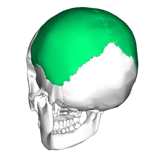
The parietal bones are two bones in the skull which, when joined at a fibrous joint known as a cranial suture, form the sides and roof of the neurocranium. In humans, each bone is roughly quadrilateral in form, and has two surfaces, four borders, and four angles. It is named from the Latin paries (-ietis), wall.
The primary sensory areas are the primary cortical regions of the five sensory systems in the brain. Except for the olfactory system, they receive sensory information from thalamic nerve projections. The term primary comes from the fact that these cortical areas are the first level in a hierarchy of sensory information processing in the brain. This should not be confused with the function of the primary motor cortex, which is the last site in the cortex for processing motor commands.

The occipital lobe is one of the four major lobes of the cerebral cortex in the brain of mammals. The name derives from its position at the back of the head, from the Latin ob, 'behind', and caput, 'head'.
Parietal is an adjective used predominantly for the parietal lobe and other relevant anatomy
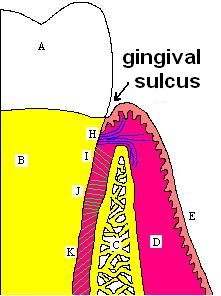
In biological morphology and anatomy, a sulcus is a furrow or fissure. It may be a groove, natural division, deep furrow, elongated cleft, or tear in the surface of a limb or an organ, most notably on the surface of the brain, but also in the lungs, certain muscles, as well as in bones, and elsewhere. Many sulci are the product of a surface fold or junction, such as in the gums, where they fold around the neck of the tooth.

The middle cerebral artery (MCA) is one of the three major paired cerebral arteries that supply blood to the cerebrum. The MCA arises from the internal carotid artery and continues into the lateral sulcus where it then branches and projects to many parts of the lateral cerebral cortex. It also supplies blood to the anterior temporal lobes and the insular cortices.

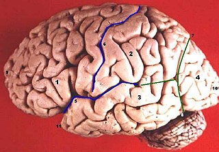
The lobes of the brain are the four major identifiable regions of the human cerebral cortex, and they comprise the surface of each hemisphere of the cerebrum. The two hemispheres are roughly symmetrical in structure, and are connected by the corpus callosum. Some sources include the insula and limbic lobe but the limbic lobe incorporates parts of the other lobes. The lobes are large areas that are anatomically distinguishable, and are also functionally distinct. Each lobe of the brain has numerous ridges, or gyri, and furrows, sulci that constitute further subzones of the cortex. The expression "lobes of the brain" usually refers only to those of the cerebrum, not to the distinct areas of the cerebellum.

The cuneus is a smaller lobe in the occipital lobe of the brain. The cuneus is bounded anteriorly by the parieto-occipital sulcus and inferiorly by the calcarine sulcus.
The posterior cerebral artery (PCA) is one of a pair of cerebral arteries that supply oxygenated blood to the occipital lobe, part of the back of the human brain. The two arteries originate from the distal end of the basilar artery, where it bifurcates into the left and right posterior cerebral arteries. These anastomose with the middle cerebral arteries and internal carotid arteries via the posterior communicating arteries.

The confluence of sinuses, torcular Herophili, or torcula is the connecting point of the superior sagittal sinus, straight sinus, and occipital sinus. It is below the internal occipital protuberance of the skull. It drains venous blood from the brain into the transverse sinuses. It may be affected by arteriovenous fistulas, a thrombus, major trauma, or surgical damage, and may be imaged with many radiology techniques.

In human brain anatomy, an operculum, may refer to the frontal, temporal, or parietal operculum, which together cover the insula as the opercula of insula. It can also refer to the occipital operculum, part of the occipital lobe.

In neuroanatomy, the parieto-occipital sulcus is a deep sulcus in the cerebral cortex that marks the boundary between the cuneus and precuneus, and also between the parietal and occipital lobes. Only a small part can be seen on the lateral surface of the hemisphere, its chief part being on the medial surface.

The superior parietal lobule is bounded in front by the upper part of the postcentral sulcus, but is usually connected with the postcentral gyrus above the end of the sulcus. The superior parietal lobule contains Brodmann's areas 5 and 7.

The squamous part of occipital bone is situated above and behind the foramen magnum, and is curved from above downward and from side to side.
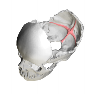
The cruciform eminence divides the deeply concave internal surface of the occipital bone into four fossae:
The sensory cortex can refer sometimes to the primary somatosensory cortex, or it can be used as a term for the primary and secondary cortices of the different senses : the visual cortex on the occipital lobes, the auditory cortex on the temporal lobes, the primary olfactory cortex on the uncus of the piriform region of the temporal lobes, the gustatory cortex on the insular lobe, and the primary somatosensory cortex on the anterior parietal lobes. Just posterior to the primary somatosensory cortex lies the somatosensory association cortex or area, which integrates sensory information from the primary somatosensory cortex to construct an understanding of the object being felt. Inferior to the frontal lobes are found the olfactory bulbs, which receive sensory input from the olfactory nerves and route those signals throughout the brain. Not all olfactory information is routed to the olfactory cortex: some neural fibers are routed to the supraorbital region of the frontal lobe, while others are routed directly to limbic structures. The direct limbic connection makes the olfactory sense unique.

In the occipital lobe, the lateral occipital sulcus, where present, divides the lateral, or middle occipital gyrus into a superior and an inferior part, which are then continuous in front with the parietal and temporal lobes. The anterior portion is often incomplete, but in some individuals it may encounter the superior temporal sulcus whilst the posterior portion originates from the middle of the curved lunate sulcus, or from a curved portion of the transverse occipital sulcus if absent.

In brain anatomy, the lunate sulcus or simian sulcus, also known as the sulcus lunatus, is a fissure in the occipital lobe variably found in humans and more often larger when present in apes and monkeys. The lunate sulcus marks the transition between V1 and V2.
About 5 centimetres (2.0 in) in front of the occipital pole of the human brain, on the infero-lateral border is an indentation or notch, named the preoccipital notch. It is considered a landmark because the occipital lobe is located just behind the line that connects that notch with the parietoccipital sulcus.

The occipital gyri (OcG) are three gyri in parallel, along the lateral portion of the occipital lobe, also referred to as a composite structure in the brain. The gyri are the superior occipital gyrus, the middle occipital gyrus, and the inferior occipital gyrus, and these are also known as the occipital face area. The superior and inferior occipital sulci separates the three occipital gyri.

