Intraparietal sulcus

Last updated
Intraparietal sulcus
Gray726 intraparietal sulcus.svg
Lateral surface of left cerebral hemisphere, viewed from the side. (Intraparietal sulcus visible at upper right, running horizontally.)
ParietCapts lateral.png
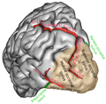
Right cerebral hemisphere, viewed from the side. The region colored in blue is parietal lobe of the human brain. Intraparietal sulcus runs horizontally at the middle of the parietal lobe.
Details
Part of Parietal lobe
Identifiers
Latin sulcus intraparietalis
Acronym(s)IPS
NeuroNames 97
NeuroLex ID birnlex_4031
TA98 A14.1.09.127
TA2 5475
FMA 83772
Anatomical terms of neuroanatomy

The intraparietal sulcus (IPS) is located on the lateral surface of the parietal lobe, and consists of an oblique and a horizontal portion. The IPS contains a series of functionally distinct subregions that have been intensively investigated using both single cell neurophysiology in primates [1] [2] and human functional neuroimaging. [3] Its principal functions are related to perceptual-motor coordination (e.g., directing eye movements and reaching) and visual attention, which allows for visually-guided pointing, grasping, and object manipulation that can produce a desired effect.

Contents

The IPS is also thought to play a role in other functions, including processing symbolic numerical information, [4] visuospatial working memory [5] , decision-making, [6] and interpreting the intent of others. [7] [ unreliable medical source? ]

Function

Five regions of the intraparietal sulcus (IPS): anterior, lateral, ventral, caudal, and medial

All of these areas have projections to the frontal lobe for executive control.

Activity in the intraparietal sulcus has also been associated with the learning of sequences of finger movements. [8]

The dorsal attention network includes the intraparietal sulcus of each hemisphere. [9] The intraparietal sulcus is activated during voluntary orientation of attention. [10]

Understanding numbers

Behavioral studies suggest that the IPS is associated with impairments of basic numerical magnitude processing and that there is a pattern of structural and functional alternations in the IPS and in the PFC in dyscalculia. [11] Children with developmental dyscalculia were found to have less gray matter in the left IPS. [12]

Studies have shown that electrical activity in a particular group of nerve cells in the intraparietal sulcus spiked when, and only when, volunteers were performing calculations. Outside experimental settings it was also found that when a patient mentioned a number—or even a quantitative reference, such as "some more", "many" or "bigger than the other one"—there was a spike of electrical activity in the same nerve-cell population of the intraparietal sulcus that was activated when the patient was doing calculations under experimental conditions. [13]

Additional images

Related Research Articles

<span class="mw-page-title-main">Cerebral cortex</span> Outer layer of the cerebrum of the mammalian brain

The cerebral cortex, also known as the cerebral mantle, is the outer layer of neural tissue of the cerebrum of the brain in humans and other mammals. It is the largest site of neural integration in the central nervous system. and plays a key role in attention, perception, awareness, thought, memory, language, and consciousness. The cerebral cortex is part of the brain responsible for cognition.

<span class="mw-page-title-main">Parietal lobe</span> Part of the brain responsible for sensory input and some language processing

The parietal lobe is one of the four major lobes of the cerebral cortex in the brain of mammals. The parietal lobe is positioned above the temporal lobe and behind the frontal lobe and central sulcus.

<span class="mw-page-title-main">Precuneus</span> Region of the parietal lobe of the brain

In neuroanatomy, the precuneus is the portion of the superior parietal lobule on the medial surface of each brain hemisphere. It is located in front of the cuneus. The precuneus is bounded in front by the marginal branch of the cingulate sulcus, at the rear by the parieto-occipital sulcus, and underneath by the subparietal sulcus. It is involved with episodic memory, visuospatial processing, reflections upon self, and aspects of consciousness.

<span class="mw-page-title-main">Frontal lobe</span> Part of the brain

The frontal lobe is the largest of the four major lobes of the brain in mammals, and is located at the front of each cerebral hemisphere. It is parted from the parietal lobe by a groove between tissues called the central sulcus and from the temporal lobe by a deeper groove called the lateral sulcus. The most anterior rounded part of the frontal lobe is known as the frontal pole, one of the three poles of the cerebrum.

Dyscalculia is a disability resulting in difficulty learning or comprehending arithmetic, such as difficulty in understanding numbers, learning how to manipulate numbers, performing mathematical calculations, and learning facts in mathematics. It is sometimes colloquially referred to as "math dyslexia", though this analogy is misleading as they are distinct syndromes.

<span class="mw-page-title-main">Brodmann area 40</span> Part of the parietal cortex in the human brain

Brodmann area 40 (BA40) is part of the parietal cortex in the human brain. The inferior part of BA40 is in the area of the supramarginal gyrus, which lies at the posterior end of the lateral fissure, in the inferior lateral part of the parietal lobe.

<span class="mw-page-title-main">Angular gyrus</span> Gyrus of the parietal lobe of the brain

The angular gyrus is a region of the brain lying mainly in the posteroinferior region of the parietal lobe, occupying the posterior part of the inferior parietal lobule. It represents the Brodmann area 39.

<span class="mw-page-title-main">Lobes of the brain</span> Parts of the cerebrum

The lobes of the brain are the major identifiable zones of the human cerebral cortex, and they comprise the surface of each hemisphere of the cerebrum. The two hemispheres are roughly symmetrical in structure, and are connected by the corpus callosum. They traditionally have been divided into four lobes, but are today considered as having six lobes each. The lobes are large areas that are anatomically distinguishable, and are also functionally distinct to some degree. Each lobe of the brain has numerous ridges, or gyri, and furrows, the sulci that constitute further subzones of the cortex. The expression "lobes of the brain" usually refers only to those of the cerebrum, not to the distinct areas of the cerebellum.

<span class="mw-page-title-main">Frontal eye fields</span> Region of the frontal cortex of the brain

The frontal eye fields (FEF) are a region located in the frontal cortex, more specifically in Brodmann area 8 or BA8, of the primate brain. In humans, it can be more accurately said to lie in a region around the intersection of the middle frontal gyrus with the precentral gyrus, consisting of a frontal and parietal portion. The FEF is responsible for saccadic eye movements for the purpose of visual field perception and awareness, as well as for voluntary eye movement. The FEF communicates with extraocular muscles indirectly via the paramedian pontine reticular formation. Destruction of the FEF causes deviation of the eyes to the ipsilateral side.

<span class="mw-page-title-main">Superior parietal lobule</span>

The superior parietal lobule is bounded in front by the upper part of the postcentral sulcus, but is usually connected with the postcentral gyrus above the end of the sulcus. The superior parietal lobule contains Brodmann's areas 5 and 7.

<span class="mw-page-title-main">Inferior parietal lobule</span> Portion of the parietal lobe of the brain

The inferior parietal lobule lies below the horizontal portion of the intraparietal sulcus, and behind the lower part of the postcentral sulcus. Also known as Geschwind's territory after Norman Geschwind, an American neurologist, who in the early 1960s recognised its importance. It is a part of the parietal lobe.

Attentional shift occurs when directing attention to a point increases the efficiency of processing of that point and includes inhibition to decrease attentional resources to unwanted or irrelevant inputs. Shifting of attention is needed to allocate attentional resources to more efficiently process information from a stimulus. Research has shown that when an object or area is attended, processing operates more efficiently. Task switching costs occur when performance on a task suffers due to the increased effort added in shifting attention. There are competing theories that attempt to explain why and how attention is shifted as well as how attention is moved through space in attentional control.

Numerical cognition is a subdiscipline of cognitive science that studies the cognitive, developmental and neural bases of numbers and mathematics. As with many cognitive science endeavors, this is a highly interdisciplinary topic, and includes researchers in cognitive psychology, developmental psychology, neuroscience and cognitive linguistics. This discipline, although it may interact with questions in the philosophy of mathematics, is primarily concerned with empirical questions.

<span class="mw-page-title-main">Superior longitudinal fasciculus</span> Association fiber tract of the brain

The superior longitudinal fasciculus (SLF) is an association tract in the brain that is composed of three separate components. It is present in both hemispheres and can be found lateral to the centrum semiovale and connects the frontal, occipital, parietal, and temporal lobes. This bundle of tracts (fasciculus) passes from the frontal lobe through the operculum to the posterior end of the lateral sulcus where they either radiate to and synapse on neurons in the occipital lobe, or turn downward and forward around the putamen and then radiate to and synapse on neurons in anterior portions of the temporal lobe.

<span class="mw-page-title-main">Posterior parietal cortex</span>

The posterior parietal cortex plays an important role in planned movements, spatial reasoning, and attention.

<span class="mw-page-title-main">Dorsal attention network</span> Large-scale brain network involved in voluntary orienting of attention

The dorsal attention network (DAN), also known anatomically as the dorsal frontoparietal network (D-FPN), is a large-scale brain network of the human brain that is primarily composed of the intraparietal sulcus (IPS) and frontal eye fields (FEF). It is named and most known for its role in voluntary orienting of visuospatial attention.

<span class="mw-page-title-main">Brain asymmetry</span> Term in human neuroanatomy referring to several things

In human neuroanatomy, brain asymmetry can refer to at least two quite distinct findings:

<span class="mw-page-title-main">Superior temporal sulcus</span> Part of the brains temporal lobe

In the human brain, the superior temporal sulcus (STS) is the sulcus separating the superior temporal gyrus from the middle temporal gyrus in the temporal lobe of the brain. A sulcus is a deep groove that curves into the largest part of the brain, the cerebrum, and a gyrus is a ridge that curves outward of the cerebrum.

The approximate number system (ANS) is a cognitive system that supports the estimation of the magnitude of a group without relying on language or symbols. The ANS is credited with the non-symbolic representation of all numbers greater than four, with lesser values being carried out by the parallel individuation system, or object tracking system. Beginning in early infancy, the ANS allows an individual to detect differences in magnitude between groups. The precision of the ANS improves throughout childhood development and reaches a final adult level of approximately 15% accuracy, meaning an adult could distinguish 100 items versus 115 items without counting. The ANS plays a crucial role in development of other numerical abilities, such as the concept of exact number and simple arithmetic. The precision level of a child's ANS has been shown to predict subsequent mathematical achievement in school. The ANS has been linked to the intraparietal sulcus of the brain.

In psychology, the numerical Stroop effect demonstrates the relationship between numerical values and physical sizes. When digits are presented visually, they can be physically large or small, irrespective of their actual values. Congruent pairs occur when size and value correspond while incongruent pairs occur when size and value are incompatible. It was found that when people are asked to compare digits, their reaction time tends to be slower in the case of incongruent pairs. This reaction time difference between congruent and incongruent pairs is termed the numerical Stroop effect

References

  1. Colby C.E.; Goldberg M.E. (1999). "Space and attention in parietal cortex". Annual Review of Neuroscience. 22: 319–349. doi:10.1146/annurev.neuro.22.1.319. PMID   10202542.
  2. Andersen R.A. (1989). "Visual and eye movement functions of the posterior parietal cortex" (PDF). Annual Review of Neuroscience. 12: 377–403. doi:10.1146/annurev.ne.12.030189.002113. PMID   2648954.
  3. Culham, J.C.; Nancy G. Kanwisher (April 2001). "Neuroimaging of cognitive functions in human parietal cortex". Current Opinion in Neurobiology . 11 (2): 157–163. doi:10.1016/S0959-4388(00)00191-4. PMID   11301234. S2CID   13907037.
  4. Cantlon J, Brannon E, Carter E, Pelphrey K (2006). "Functional imaging of numerical processing in adults and 4-y-old children". PLOS Biol. 4 (5): e125. doi: 10.1371/journal.pbio.0040125 . PMC   1431577 . PMID   16594732.
  5. Todd JJ, Marois R (2004). "Capacity limit of visual short-term memory in human posterior parietal cortex". Nature. 428 (6984): 751–754. Bibcode:2004Natur.428..751T. doi:10.1038/nature02466. PMID   15085133. S2CID   4415712.
  6. Valdebenito-Oyarzo, Gabriela; Martínez-Molina, María Paz; Soto-Icaza, Patricia; Zamorano, Francisco; Figueroa-Vargas, Alejandra; Larraín-Valenzuela, Josefina; Stecher, Ximena; Salinas, César; Bastin, Julien; Valero-Cabré, Antoni; Polania, Rafael; Billeke, Pablo (10 January 2024). "The parietal cortex has a causal role in ambiguity computations in humans". PLOS Biology. 22 (1): e3002452. doi: 10.1371/journal.pbio.3002452 . PMC   10824459 . PMID   38198502.
  7. Grafton, Hamilton (2006). "Dartmouth Study Finds How The Brain Interprets The Intent Of Others". Science Daily.
  8. Sakai, K.; Ramnani, N.; Passingham, R. E. (2002). "Learning of sequences of finger movements and timing: Frontal lobe and action-oriented representation". Journal of Neurophysiology. 88 (4): 2035–2046. doi:10.1152/jn.2002.88.4.2035. PMID   12364526.
  9. Fox, M.D.; Corbetta, M.; Snyder, A.Z.; Vincent, J.L.; Raichle, M.E. (2006). "Spontaneous neuronal activity distinguishes human dorsal and ventral attention systems". Proceedings of the National Academy of Sciences . 103 (26): 10046–10051. Bibcode:2006PNAS..10310046F. doi: 10.1073/pnas.0604187103 . PMC   1480402 . PMID   16788060.
  10. Corbetta, M.; Kincade, J. M.; Ollinger, J. M.; McAvoy, M. P.; Shulman, G. L. (2000). "Voluntary orienting is dissociated from target detection in human posterior parietal cortex". Nature Neuroscience. 3 (3): 292–297. doi:10.1038/73009. PMID   10700263. S2CID   52807698.
  11. Ansari D.; Karmiloff-Smith A. (2002). "Atypical trajectories of number development: a neuroconstructivist perspective". Trends in Cognitive Sciences. 6 (12): 511–516. doi:10.1016/S1364-6613(02)02040-5. PMID   12475711. S2CID   1228615.
  12. Kucian K, et al. (2006). "Impaired neural networks for approximate calculation in dyscalculic children: a functional MRI study". Behavioral and Brain Functions. 2: 31. doi: 10.1186/1744-9081-2-31 . PMC   1574332 . PMID   16953876.
  13. Dastjerdi, M.; Ozker, M.; Foster, B. L.; Rangarajan, V.; Parvizi, J. (2013). "Numerical processing in the human parietal cortex during experimental and natural conditions". Nature Communications. 4: 2528. Bibcode:2013NatCo...4.2528D. doi:10.1038/ncomms3528. PMC   3826627 . PMID   24129341.