A Brodmann area is a region of the cerebral cortex, in the human or other primate brain, defined by its cytoarchitecture, or histological structure and organization of cells. The concept was first introduced by the German anatomist Korbinian Brodmann in the early 20th century. Brodmann mapped the human brain based on the varied cellular structure across the cortex and identified 52 distinct regions, which he numbered 1 to 52. These regions, or Brodmann areas, correspond with diverse functions including sensation, motor control, and cognition.

The parietal lobe is one of the four major lobes of the cerebral cortex in the brain of mammals. The parietal lobe is positioned above the temporal lobe and behind the frontal lobe and central sulcus.

In neuroanatomy, the precuneus is the portion of the superior parietal lobule on the medial surface of each brain hemisphere. It is located in front of the cuneus. The precuneus is bounded in front by the marginal branch of the cingulate sulcus, at the rear by the parieto-occipital sulcus, and underneath by the subparietal sulcus. It is involved with episodic memory, visuospatial processing, reflections upon self, and aspects of consciousness.
The primary sensory areas are the primary cortical regions of the five sensory systems in the brain. Except for the olfactory system, they receive sensory information from thalamic nerve projections. The term primary comes from the fact that these cortical areas are the first level in a hierarchy of sensory information processing in the brain. This should not be confused with the function of the primary motor cortex, which is the last site in the cortex for processing motor commands.
Brodmann area 6 (BA6) is part of the frontal cortex in the human brain. Situated just anterior to the primary motor cortex (BA4), it is composed of the premotor cortex and, medially, the supplementary motor area (SMA). This large area of the frontal cortex is believed to play a role in planning complex, coordinated movements.

Brodmann area 10 is the anterior-most portion of the prefrontal cortex in the human brain. BA10 was originally defined broadly in terms of its cytoarchitectonic traits as they were observed in the brains of cadavers, but because modern functional imaging cannot precisely identify these boundaries, the terms anterior prefrontal cortex, rostral prefrontal cortex and frontopolar prefrontal cortex are used to refer to the area in the most anterior part of the frontal cortex that approximately covers BA10—simply to emphasize the fact that BA10 does not include all parts of the prefrontal cortex.
Brodmann area 7 is one of Brodmann's cytologically defined regions of the brain corresponding to precuneus and superior parietal lobule (SPL). It is involved in locating objects in space. It serves as a point of convergence between vision and proprioception to determine where objects are in relation to parts of the body.

Brodmann area 40 (BA40) is part of the parietal cortex in the human brain. The inferior part of BA40 is in the area of the supramarginal gyrus, which lies at the posterior end of the lateral fissure, in the inferior lateral part of the parietal lobe.

The inferior frontal gyrus(IFG), (gyrus frontalis inferior), is the lowest positioned gyrus of the frontal gyri, of the frontal lobe, and is part of the prefrontal cortex.
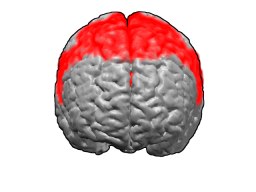
In neuroanatomy, the superior frontal gyrus is a gyrus – a ridge on the brain's cerebral cortex – which makes up about one third of the frontal lobe. It is bounded laterally by the superior frontal sulcus.

The transverse temporal gyri, also called Heschl's gyri or Heschl's convolutions, are gyri found in the area of primary auditory cortex buried within the lateral sulcus of the human brain, occupying Brodmann areas 41 and 42. Transverse temporal gyri are superior to and separated from the planum temporale by Heschl's sulcus. Transverse temporal gyri are found in varying numbers in both the right and left hemispheres of the brain and one study found that this number is not related to the hemisphere or dominance of hemisphere studied in subjects. Transverse temporal gyri can be viewed in the sagittal plane as either an omega shape or a heart shape.

The superior temporal gyrus (STG) is one of three gyri in the temporal lobe of the human brain, which is located laterally to the head, situated somewhat above the external ear.

In neuroanatomy, the primary somatosensory cortex is located in the postcentral gyrus of the brain's parietal lobe, and is part of the somatosensory system. It was initially defined from surface stimulation studies of Wilder Penfield, and parallel surface potential studies of Bard, Woolsey, and Marshall. Although initially defined to be roughly the same as Brodmann areas 3, 1 and 2, more recent work by Kaas has suggested that for homogeny with other sensory fields only area 3 should be referred to as "primary somatosensory cortex", as it receives the bulk of the thalamocortical projections from the sensory input fields.

The angular gyrus is a region of the brain lying mainly in the posteroinferior region of the parietal lobe, occupying the posterior part of the inferior parietal lobule. It represents the Brodmann area 39.
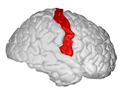
The precentral gyrus is a prominent gyrus on the surface of the posterior frontal lobe of the brain. It is the site of the primary motor cortex that in humans is cytoarchitecturally defined as Brodmann area 4.

The lobes of the brain are the major identifiable zones of the human cerebral cortex, and they comprise the surface of each hemisphere of the cerebrum. The two hemispheres are roughly symmetrical in structure, and are connected by the corpus callosum. They traditionally have been divided into four lobes, but are today considered as having six lobes each. The lobes are large areas that are anatomically distinguishable, and are also functionally distinct to some degree. Each lobe of the brain has numerous ridges, or gyri, and furrows, the sulci that constitute further subzones of the cortex. The expression "lobes of the brain" usually refers only to those of the cerebrum, not to the distinct areas of the cerebellum.

Brodmann area 43, the subcentral area, is a structurally distinct area of the cerebral cortex defined on the basis of cytoarchitecture. Along with Brodmann Area 1, 2, and 3, Brodmann area 43 is a subdivision of the postcentral region of the brain, suggesting a somatosensory function. The histological structure of Area 43 was initially described by Korbinian Brodmann, but it was not labeled on his map of cortical areas.

Middle temporal gyrus is a gyrus in the brain on the temporal lobe. It is located between the superior temporal gyrus and inferior temporal gyrus. It corresponds largely to Brodmann area 21.

The superior parietal lobule is bounded in front by the upper part of the postcentral sulcus, but is usually connected with the postcentral gyrus above the end of the sulcus. The superior parietal lobule contains Brodmann's areas 5 and 7.

The inferior parietal lobule lies below the horizontal portion of the intraparietal sulcus, and behind the lower part of the postcentral sulcus. Also known as Geschwind's territory after Norman Geschwind, an American neurologist, who in the early 1960s recognised its importance. It is a part of the parietal lobe.