| Cerebellar hemisphere | |
|---|---|
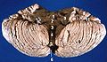
 Superior view of the cerebellum Left cerebellar hemisphere Right cerebellar hemisphere | |
 Schematic representation of the major anatomical subdivisions of the cerebellum. Superior view of an "unrolled" cerebellum, placing the vermis in one plane. | |
| Details | |
| Identifiers | |
| Latin | hemisphaerium cerebelli |
| NeuroNames | 1214 |
| NeuroLex ID | birnlex_1575 |
| TA98 | A14.1.07.004 |
| TA2 | 5804 |
| FMA | 76925 |
| Anatomical terms of neuroanatomy | |
The cerebellar hemispheres are the two lateral halves of the cerebellum. They are joined by the vermis.