Cerebral aqueduct

Last updated
Cerebral aqueduct
Cn3nucleus-en.svg
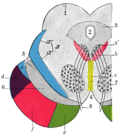
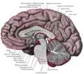
Section through superior colliculus showing path of oculomotor nerve.
Gray736.png
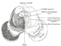
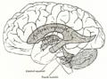
Drawing of a cast of the ventricular cavities, viewed from the side.
Details
Part of Ventricular system
Identifiers
Latin aqueductus mesencephali (cerebri).
aqueductus Sylvii
MeSH D002535
NeuroNames 509
NeuroLex ID birnlex_1261
TA98 A14.1.06.501
TA2 5910
FMA 78467
Anatomical terms of neuroanatomy

The cerebral aqueduct (aque ductus mesencephali, mesencephalic duct, sylvian aqueduct or aqueduct of Sylvius) is a narrow 15 mm conduit for cerebrospinal fluid (CSF) that connects the third ventricle to the fourth ventricle of the ventricular system of the brain. It is located in the midbrain dorsal to the pons and ventral to the cerebellum. [1] The cerebral aqueduct is surrounded by an enclosing area of gray matter called the periaqueductal gray, or central gray. It was first named after Franciscus Sylvius.

Contents

Structure

Development

The cerebral aqueduct, as other parts of the ventricular system of the brain, develops from the central canal of the neural tube, and it originates from the portion of the neural tube that is present in the developing mesencephalon, hence the name "mesencephalic duct." [2]

Function

The cerebral aqueduct acts like a canal that passes through the midbrain. It connects the third ventricle with the fourth ventricle so that cerebrospinal fluid (CSF) moves between the cerebral ventricles and the canal connecting these ventricles. [1]

Clinical significance

Aqueductal stenosis, a narrowing of the cerebral aqueduct, obstructs the flow of CSF and has been associated with non-communicating hydrocephalus. Such narrowing can be congenital, arise via tumor compression (e.g. pinealoblastoma), or through cyclical gliosis secondary to an initial partial obstruction. [1]

History

The cerebral aqueduct was first named after Franciscus Sylvius. [3]

Additional images

See also

Related Research Articles

<span class="mw-page-title-main">Central nervous system</span> Brain and spinal cord

The central nervous system (CNS) is the part of the nervous system consisting of the brain and spinal cord, the retina and optic nerve, and the olfactory nerve and epithelia. The CNS is so named because the brain integrates the received information and coordinates and influences the activity of all parts of the bodies of bilaterally symmetric and triploblastic animals—that is, all multicellular animals except sponges and diploblasts. It is a structure composed of nervous tissue positioned along the rostral to caudal axis of the body and may have an enlarged section at the rostral end which is a brain. Only arthropods, cephalopods and vertebrates have a true brain, though precursor structures exist in onychophorans, gastropods and lancelets.

<span class="mw-page-title-main">Cerebrospinal fluid</span> Clear, colorless bodily fluid found in the brain and spinal cord

Cerebrospinal fluid (CSF) is a clear, colorless body fluid found within the tissue that surrounds the brain and spinal cord of all vertebrates.

Articles related to anatomy include:

<span class="mw-page-title-main">Brainstem</span> Posterior part of the brain, adjoining and structurally continuous

The brainstem is the stalk-like part of the brain that interconnects the cerebrum and diencephalon with the spinal cord. In the human brain, the brainstem is composed of the midbrain, the pons, and the medulla oblongata. The midbrain is continuous with the thalamus of the diencephalon through the tentorial notch.

<span class="mw-page-title-main">Ventricular system</span> Set of structures containing cerebrospinal fluid in the brain

In neuroanatomy, the ventricular system is a set of four interconnected cavities known as cerebral ventricles in the brain. Within each ventricle is a region of choroid plexus which produces the circulating cerebrospinal fluid (CSF). The ventricular system is continuous with the central canal of the spinal cord from the fourth ventricle, allowing for the flow of CSF to circulate.

<span class="mw-page-title-main">Third ventricle</span> Ventricle of the brain located between the two thalami

The third ventricle is one of the four connected ventricles of the ventricular system within the mammalian brain. It is a slit-like cavity formed in the diencephalon between the two thalami, in the midline between the right and left lateral ventricles, and is filled with cerebrospinal fluid (CSF).

<span class="mw-page-title-main">Midbrain</span> Forward-most portion of the brainstem

The midbrain or mesencephalon is the rostral-most portion of the brainstem connecting the diencephalon and cerebrum with the pons. It consists of the cerebral peduncles, tegmentum, and tectum.

<span class="mw-page-title-main">Oculomotor nucleus</span>

The fibers of the oculomotor nerve arise from a nucleus in the midbrain, which lies in the gray substance of the floor of the cerebral aqueduct and extends in front of the aqueduct for a short distance into the floor of the third ventricle. From this nucleus the fibers pass forward through the tegmentum, the red nucleus, and the medial part of the substantia nigra, forming a series of curves with a lateral convexity, and emerge from the oculomotor sulcus on the medial side of the cerebral peduncle.

<span class="mw-page-title-main">Optic tract</span> Neural pathway within the human visual system

In neuroanatomy, the optic tract is a part of the visual system in the brain. It is a continuation of the optic nerve that relays information from the optic chiasm to the ipsilateral lateral geniculate nucleus (LGN), pretectal nuclei, and superior colliculus.

<span class="mw-page-title-main">Fourth ventricle</span> Ventricle in front of the cerebellum

The fourth ventricle is one of the four connected fluid-filled cavities within the human brain. These cavities, known collectively as the ventricular system, consist of the left and right lateral ventricles, the third ventricle, and the fourth ventricle. The fourth ventricle extends from the cerebral aqueduct to the obex, and is filled with cerebrospinal fluid (CSF).

<span class="mw-page-title-main">Lateral ventricles</span> Two largest ventricles in each cerebral hemisphere

The lateral ventricles are the two largest ventricles of the brain and contain cerebrospinal fluid (CSF). Each cerebral hemisphere contains a lateral ventricle, known as the left or right lateral ventricle, respectively.

<span class="mw-page-title-main">Red nucleus</span> Structure in the human brain

The red nucleus or nucleus ruber is a structure in the rostral midbrain involved in motor coordination. The red nucleus is pale pink, which is believed to be due to the presence of iron in at least two different forms: hemoglobin and ferritin. The structure is located in the tegmentum of the midbrain next to the substantia nigra and comprises caudal magnocellular and rostral parvocellular components. The red nucleus and substantia nigra are subcortical centers of the extrapyramidal motor system.

<span class="mw-page-title-main">Subarachnoid cisterns</span> Spaces around the brain filled with cerebrospinal fluid

The subarachnoid cisterns are spaces formed by openings in the subarachnoid space, an anatomic space in the meninges of the brain. The space is situated between the two meninges, the arachnoid mater and the pia mater. These cisterns are filled with cerebrospinal fluid (CSF).

<span class="mw-page-title-main">Corpora quadrigemina</span> Part of the brain

In the brain, the corpora quadrigemina are the four colliculi—two inferior, two superior—located on the tectum of the dorsal aspect of the midbrain. They are respectively named the inferior and superior colliculus.

<span class="mw-page-title-main">Rhomboid fossa</span> Depression in the human brain

The rhomboid fossa is a rhombus-shaped depression that is the anterior part of the fourth ventricle. Its anterior wall, formed by the back of the pons and the medulla oblongata, constitutes the floor of the fourth ventricle.

<span class="mw-page-title-main">Superior cerebellar peduncle</span>

In the human brain, the superior cerebellar peduncle is a paired structure of white matter that connects the cerebellum to the midbrain. It consists mainly of efferent fibers, the cerebellothalamic tract that runs from a cerebellar hemisphere to the contralateral thalamus, and the cerebellorubral tract that runs from a cerebellar hemisphere to the red nucleus. It also contains afferent tracts, most prominent of which is the ventral spinocerebellar tract. Other afferent tracts are the trigeminothalamic fibers, tectocerebellar fibers, and noradrenergic fibers from the locus coeruleus. The superior peduncle emerges from the upper and medial parts of the white matter of each hemisphere and is placed under cover of the upper part of the cerebellum.

<span class="mw-page-title-main">Middle cerebellar peduncle</span> Structure in the brain connecting the pons to the cerebellum

The middle cerebellar peduncle is a paired structure of the brain. It connects the pons to the cerebellum, with fibres originating from the pontine nucleus and travelling to the opposite hemisphere of the cerebellar cortex. It is supplied by the anterior inferior cerebellar artery (AICA) and branches from the basilar artery. It conveys information from the cerebrum and the pons to the cerebellum.

<span class="mw-page-title-main">Superior medullary velum</span> Thin layer between the superior cerebellar peduncles

The superior medullary velum is a thin, transparent lamina of white matter which - together with the inferior medullary velum - forms the roof of the fourth ventricle. It extends between the two superior cerebellar peduncles. The lingula of cerebellum covers - and adheres to - its dorsal surface.

<span class="mw-page-title-main">Aqueductal stenosis</span> Narrowing of the aqueduct of Sylvius

Aqueductal stenosis is a narrowing of the aqueduct of Sylvius which blocks the flow of cerebrospinal fluid (CSF) in the ventricular system. Blockage of the aqueduct can lead to hydrocephalus, specifically as a common cause of congenital and/or obstructive hydrocephalus.

References

  1. 1 2 3 Rubino, Jessica; Hogg, Jeffery (31 July 2020). Neuroanatomy, cerebral aqueduct (Sylvian). Treasure Island, FL: StatPearls Publishing.
  2. Le, Tao; Bhushan, Vikas; Vasan, Neil (2010). First Aid for the USMLE Step 1: 2010 20th Anniversary Edition. USA: The McGraw-Hill Companies, Inc. pp.  126. ISBN   978-0-07-163340-6.
  3. Kompanje, Erwin J. O. (1 January 2005). "An historical homage from Denmark: the aqueduct of Sylvius". Neurosurgical Review. 28 (1): 77–78. doi:10.1007/s10143-004-0361-6. ISSN   1437-2320.