Articles related to anatomy include:

The third ventricle is one of the four connected cerebral ventricles of the ventricular system within the mammalian brain. It is a slit-like cavity formed in the diencephalon between the two thalami, in the midline between the right and left lateral ventricles, and is filled with cerebrospinal fluid (CSF).

The fornix is a C-shaped bundle of nerve fibers in the brain that acts as the major output tract of the hippocampus. The fornix also carries some afferent fibers to the hippocampus from structures in the diencephalon and basal forebrain. The fornix is part of the limbic system. While its exact function and importance in the physiology of the brain are still not entirely clear, it has been demonstrated in humans that surgical transection—the cutting of the fornix along its body—can cause memory loss. There is some debate over what type of memory is affected by this damage, but it has been found to most closely correlate with recall memory rather than recognition memory. This means that damage to the fornix can cause difficulty in recalling long-term information such as details of past events, but it has little effect on the ability to recognize objects or familiar situations.

The fourth ventricle is one of the four connected fluid-filled cavities within the human brain. These cavities, known collectively as the ventricular system, consist of the left and right lateral ventricles, the third ventricle, and the fourth ventricle. The fourth ventricle extends from the cerebral aqueduct to the obex, and is filled with cerebrospinal fluid (CSF).

The lateral ventricles are the two largest ventricles of the brain and contain cerebrospinal fluid. Each cerebral hemisphere contains a lateral ventricle, known as the left or right lateral ventricle, respectively.

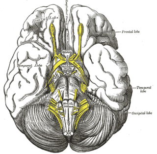
The olivary bodies or simply olives are a pair of prominent oval structures on either side of the medullary pyramids in the medulla, the lower portion of the brainstem. They contain the olivary nuclei.

In the brain, the interventricular foramina are channels that connect the paired lateral ventricles with the third ventricle at the midline of the brain. As channels, they allow cerebrospinal fluid (CSF) produced in the lateral ventricles to reach the third ventricle and then the rest of the brain's ventricular system. The walls of the interventricular foramina also contain choroid plexus, a specialized CSF-producing structure, that is continuous with that of the lateral and third ventricles above and below it.

The lentiform nucleus are the putamen (laterally) and the globus pallidus (medially), collectively. Due to their proximity, these two structures were formerly considered one, however, the two are separated by a thin layer of white matter - the external medullary lamina - and are functionally and connectionally distinct.

The posterior cerebral artery (PCA) is one of a pair of cerebral arteries that supply oxygenated blood to the occipital lobe, part of the back of the human brain. The two arteries originate from the distal end of the basilar artery, where it bifurcates into the left and right posterior cerebral arteries. These anastomose with the middle cerebral arteries and internal carotid arteries via the posterior communicating arteries.

The coronary sinus is the largest vein of the heart. It drains over half of the deoxygenated blood from the heart muscle into the right atrium. It begins on the backside of the heart, in between the left atrium, and left ventricle; it begins at the junction of the great cardiac vein, and oblique vein of the left atrium. It receives multiple tributaries. It passes across the backside of the heart along a groove between left atrium and left ventricle, then drains into the right atrium at the orifice of the coronary sinus.

The thyroarytenoid muscle is a broad, thin muscle that forms the body of the vocal fold and that supports the wall of the ventricle and its appendix. It functions to shorten the vocal folds.

The calcarine sulcus is an anatomical landmark located at the caudal end of the medial surface of the brain of humans and other primates. Its name comes from the Latin "calcar" meaning "spur". It is very deep, and known as a complete sulcus.

Circumventricular organs (CVOs) are structures in the brain characterized by their extensive and highly permeable capillaries, unlike those in the rest of the brain where there exists a blood–brain barrier (BBB) at the capillary level. Although the term "circumventricular organs" was originally proposed in 1958 by Austrian anatomist Helmut O. Hofer concerning structures around the brain ventricular system, the penetration of blood-borne dyes into small specific CVO regions was discovered in the early 20th century. The permeable CVOs enabling rapid neurohumoral exchange include the subfornical organ (SFO), the area postrema (AP), the vascular organ of lamina terminalis, the median eminence, the pituitary neural lobe, and the pineal gland.
The tuber cinereum is the portion of hypothalamus forming the floor of the third ventricle situated between the optic chiasm, and the mammillary bodies. The tuberal region is one of the three regions of the hypothalamus, the other two being the chiasmatic region and the mamillary region.

The circumflex branch of left coronary artery is a branch of the left coronary artery. It winds around the left side of the heart along the atrioventricular groove. It supplies the posterolateral portion of the left ventricle.

The rhomboid fossa is a rhombus-shaped depression that is the anterior part of the fourth ventricle. Its anterior wall, formed by the back of the pons and the medulla oblongata, constitutes the floor of the fourth ventricle.

The collateral fissure is a large sulcus on the tentorial surface of the cerebral hemisphere and extends from near the occipital pole to within a short distance of the temporal pole. It is also known as the medial occipitotemporal sulcus.

Lamina affixa is a layer of epithelium growing on the surface of the thalamus and forming the floor of the central part of lateral ventricle, on whose medial margin is attached the choroid plexus of the lateral ventricle; it covers the superior thalamostriate vein and the superior choroid vein. The torn edge of this plexus is called the tela choroidea.
The cistern of lamina terminalis is one of the subarachnoid cisterns. It is situated either superior to the lamina terminalis, or rostral/anterior to the lamina terminalis and anterior commissure between the two frontal lobes of the cerebrum. It is situated rostral/anterior to the third ventricle. The cistern is an extension of interpeduncular cistern. The cistern of lamina terminalis interconnects the chiasmatic cistern and pericallosal cistern.
