Cerebrospinal fluid (CSF) is a clear, colorless body fluid found within the tissue that surrounds the brain and spinal cord of all vertebrates. It replaces the body fluid found outside the cells of all bilateral animals. This article discusses the CSF as found in human anatomy.
Articles related to anatomy include:
The ventricular system is a set of four interconnected cavities known as ventricles in the brain. Within each ventricle is a region of choroid plexus which produces the circulating cerebrospinal fluid (CSF). The ventricular system is continuous with the central canal of the spinal cord from the fourth ventricle, allowing for the flow of CSF to circulate.

Pia mater, often referred to as simply the pia, is the delicate innermost layer of the meninges, the membranes surrounding the brain and spinal cord. Pia mater is medieval Latin meaning "tender mother". The other two meningeal membranes are the dura mater and the arachnoid mater. Both the pia and arachnoid mater are derivatives of the neural crest while the dura is derived from embryonic mesoderm. The pia mater is a thin fibrous tissue that is permeable to water and small solutes. The pia mater allows blood vessels to pass through and nourish the brain. The perivascular space between blood vessels and pia mater is proposed to be part of a pseudolymphatic system for the brain. When the pia mater becomes irritated and inflamed the result is meningitis.

The third ventricle is one of the four connected ventricles of the ventricular system within the mammalian brain. It is a slit-like cavity formed in the diencephalon between the two thalami, in the midline between the right and left lateral ventricles, and is filled with cerebrospinal fluid (CSF).
The choroid plexus or plica choroidea, is a plexus of cells that arises from the tela choroidea in each of the ventricles of the brain. The choroid plexus produces most of the cerebrospinal fluid (CSF) of the central nervous system. CSF is produced and secreted by the regions of choroid plexus. The choroid plexus consists of modified ependymal cells surrounding a core of capillaries and loose connective tissue.

The fourth ventricle is one of the four connected fluid-filled cavities within the human brain. These cavities, known collectively as the ventricular system, consist of the left and right lateral ventricles, the third ventricle, and the fourth ventricle. The fourth ventricle extends from the cerebral aqueduct to the obex, and is filled with cerebrospinal fluid (CSF).
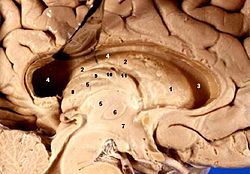
The median aperture drains cerebrospinal fluid (CSF) from the fourth ventricle into the cisterna magna. The two other openings of the fourth ventricle are the lateral apertures, one on the left and one on the right, which drain cerebrospinal fluid into the cerebellopontine angle cistern. The median foramen on axial images is posterior to the pons and anterior to the caudal cerebellum. It is surrounded by the obex and gracile tubercles of the medulla, tela choroidea of the fourth ventricle and its choroid plexus, which is attached to the cerebellar vermis.

The lateral ventricles are the two largest ventricles of the brain and contain cerebrospinal fluid (CSF). Each cerebral hemisphere contains a lateral ventricle, known as the left or right ventricle, respectively.

In the brain, the interventricular foramina are channels that connect the paired lateral ventricles with the third ventricle at the midline of the brain. As channels, they allow cerebrospinal fluid (CSF) produced in the lateral ventricles to reach the third ventricle and then the rest of the brain's ventricular system. The walls of the interventricular foramina also contain choroid plexus, a specialized CSF-producing structure, that is continuous with that of the lateral and third ventricles above and below it.

The posterior cerebral artery (PCA) is one of a pair of arteries that supply oxygenated blood to the occipital lobe, part of the back of the human brain. The two arteries originate from the distal end of the basilar artery, where it bifurcates into the left and right posterior cerebral arteries. These anastomose with the middle cerebral arteries and internal carotid arteries via the posterior communicating arteries.

The posterior inferior cerebellar artery (PICA) is the largest branch of the vertebral artery. It is one of the three main arteries that supply blood to the cerebellum, a part of the brain. Blockage of the posterior inferior cerebellar artery can result in a type of stroke called lateral medullary syndrome.

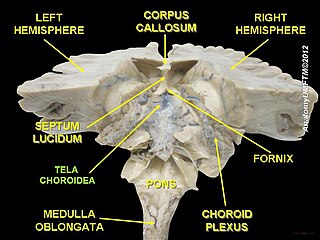
The tela choroidea is a region of meningeal pia mater that adheres to the underlying ependyma, and gives rise to the choroid plexus in each of the brain’s four ventricles. Tela is Latin for woven and is used to describe a web-like membrane or layer. The tela choroidea is a very thin part of the loose connective tissue of pia mater overlying and closely adhering to the ependyma. It has a rich blood supply. The ependyma and vascular pia mater – the tela choroidea, form regions of minute projections known as a choroid plexus that projects into each ventricle. The choroid plexus produces most of the cerebrospinal fluid of the central nervous system that circulates through the ventricles of the brain, the central canal of the spinal cord, and the subarachnoid space. The tela choroidea in the ventricles forms from different parts of the roof plate in the development of the embryo.

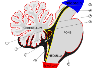
The superior medullary velum is a thin, transparent lamina of white matter, which stretches between the superior cerebellar peduncles; on the dorsal surface of its lower half the folia and lingula are prolonged.
The internal cerebral veins drain the deep parts of the hemisphere and are two in number; each internal cerebral vein is formed near the interventricular foramina by the union of the superior thalamostriate vein and the superior choroid vein.

The vorticose veins, referred to clinically as the vortex veins, drain the ocular choroid. The number of vortex veins is known to vary from 4 to 8 with about 65% of the normal population having 4 or 5. In most cases, there is at least one vortex vein in each quadrant. Typically, the entrances to the vortex veins in the outer layer of the choroid can be observed funduscopically and provide an important clinical landmarks identifying the ocular equator. However, the veins run posteriorly in the sclera exiting the eye well posterior to the equator.
The choroid consists mainly of a dense capillary plexus, and of small arteries and veins carrying blood to and returning it from this plexus.

Choroid plexus papilloma, also known as papilloma of the choroid plexus, is a rare benign neuroepithelial intraventricular WHO grade I lesion found in the choroid plexus. It leads to increased cerebrospinal fluid production, thus causing increased intracranial pressure and hydrocephalus.

A choroid plexus carcinoma is a type of choroid plexus tumor that affects the choroid plexus of the brain. It is considered the worst of the three grades of chord plexus tumors, having a much poorer prognosis than choroid atypical plexus papilloma and choroid plexus papilloma. The disease creates lesions in the brain and increases cerebrospinal fluid volume, resulting in hydrocephalus.

The choroid veins are the superior choroid vein and the inferior choroid vein of the lateral ventricle. Both veins drain different parts of the choroid plexus.