Related Research Articles

The putamen is a round structure located at the base of the forebrain (telencephalon). The putamen and caudate nucleus together form the dorsal striatum. It is also one of the structures that compose the basal nuclei. Through various pathways, the putamen is connected to the substantia nigra, the globus pallidus, the claustrum, and the thalamus, in addition to many regions of the cerebral cortex. A primary function of the putamen is to regulate movements at various stages and influence various types of learning. It employs GABA, acetylcholine, and enkephalin to perform its functions. The putamen also plays a role in degenerative neurological disorders, such as Parkinson's disease.

The striatum or corpus striatum is a nucleus in the subcortical basal ganglia of the forebrain. The striatum is a critical component of the motor and reward systems; receives glutamatergic and dopaminergic inputs from different sources; and serves as the primary input to the rest of the basal ganglia.
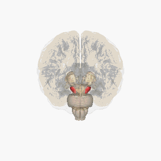
The substantia nigra (SN) is a basal ganglia structure located in the midbrain that plays an important role in reward and movement. Substantia nigra is Latin for "black substance", reflecting the fact that parts of the substantia nigra appear darker than neighboring areas due to high levels of neuromelanin in dopaminergic neurons. Parkinson's disease is characterized by the loss of dopaminergic neurons in the substantia nigra pars compacta.

The basal ganglia (BG) or basal nuclei are a group of subcortical nuclei found in the brains of vertebrates. In humans and other primates, differences exist, primarily in the division of the globus pallidus into external and internal regions, and in the division of the striatum. Positioned at the base of the forebrain and the top of the midbrain, they have strong connections with the cerebral cortex, thalamus, brainstem and other brain areas. The basal ganglia are associated with a variety of functions, including regulating voluntary motor movements, procedural learning, habit formation, conditional learning, eye movements, cognition, and emotion.

The globus pallidus (GP), also known as paleostriatum or dorsal pallidum, is a subcortical structure of the brain. It consists of two adjacent segments, one external, known in rodents simply as the globus pallidus, and one internal, known in rodents as the entopeduncular nucleus. It is part of the telencephalon, but retains close functional ties with the subthalamus in the diencephalon – both of which are part of the extrapyramidal motor system. The globus pallidus is a major component of the basal ganglia, with principal inputs from the striatum, and principal direct outputs to the thalamus and the substantia nigra. The latter is made up of similar neuronal elements, has similar afferents from the striatum, similar projections to the thalamus, and has a similar synaptology. Neither receives direct cortical afferents, and both receive substantial additional inputs from the intralaminar thalamus.

The midbrain or mesencephalon is the rostral-most portion of the brainstem connecting the diencephalon and cerebrum with the pons. It consists of the cerebral peduncles, tegmentum, and tectum.

Dopaminergic pathways in the human brain are involved in both physiological and behavioral processes including movement, cognition, executive functions, reward, motivation, and neuroendocrine control. Each pathway is a set of projection neurons, consisting of individual dopaminergic neurons.

The nigrostriatal pathway is a bilateral dopaminergic pathway in the brain that connects the substantia nigra pars compacta (SNc) in the midbrain with the dorsal striatum in the forebrain. It is one of the four major dopamine pathways in the brain, and is critical in the production of movement as part of a system called the basal ganglia motor loop. Dopaminergic neurons of this pathway release dopamine from axon terminals that synapse onto GABAergic medium spiny neurons (MSNs), also known as spiny projection neurons (SPNs), located in the striatum.
In neuroanatomy, the superior colliculus is a structure lying on the roof of the mammalian midbrain. In non-mammalian vertebrates, the homologous structure is known as the optic tectum or optic lobe. The adjective form tectal is commonly used for both structures.

The subthalamic nucleus (STN) is a small lens-shaped nucleus in the brain where it is, from a functional point of view, part of the basal ganglia system. In terms of anatomy, it is the major part of the subthalamus. As suggested by its name, the subthalamic nucleus is located ventral to the thalamus. It is also dorsal to the substantia nigra and medial to the internal capsule. It was first described by Jules Bernard Luys in 1865, and the term corpus Luysi or Luys' body is still sometimes used.
The pedunculopontine nucleus (PPN) or pedunculopontine tegmental nucleus is a collection of neurons located in the upper pons in the brainstem. It is involved in voluntary movements, arousal, and provides sensory feedback to the cerebral cortex and one of the main components of the reticular activating system. It is a potential target for deep brain stimulation treatment for Parkinson's disease. It was first described in 1909 by Louis Jacobsohn-Lask, a German neuroanatomist.

In the scientific study of vision, smooth pursuit describes a type of eye movement in which the eyes remain fixated on a moving object. It is one of two ways that visual animals can voluntarily shift gaze, the other being saccadic eye movements. Pursuit differs from the vestibulo-ocular reflex, which only occurs during movements of the head and serves to stabilize gaze on a stationary object. Most people are unable to initiate pursuit without a moving visual signal. The pursuit of targets moving with velocities of greater than 30°/s tends to require catch-up saccades. Smooth pursuit is asymmetric: most humans and primates tend to be better at horizontal than vertical smooth pursuit, as defined by their ability to pursue smoothly without making catch-up saccades. Most humans are also better at downward than upward pursuit. Pursuit is modified by ongoing visual feedback.
The zona incerta (ZI) is a horizontally elongated region of gray matter in the subthalamus below the thalamus. Its connections project extensively over the brain from the cerebral cortex down into the spinal cord.

The basal ganglia form a major brain system in all species of vertebrates, but in primates there are special features that justify a separate consideration. As in other vertebrates, the primate basal ganglia can be divided into striatal, pallidal, nigral, and subthalamic components. In primates, however, there are two pallidal subdivisions called the external globus pallidus (GPe) and internal globus pallidus (GPi). Also in primates, the dorsal striatum is divided by a large tract called the internal capsule into two masses named the caudate nucleus and the putamen—in most other species no such division exists, and only the striatum as a whole is recognized. Beyond this, there is a complex circuitry of connections between the striatum and cortex that is specific to primates. This complexity reflects the difference in functioning of different cortical areas in the primate brain.
The pars compacta (SNpc) is one of two subdivisions of the substantia nigra of the midbrain ; it is situated medial to the pars reticulata. It is formed by dopaminergic neurons. It projects to the striatum and portions of the cerebral cortex. It is functionally involved in fine motor control.

Medium spiny neurons (MSNs), also known as spiny projection neurons (SPNs), are a special type of GABAergic inhibitory cell representing 95% of neurons within the human striatum, a basal ganglia structure. Medium spiny neurons have two primary phenotypes : D1-type MSNs of the direct pathway and D2-type MSNs of the indirect pathway. Most striatal MSNs contain only D1-type or D2-type dopamine receptors, but a subpopulation of MSNs exhibit both phenotypes.
The isothalamus is a division used by some researchers in describing the thalamus.

The external globus pallidus combines with the internal globus pallidus (GPi) to form the globus pallidus, an anatomical subset of the basal ganglia. Globus pallidus means "pale globe" in Latin, indicating its appearance. The external globus pallidus is the segment of the globus pallidus that is relatively further (lateral) from the midline of the brain.
Basal ganglia disease is a group of physical problems that occur when the group of nuclei in the brain known as the basal ganglia fail to properly suppress unwanted movements or to properly prime upper motor neuron circuits to initiate motor function. Research indicates that increased output of the basal ganglia inhibits thalamocortical projection neurons. Proper activation or deactivation of these neurons is an integral component for proper movement. If something causes too much basal ganglia output, then the ventral anterior (VA) and ventral lateral (VL) thalamocortical projection neurons become too inhibited, and one cannot initiate voluntary movement. These disorders are known as hypokinetic disorders. However, a disorder leading to abnormally low output of the basal ganglia leads to reduced inhibition, and thus excitation, of the thalamocortical projection neurons which synapse onto the cortex. This situation leads to an inability to suppress unwanted movements. These disorders are known as hyperkinetic disorders.
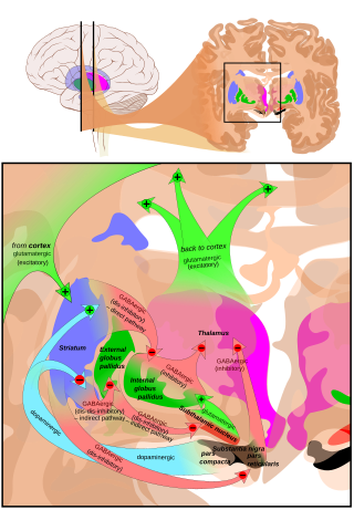
The cortico-basal ganglia-thalamo-cortical loop is a system of neural circuits in the brain. The loop involves connections between the cortex, the basal ganglia, the thalamus, and back to the cortex. It is of particular relevance to hyperkinetic and hypokinetic movement disorders, such as Parkinson's disease and Huntington's disease, as well as to mental disorders of control, such as attention deficit hyperactivity disorder (ADHD), obsessive–compulsive disorder (OCD), and Tourette syndrome.
References
- ↑ François, C.; Yelnik, J. & Percheron, G. (1987). "Golgi study of the primate substantia nigra. II. Spatial organization of dendritic arborizations in relation to the cytoarchitectonic boundaries and to the striatonigral bundle". Journal of Comparative Neurology. 265 (4): 473–493. doi:10.1002/cne.902650403. PMID 3123530.
- ↑ François, C.; Percheron, G. & Yelnik, J. (1984). "Localization of nigrostriatal, nigrothalamic and nigrotectal neurons in ventricular coordinates in macaques". Neuroscience. 13 (1): 61–76. doi:10.1016/0306-4522(84)90259-8. PMID 6387531.
- ↑ Atherton, J.F. & Bevan, M.D. (2005). "Ionic mechanisms underlying autonomous action potential generation in the somata and dendrites of GABAergic substantia nigra pars reticulata neurons in vitro". Journal of Neuroscience. 25 (36): 8272–8281. doi:10.1523/JNEUROSCI.1475-05.2005. PMC 6725542 . PMID 16148235.
- ↑ Schultz, W. (1986). "Activity of pars reticulata neurons of monkey substantia nigra in relation to motor, sensory and complex events". Journal of Neurophysiology. 55 (4): 660–677. doi:10.1152/jn.1986.55.4.660. PMID 3701399.
- ↑ Hikosaka, O; Wurtz, RH (1983). "Visual and oculomotor functions of monkey substantia nigra pars reticulata. I. Relation of visual and auditory responses to saccades". Journal of Neurophysiology. 49 (5): 1230–53. doi:10.1152/jn.1983.49.5.1230. PMID 6864248.
- ↑ Hikosaka, O; Wurtz, RH (1983). "Visual and oculomotor functions of monkey substantia nigra pars reticulata. II. Visual responses related to fixation of gaze". Journal of Neurophysiology. 49 (5): 1254–67. doi:10.1152/jn.1983.49.5.1254. PMID 6864249.
- ↑ Hikosaka, O; Wurtz, RH (1983). "Visual and oculomotor functions of monkey substantia nigra pars reticulata. III. Memory-contingent visual and saccade responses". Journal of Neurophysiology. 49 (5): 1268–84. doi:10.1152/jn.1983.49.5.1268. PMID 6864250.
- ↑ Hikosaka, O; Wurtz, RH (1983). "Visual and oculomotor functions of monkey substantia nigra pars reticulata. IV. Relation of substantia nigra to superior colliculus". Journal of Neurophysiology. 49 (5): 1285–301. doi:10.1152/jn.1983.49.5.1285. PMID 6306173.
- ↑ Principles of neural science. Kandel, Eric R. (5th ed.). New York: McGraw-Hill Medical. 2012. ISBN 978-0-07-139011-8. OCLC 820110349.
{{cite book}}: CS1 maint: others (link)