Atypical ductal hyperplasia

Last updated
Atypical ductal hyperplasia
Atypical ductal hyperplasia - very low mag.jpg
Very low magnification micrograph of atypical ductal hyperplasia (ADH). The piece with ADH was circled by the pathologist with a marker, as it is so small, and sent for an additional opinion. H&E stain.
Specialty Gynecology, pathology

Atypical ductal hyperplasia (ADH) is the term used for a benign lesion of the breast that indicates an increased risk of breast cancer. [1]

Contents

The name of the entity is descriptive of the lesion; ADH is characterized by cellular proliferation (hyperplasia) within one or two breast ducts and (histomorphologic) architectural abnormalities, i.e. the cells are arranged in an abnormal or atypical way, more so than usual ductal hyperplasia.

In the context of a core (needle) biopsy, ADH is considered an indication for a breast lumpectomy, also known as a surgical (excisional) biopsy, to exclude the presence of breast cancer. [2]

Signs and symptoms

ADH, generally, is asymptomatic. It usually comes to medical attention on a screening mammogram, as a non-specific suspicious abnormality that requires a biopsy.

Pathology

ADH, cytologically, architecturally and on a molecular basis, is identical to a low-grade ductal carcinoma in situ (DCIS); [3] however, it has a limited extent, i.e. is present in a very small amount (< 2 mm).

Relation to low-grade ductal carcinoma in situ

While the histopathologic features and molecular features of ADH are that of (low-grade) DCIS, its clinical behaviour, unlike low-grade DCIS, is substantially better; thus, the more aggressive treatment for DCIS is not justified.

Diagnosis

Histological appearance of atypical ductal hyperplasia (ADH) and immunohistochemical phenotype:
- A - One focus (< 2 mm) of two architecturally disarranged cross sections of tubuli showing a monotonous intraductal proliferation with secondary intraluminal architecture. Hematoxylin and Eosin stain.
- B - One area of an ADH with associated calcifications intraluminal. Hematoxylin and Eosin stain.
- C - Higher magnification of ADH shows low-grade nuclear atypia and monotonous cell proliferation along with secondary intraluminal architecture. Hematoxylin and Eosin stain.
- D - Strong and uniform expression of estrogen receptors (ER). ER immunohistochemistry.
- E - Lack of basal cytokeratins (CK5/6). CK5/6 immunohistochemistry. Atypical ductal hyperplasia with immunotyping.jpg
Histological appearance of atypical ductal hyperplasia (ADH) and immunohistochemical phenotype:
- A - One focus (< 2 mm) of two architecturally disarranged cross sections of tubuli showing a monotonous intraductal proliferation with secondary intraluminal architecture. Hematoxylin and Eosin stain.
- B - One area of an ADH with associated calcifications intraluminal. Hematoxylin and Eosin stain.
- C - Higher magnification of ADH shows low-grade nuclear atypia and monotonous cell proliferation along with secondary intraluminal architecture. Hematoxylin and Eosin stain.
- D - Strong and uniform expression of estrogen receptors (ER). ER immunohistochemistry.
- E - Lack of basal cytokeratins (CK5/6). CK5/6 immunohistochemistry.

It is diagnosed based on tissue, e.g. a biopsy, [5] showing ductal hyperplasia.

There is no single definite cutoff that separates atypical ductal hyperplasia from ductal carcinoma in situ, but the following are important distinctive features of atypical ductal hyperplasia, with suggested cutoffs: [6]

Treatment

ADH, if found on a surgical (excisional) biopsy of a mammographic abnormality, does not require any further treatment, only mammographic follow-up.

If ADH is found on a core (needle) biopsy (a procedure which generally does not excise a suspicious mammographic abnormality), a surgical biopsy, i.e. a breast lumpectomy, to completely excise the abnormality and exclude breast cancer is the typical recommendation.

Prognosis

Cancer risk for ADH on a core biopsy

The rate at which breast cancer (ductal carcinoma in situ or invasive mammary carcinoma (all breast cancer except DCIS and LCIS)) is found at the time of a surgical (excisional) biopsy, following the diagnosis of ADH on a core (needle) biopsy varies considerably from hospital-to-hospital (range 4-54%). [7] In two large studies, the conversion of an ADH on core biopsy to breast cancer on surgical excision, known as "up-grading", is approximately 30%. [7] [8]

Cancer risk based on follow-up

The relative risk of breast cancer based on a median follow-up of 8 years, in a case control study of US registered nurses, is 3.7. [9]

See also

Related Research Articles

<span class="mw-page-title-main">Nipple discharge</span> Medical condition

Nipple discharge is fluid from the nipple, with or without squeezing the breast. The discharge can be milky, clear, green, purulent, bloody, or faintly yellow. The consistency can be thick, thin, sticky, or watery.

Carcinoma <i>in situ</i> Medical condition

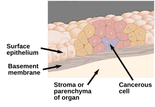
Carcinoma in situ (CIS) is a group of abnormal cells. While they are a form of neoplasm, there is disagreement over whether CIS should be classified as cancer. This controversy also depends on the exact CIS in question. Some authors do not classify them as cancer, however, recognizing that they can potentially become cancer. Others classify certain types as a non-invasive form of cancer. The term "pre-cancer" has also been used.

<span class="mw-page-title-main">Lumpectomy</span> Limited surgical removal of breast tissue

Lumpectomy is a surgical removal of a discrete portion or "lump" of breast tissue, usually in the treatment of a malignant tumor or breast cancer. It is considered a viable breast conservation therapy, as the amount of tissue removed is limited compared to a full-breast mastectomy, and thus may have physical and emotional advantages over more disfiguring treatment. Sometimes a lumpectomy may be used to either confirm or rule out that cancer has actually been detected. A lumpectomy is usually recommended to patients whose cancer has been detected early and who do not have enlarged tumors. Although a lumpectomy is used to allow for most of the breast to remain intact, the procedure may result in adverse affects that can include sensitivity and result in scar tissue, pain, and possible disfiguration of the breast if the lump taken out is significant. According to National Comprehensive Cancer Network guidelines, lumpectomy may be performed for ductal carcinoma in situ (DCIS), invasive ductal carcinoma, or other conditions.

<span class="mw-page-title-main">Invasive carcinoma of no special type</span> Medical condition

Invasive carcinoma of no special type, invasive breast carcinoma of no special type (IBC-NST), invasive ductal carcinoma (IDC), infiltrating ductal carcinoma (IDC) or invasive ductal carcinoma, not otherwise specified (NOS) is a disease. For international audiences this article will use "invasive carcinoma NST" because it is the preferred term of the World Health Organization (WHO).

<span class="mw-page-title-main">Ductal carcinoma in situ</span> Medical condition

Ductal carcinoma in situ (DCIS), also known as intraductal carcinoma, is a pre-cancerous or non-invasive cancerous lesion of the breast. DCIS is classified as Stage 0. It rarely produces symptoms or a breast lump one can feel, typically being detected through screening mammography. It has been diagnosed in a significant percentage of men.

<span class="mw-page-title-main">Fibrocystic breast changes</span> Medical condition

Fibrocystic breast changes is a condition of the breasts where there may be pain, breast cysts, and breast masses. The breasts may be described as "lumpy" or "doughy". Symptoms may worsen during certain parts of the menstrual cycle due to hormonal stimulation. These are normal breast changes, not associated with cancer.

Atypical hyperplasia is a benign (noncancerous) cellular hyperplasia in which cells show some atypia. In this condition, cells look abnormal under a microscope and are increased in number.

Stereotactic biopsy, also known as stereotactic core biopsy, is a biopsy procedure that uses a computer and imaging performed in at least two planes to localize a target lesion in three-dimensional space and guide the removal of tissue for examination by a pathologist under a microscope. Stereotactic core biopsy makes use of the underlying principle of parallax to determine the depth or "Z-dimension" of the target lesion.

Comedocarcinoma is a kind of breast cancer that demonstrates comedonecrosis, which is the central necrosis of cancer cells within involved ducts. Comedocarcinomas are usually non-infiltrating and intraductal tumors, characterized as a comedo-type, high-grade ductal carcinoma in situ (DCIS). However, there have been accounts of comedocarcinoma which has then diversified into other cell types and developed into infiltrating (invasive) ductal carcinoma. Recurrence and survival rates differ for invasive breast cancer which has originated as comedocarcinoma compared with other types of cancer cells.

<span class="mw-page-title-main">Lobular carcinoma in situ</span> Medical condition

Lobular carcinoma in situ (LCIS) is an incidental microscopic finding with characteristic cellular morphology and multifocal tissue patterns. The condition is a laboratory diagnosis and refers to unusual cells in the lobules of the breast. The lobules and acini of the terminal duct-lobular unit (TDLU), the basic functional unit of the breast, may become distorted and undergo expansion due to the abnormal proliferation of cells comprising the structure. These changes represent a spectrum of atypical epithelial lesions that are broadly referred to as lobular neoplasia (LN).

<span class="mw-page-title-main">Fat necrosis</span> Medical condition

Fat necrosis is a form of necrosis that is caused by the action of lipases on adipocytes.

<span class="mw-page-title-main">High-grade prostatic intraepithelial neoplasia</span> Medical condition

High-grade prostatic intraepithelial neoplasia (HGPIN) is an abnormality of prostatic glands and believed to precede the development of prostate adenocarcinoma.

The Bethesda system (TBS), officially called The Bethesda System for Reporting Cervical Cytology, is a system for reporting cervical or vaginal cytologic diagnoses, used for reporting Pap smear results. It was introduced in 1988 and revised in 1991, 2001, and 2014. The name comes from the location of the conference, sponsored by the National Institutes of Health, that established the system.

<span class="mw-page-title-main">Intraductal papilloma</span> Medical condition

Intraductal papillomas of the breast are benign lesions with an incidence of approximately 2-3% in humans. They result from abnormal proliferation of the epithelial cells lining the breast ducts.

<span class="mw-page-title-main">Radial scar</span> Medical condition

A radial scar, formally radial scar of the breast, is a benign breast lesion that can radiologically mimic malignancy, i.e. cancer.

<span class="mw-page-title-main">Nipple adenoma</span> Medical condition

A nipple adenoma is a rare benign tumour of the breast.

<span class="mw-page-title-main">Breast biopsy</span> Surgical diagnostic procedure for breast tumours

A breast biopsy is usually done after a suspicious lesion is discovered on either mammography or ultrasound to get tissue for pathological diagnosis. Several methods for a breast biopsy now exist. The most appropriate method of biopsy for a patient depends upon a variety of factors, including the size, location, appearance and characteristics of the abnormality. The different types of breast biopsies include fine-needle aspiration (FNA), vacuum-assisted biopsy, core needle biopsy, and surgical excision biopsy. Breast biopsies can be done utilizing ultrasound, MRI or a stereotactic biopsy imaging guidance. Vacuum assisted biopsies are typically done using stereotactic techniques when the suspicious lesion can only be seen on mammography. On average, 5–10 biopsies of a suspicious breast lesion will lead to the diagnosis of one case of breast cancer. Needle biopsies have largely replaced open surgical biopsies in the initial assessment of imaging as well as palpable abnormalities in the breast.

<span class="mw-page-title-main">Collagenous spherulosis</span> Medical condition

Collagenous spherulosis, or simple spherulosis, is a benign finding in breast pathology. It is almost always an incidental finding, though it is occasionally associated with calcifications, which may lead to a biopsy.

Papillomatosis of the breast (PB) is a rare, benign, epitheliosis-like lesion, i.e. an overgrowth of the cells lining the ducts of glands that resembles a papilla or nipple-like nodule/tumor. PB tumors develop in the apocrine glands of the breast. PB is also termed juvenile papillomatosis because of its frequent occurrence in younger women and Swiss cheese disease because of its microscopic appearance. Rarely, PB has also been diagnosed in very young, adolescent, and adult males.

Papillary carcinomas of the breast (PCB), also termed malignant papillary carcinomas of the breast, are rare forms of the breast cancers. The World Health Organization (2019) classified papillary neoplasms of the breast into 5 types: intraductal papilloma, papillary ductal carcinoma in situ (PDCIS), encapsulated papillary carcinoma (EPC), solid-papillary carcinoma (SPC), and invasive papillary carcinoma (IPC). The latter four carcinomas are considered here; intraductal papilloma is a benign neoplasm. The World Health Organization regarded solid papillary carcinoma as having two subtypes: in situ and invasive SPC.

References

  1. "Understanding Breast Changes - National Cancer Institute". Archived from the original on May 27, 2010.
  2. Liberman L, Cohen MA, Dershaw DD, Abramson AF, Hann LE, Rosen PP (May 1995). "Atypical ductal hyperplasia diagnosed at stereotaxic core biopsy of breast lesions: an indication for surgical biopsy". AJR Am J Roentgenol. 164 (5): 1111–3. doi: 10.2214/ajr.164.5.7717215 . PMID   7717215.
  3. Ghofrani, M.; Tapia, B.; Tavassoli, FA. (Dec 2006). "Discrepancies in the diagnosis of intraductal proliferative lesions of the breast and its management implications: results of a multinational survey". Virchows Arch. 449 (6): 609–16. doi:10.1007/s00428-006-0245-y. PMC   1888715 . PMID   17058097.
  4. Rageth, Christoph J.; Rubenov, Ravit; Bronz, Cristian; Dietrich, Daniel; Tausch, Christoph; Rodewald, Ann-Katrin; Varga, Zsuzsanna (2018). "Atypical ductal hyperplasia and the risk of underestimation: tissue sampling method, multifocality, and associated calcification significantly influence the diagnostic upgrade rate based on subsequent surgical specimens". Breast Cancer. 26 (4): 452–458. doi: 10.1007/s12282-018-00943-2 . ISSN   1340-6868. PMC   6570781 . PMID   30591993. This article is distributed under the terms of the Creative Commons Attribution 4.0 International License (http://creativecommons.org/licenses/by/4.0/)
  5. Eby PR, Ochsner JE, DeMartini WB, Allison KH, Peacock S, Lehman CD (January 2009). "Frequency and upgrade rates of atypical ductal hyperplasia diagnosed at stereotactic vacuum-assisted breast biopsy: 9-versus 11-gauge". AJR Am J Roentgenol. 192 (1): 229–34. doi:10.2214/AJR.08.1342. PMID   19098204.
  6. Tozbikian, Gary; Brogi, Edi; Vallejo, Christina E.; Giri, Dilip; Murray, Melissa; Catalano, Jeffrey; Olcese, Cristina; Van Zee, Kimberly J.; Wen, Hannah Yong (2016). "Atypical Ductal Hyperplasia Bordering on Ductal Carcinoma In Situ". International Journal of Surgical Pathology. 25 (2): 100–107. doi:10.1177/1066896916662154. ISSN   1066-8969. PMC   5285492 . PMID   27481892.
  7. 1 2 Deshaies, I.; Provencher, L.; Jacob, S.; Côté, G.; Robert, J.; Desbiens, C.; Poirier, B.; Hogue, JC.; Vachon, E. (Feb 2011). "Factors associated with upgrading to malignancy at surgery of atypical ductal hyperplasia diagnosed on core biopsy". Breast. 20 (1): 50–5. doi: 10.1016/j.breast.2010.06.004 . PMID   20619647.
  8. Margenthaler, JA.; Duke, D.; Monsees, BS.; Barton, PT.; Clark, C.; Dietz, JR. (Oct 2006). "Correlation between core biopsy and excisional biopsy in breast high-risk lesions". Am J Surg. 192 (4): 534–7. doi:10.1016/j.amjsurg.2006.06.003. PMID   16978969.
  9. London, SJ.; Connolly, JL.; Schnitt, SJ.; Colditz, GA. (Feb 1992). "A prospective study of benign breast disease and the risk of breast cancer". JAMA. 267 (7): 941–4. doi:10.1001/jama.1992.03480070057030. PMID   1734106.