Axon hillock

Last updated
Axon hillock
Axon Hillock.png
Red labeled is pointing directly at the axon hillock.
Details
Part of Axon of a nerve
System Nervous system
Identifiers
Latin colliculus axonis
TH H2.00.06.1.00006
Anatomical terminology

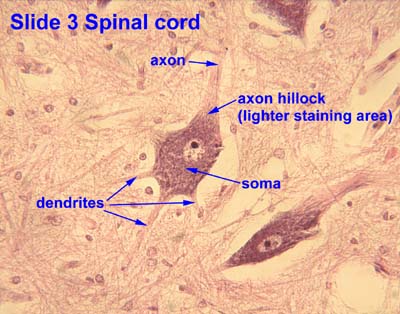
The axon hillock is a specialized part of the cell body (or soma) of a neuron that connects to the axon. It can be identified using light microscopy from its appearance and location in a neuron and from its sparse distribution of Nissl substance. [1]

Contents

The axon hillock is the last site in the soma where membrane potentials propagated from synaptic inputs are summated before being transmitted to the axon. [2] For many years, it was believed that the axon hillock was the usual site of initiation of action potentials—the trigger zone. It is now thought that the earliest site of action potential initiation is at the axonal initial segment: just between the peak of the axon hillock and the initial (unmyelinated) segment of the axon. [3] However, the positive point, at which the action potential starts, varies between cells.[ citation needed ] It can also be altered by hormonal stimulation of the neuron, or by second messenger effects of neurotransmitters.[ citation needed ]

The axon hillock also delineates separate membrane domains between the cell body and axon. [4] This allows for localization of membrane proteins to either the axonal or somal side of the cell.

Structure

The axon hillock and initial segment have a number of specialized properties that make them capable of action potential generation, including adjacency to the axon and a much higher density of voltage-gated ion channels than is found in the rest of the cell body. [5] In dorsal root ganglion cells, the cell body is thought to have approximately 1 voltage-gated sodium channel per square micrometre, while the axon hillock and initial segment of the axon have about ~100–200 voltage-gated sodium channels per square micrometre; in comparison, the nodes of Ranvier along the axon are thought to have ~1000–2000 such channels per square micrometre. [6] This clustering of voltage-gated ion channels is a consequence of plasma-membrane and cytoskeletal associating proteins such as ankyrin. [7]

In electrophysiological models, the axon hillock is included with the initial segment of the axon where membrane potentials propagated from synaptic inputs to the dendrites or cell body are summed.[ citation needed ]

Function

Both inhibitory postsynaptic potentials (IPSPs) and excitatory postsynaptic potentials (EPSPs) are summed in the axon hillock and once a triggering threshold is exceeded, an action potential propagates through the rest of the axon (and "backwards" towards the dendrites as seen in neural backpropagation). The triggering is due to positive feedback between highly crowded voltage-gated sodium channels, which are present at the critical density at the axon hillock (and nodes of ranvier) but not in the soma.

In its resting state, a neuron is polarized, with its inside at about −70 mV relative to its surroundings. When an excitatory neurotransmitter is released by the presynaptic neuron and binds to the postsynaptic dendritic spines, ligand-gated ion channels open, allowing sodium ions to enter the cell. This may make the postsynaptic membrane depolarized (less negative). This depolarization will travel towards the axon hillock, diminishing exponentially with time and distance. If several such events occur in a short time, the axon hillock may become sufficiently depolarized for the voltage-gated sodium channels to open. This initiates an action potential that then propagates down the axon.

As sodium enters the cell, the cell membrane potential becomes more positive, which activates even more sodium channels in the membrane. The sodium influx eventually overtakes the potassium efflux (via the two-pore-domain potassium channels or leak channels, initiating a positive feedback loop (rising phase). At around +40 mV, the voltage-gated sodium channels begin to close (peak phase) and the voltage-gated potassium channels begin to open, moving potassium down its electrochemical gradient and out of the cell (falling phase).

The potassium channels exhibit a delayed reaction to the membrane repolarisation, and, even after the resting potential is achieved, some potassium continues to flow out, resulting in an intracellular fluid that is more negative than the resting potential, and during which no action potential can begin (undershoot phase/refractory period). This undershoot phase ensures that the action potential propagates down the axon and not back up it.

Once this initial action potential is initiated, principally at the axon hillock, it propagates down the length of the axon. Under normal conditions, the action potential would attenuate very quickly due to the porous nature of the cell membrane. To ensure faster and more efficient propagation of action potentials, the axon is myelinated. Myelin, a derivative of cholesterol, acts as an insulating sheath and ensures that the signal cannot escape through the ion or leak channels. There are, nevertheless, gaps in the insulation (nodes of Ranvier), which boost the signal strength. As the action potential reaches a node of Ranvier, it depolarises the cell membrane. As the cell membrane is depolarised, the voltage-gated sodium channels open and sodium rushes in, triggering a fresh new action potential.

Related Research Articles

<span class="mw-page-title-main">Axon</span> Long projection on a neuron that conducts signals to other neurons

An axon or nerve fiber is a long, slender projection of a nerve cell, or neuron, in vertebrates, that typically conducts electrical impulses known as action potentials away from the nerve cell body. The function of the axon is to transmit information to different neurons, muscles, and glands. In certain sensory neurons, such as those for touch and warmth, the axons are called afferent nerve fibers and the electrical impulse travels along these from the periphery to the cell body and from the cell body to the spinal cord along another branch of the same axon. Axon dysfunction can be the cause of many inherited and acquired neurological disorders that affect both the peripheral and central neurons. Nerve fibers are classed into three types – group A nerve fibers, group B nerve fibers, and group C nerve fibers. Groups A and B are myelinated, and group C are unmyelinated. These groups include both sensory fibers and motor fibers. Another classification groups only the sensory fibers as Type I, Type II, Type III, and Type IV.

<span class="mw-page-title-main">Dendrite</span> Small projection on a neuron that receives signals

A dendrite or dendron is a branched protoplasmic extension of a nerve cell that propagates the electrochemical stimulation received from other neural cells to the cell body, or soma, of the neuron from which the dendrites project. Electrical stimulation is transmitted onto dendrites by upstream neurons via synapses which are located at various points throughout the dendritic tree.

<span class="mw-page-title-main">Myelin</span> Fatty substance that surrounds nerve cell axons to insulate them and increase transmission speed

Myelin is a lipid-rich material that surrounds nerve cell axons to insulate them and increase the rate at which electrical impulses pass along the axon. The myelinated axon can be likened to an electrical wire with insulating material (myelin) around it. However, unlike the plastic covering on an electrical wire, myelin does not form a single long sheath over the entire length of the axon. Rather, myelin ensheaths the axon segmentally: in general, each axon is encased in multiple long sheaths with short gaps between, called nodes of Ranvier. At the nodes of Ranvier, which are approximately one thousandth of a mm in length, the axon's membrane is bare of myelin.

<span class="mw-page-title-main">Neuron</span> Electrically excitable cell found in the nervous system of animals

Within a nervous system, a neuron, neurone, or nerve cell is an electrically excitable cell that fires electric signals called action potentials across a neural network. Neurons communicate with other cells via synapses, which are specialized connections that commonly use minute amounts of chemical neurotransmitters to pass the electric signal from the presynaptic neuron to the target cell through the synaptic gap.

<span class="mw-page-title-main">Action potential</span> Neuron communication by electric impulses

An action potential occurs when the membrane potential of a specific cell rapidly rises and falls. This depolarization then causes adjacent locations to similarly depolarize. Action potentials occur in several types of animal cells, called excitable cells, which include neurons, muscle cells, and in some plant cells. Certain endocrine cells such as pancreatic beta cells, and certain cells of the anterior pituitary gland are also excitable cells.

<span class="mw-page-title-main">Depolarization</span> Change in a cells electric charge distribution

In biology, depolarization or hypopolarization is a change within a cell, during which the cell undergoes a shift in electric charge distribution, resulting in less negative charge inside the cell compared to the outside. Depolarization is essential to the function of many cells, communication between cells, and the overall physiology of an organism.

<span class="mw-page-title-main">Membrane potential</span> Type of physical quantity

Membrane potential is the difference in electric potential between the interior and the exterior of a biological cell. That is, there is a difference in the energy required for electric charges to move from the internal to exterior cellular environments and vice versa, as long as there is no acquisition of kinetic energy or the production of radiation. The concentration gradients of the charges directly determine this energy requirement. For the exterior of the cell, typical values of membrane potential, normally given in units of milli volts and denoted as mV, range from –80 mV to –40 mV.

<span class="mw-page-title-main">Threshold potential</span> Critical potential value

In electrophysiology, the threshold potential is the critical level to which a membrane potential must be depolarized to initiate an action potential. In neuroscience, threshold potentials are necessary to regulate and propagate signaling in both the central nervous system (CNS) and the peripheral nervous system (PNS).

<span class="mw-page-title-main">Node of Ranvier</span> Gaps between myelin sheaths on the axon of a neuron

In neuroscience and anatomy, nodes of Ranvier, also known as myelin-sheath gaps, occur along a myelinated axon where the axolemma is exposed to the extracellular space. Nodes of Ranvier are uninsulated and highly enriched in ion channels, allowing them to participate in the exchange of ions required to regenerate the action potential. Nerve conduction in myelinated axons is referred to as saltatory conduction due to the manner in which the action potential seems to "jump" from one node to the next along the axon. This results in faster conduction of the action potential.

<span class="mw-page-title-main">End-plate potential</span>

End plate potentials (EPPs) are the voltages which cause depolarization of skeletal muscle fibers caused by neurotransmitters binding to the postsynaptic membrane in the neuromuscular junction. They are called "end plates" because the postsynaptic terminals of muscle fibers have a large, saucer-like appearance. When an action potential reaches the axon terminal of a motor neuron, vesicles carrying neurotransmitters are exocytosed and the contents are released into the neuromuscular junction. These neurotransmitters bind to receptors on the postsynaptic membrane and lead to its depolarization. In the absence of an action potential, acetylcholine vesicles spontaneously leak into the neuromuscular junction and cause very small depolarizations in the postsynaptic membrane. This small response (~0.4mV) is called a miniature end plate potential (MEPP) and is generated by one acetylcholine-containing vesicle. It represents the smallest possible depolarization which can be induced in a muscle.

<span class="mw-page-title-main">Voltage-gated ion channel</span> Type of ion channel transmembrane protein

Voltage-gated ion channels are a class of transmembrane proteins that form ion channels that are activated by changes in the electrical membrane potential near the channel. The membrane potential alters the conformation of the channel proteins, regulating their opening and closing. Cell membranes are generally impermeable to ions, thus they must diffuse through the membrane through transmembrane protein channels. They have a crucial role in excitable cells such as neuronal and muscle tissues, allowing a rapid and co-ordinated depolarization in response to triggering voltage change. Found along the axon and at the synapse, voltage-gated ion channels directionally propagate electrical signals. Voltage-gated ion-channels are usually ion-specific, and channels specific to sodium (Na+), potassium (K+), calcium (Ca2+), and chloride (Cl) ions have been identified. The opening and closing of the channels are triggered by changing ion concentration, and hence charge gradient, between the sides of the cell membrane.

Molecular neuroscience is a branch of neuroscience that observes concepts in molecular biology applied to the nervous systems of animals. The scope of this subject covers topics such as molecular neuroanatomy, mechanisms of molecular signaling in the nervous system, the effects of genetics and epigenetics on neuronal development, and the molecular basis for neuroplasticity and neurodegenerative diseases. As with molecular biology, molecular neuroscience is a relatively new field that is considerably dynamic.

Sodium channels are integral membrane proteins that form ion channels, conducting sodium ions (Na+) through a cell's membrane. They belong to the superfamily of cation channels.

Neural backpropagation is the phenomenon in which, after the action potential of a neuron creates a voltage spike down the axon, another impulse is generated from the soma and propagates towards the apical portions of the dendritic arbor or dendrites. In addition to active backpropagation of the action potential, there is also passive electrotonic spread. While there is ample evidence to prove the existence of backpropagating action potentials, the function of such action potentials and the extent to which they invade the most distal dendrites remain highly controversial.

<span class="mw-page-title-main">SCN8A</span> Protein-coding gene in the species Homo sapiens

Sodium channel protein type 8 subunit alpha also known as Nav1.6 is a membrane protein encoded by the SCN8A gene. Nav1.6 is one sodium channel isoform and is the primary voltage-gated sodium channel at each node of Ranvier. The channels are highly concentrated in sensory and motor axons in the peripheral nervous system and cluster at the nodes in the central nervous system.

<span class="mw-page-title-main">Ankyrin-3</span> Protein-coding gene in the species Homo sapiens

Ankyrin-3 (ANK-3), also known as ankyrin-G, is a protein from ankyrin family that in humans is encoded by the ANK3 gene.

<span class="mw-page-title-main">Dendritic spike</span> Action potential generated in the dendrite of a neuron

In neurophysiology, a dendritic spike refers to an action potential generated in the dendrite of a neuron. Dendrites are branched extensions of a neuron. They receive electrical signals emitted from projecting neurons and transfer these signals to the cell body, or soma. Dendritic signaling has traditionally been viewed as a passive mode of electrical signaling. Unlike its axon counterpart which can generate signals through action potentials, dendrites were believed to only have the ability to propagate electrical signals by physical means: changes in conductance, length, cross sectional area, etc. However, the existence of dendritic spikes was proposed and demonstrated by W. Alden Spencer, Eric Kandel, Rodolfo Llinás and coworkers in the 1960s and a large body of evidence now makes it clear that dendrites are active neuronal structures. Dendrites contain voltage-gated ion channels giving them the ability to generate action potentials. Dendritic spikes have been recorded in numerous types of neurons in the brain and are thought to have great implications in neuronal communication, memory, and learning. They are one of the major factors in long-term potentiation.

Cellular neuroscience is a branch of neuroscience concerned with the study of neurons at a cellular level. This includes morphology and physiological properties of single neurons. Several techniques such as intracellular recording, patch-clamp, and voltage-clamp technique, pharmacology, confocal imaging, molecular biology, two photon laser scanning microscopy and Ca2+ imaging have been used to study activity at the cellular level. Cellular neuroscience examines the various types of neurons, the functions of different neurons, the influence of neurons upon each other, and how neurons work together.

<span class="mw-page-title-main">Nonsynaptic plasticity</span> Form of neuroplasticity

Nonsynaptic plasticity is a form of neuroplasticity that involves modification of ion channel function in the axon, dendrites, and cell body that results in specific changes in the integration of excitatory postsynaptic potentials and inhibitory postsynaptic potentials. Nonsynaptic plasticity is a modification of the intrinsic excitability of the neuron. It interacts with synaptic plasticity, but it is considered a separate entity from synaptic plasticity. Intrinsic modification of the electrical properties of neurons plays a role in many aspects of plasticity from homeostatic plasticity to learning and memory itself. Nonsynaptic plasticity affects synaptic integration, subthreshold propagation, spike generation, and other fundamental mechanisms of neurons at the cellular level. These individual neuronal alterations can result in changes in higher brain function, especially learning and memory. However, as an emerging field in neuroscience, much of the knowledge about nonsynaptic plasticity is uncertain and still requires further investigation to better define its role in brain function and behavior.

In molecular biology, the ankyrin-G binding motif of KCNQ2-3 is a protein motif found in the potassium channels KCNQ2 and KCNQ3.

References

  1. Palay, Sanford L.; Sotelo, Constantino; Peters, Alan; Orkand, Paula M. (1968). "The Axon Hillock and the Initial Segment". The Journal of Cell Biology. 38 (1): 193–201. doi:10.1083/jcb.38.1.193. PMC   2107452 . PMID   5691973.
  2. Hemmings, Hugh C.; Egan, Talmage D. (2012-12-06). Pharmacology and Physiology for Anesthesia E-Book: Foundations and Clinical Application. Elsevier Health Sciences. ISBN   9781455737932.
  3. Clark BD, Goldberg EM, Rudy B (December 2009). "Electrogenic Tuning of the Axon Initial Segment". Neuroscientist. 15 (6): 651–668. doi:10.1177/1073858409341973. PMC   2951114 . PMID   20007821.
  4. Kobayashi, Toshihide; Storrie, Brian; Simons, Kai; Dotti, Carlos (15 October 1992). "A functional barrier to movement of lipids in polarized neurons". Nature. 359 (6396): 647–650. Bibcode:1992Natur.359..647K. doi:10.1038/359647a0. PMID   1406997. S2CID   4325727.
  5. Wollner D, Catterall WA (November 1986). "Localization of sodium channels in axon hillocks and initial segments of retinal ganglion cells". Proceedings of the National Academy of Sciences of the United States of America. 83 (21): 8424–28. Bibcode:1986PNAS...83.8424W. doi: 10.1073/pnas.83.21.8424 . PMC   386941 . PMID   2430289.
  6. Safronov BV, Wolff M, Vogel W (February 1, 1999). "Axonal expression of sodium channels in rat spinal neurones during postnatal development". J. Physiol. 514 (3): 729–34. doi:10.1111/j.1469-7793.1999.729ad.x. PMC   2269106 . PMID   9882745.
  7. Zhou D, Lambert S, Malen PL, Carpenter S, Boland LM, Bennett V (November 30, 1998). "AnkyrinG Is Required for Clustering of Voltage-gated Na Channels at Axon Initial Segments and for Normal Action Potential Firing". The Journal of Cell Biology. 143 (5): 1295–304. doi:10.1083/jcb.143.5.1295. PMC   2133082 . PMID   9832557.