Ball and chain inactivation

Last updated
Diagram of a voltage-gated ion channel, showing the three states: closed, open, and inactivated. Ball and chain inactivation can only happen if the channel is open. Inactivation diagram.jpg
Diagram of a voltage-gated ion channel, showing the three states: closed, open, and inactivated. Ball and chain inactivation can only happen if the channel is open.

In neuroscience, ball and chain inactivation is a model to explain the fast inactivation mechanism of voltage-gated ion channels. The process is also called hinged-lid inactivation or N-type inactivation. A voltage-gated ion channel can be in three states: open, closed, or inactivated. The inactivated state is mainly achieved through fast inactivation, by which a channel transitions rapidly from an open to an inactivated state. The model proposes that the inactivated state, which is stable and non-conducting, is caused by the physical blockage of the pore. The blockage is caused by a "ball" of amino acids connected to the main protein by a string of residues on the cytoplasmic side of the membrane. The ball enters the open channel and binds to the hydrophobic inner vestibule within the channel. This blockage causes inactivation of the channel by stopping the flow of ions. [1] [2] This phenomenon has mainly been studied in potassium channels and sodium channels. [3]

Contents

Discovery

Electrophysiological evidence

The initial evidence for a ball and chain inactivation came in 1977 with Clay Armstrong and Francisco Bezanilla's work. [4] The suggestion of a physical basis for non-conductance came from experiments in squid giant axons, showing that internal treatment with pronase disrupted the inactivation phenomenon. This suggested a physical, tethered mechanism for inactivation as the pronase was inferred to degrade the channel blocker and abolish the inactivation process. These experiments also showed that inactivation can only occur after the opening of the channel. This was done by hyperpolarising the membrane, causing the channel to open, and observing a delay in inactivation. Inactivation was not observed when the membrane was depolarised (closed). Introducing tetraethylammonium (TEA) on the intracellular side of the channel was found to mimic inactivation in non-inactivating channels. [5] Blockage of the channel by TEA is mutually exclusive with peptide-mediate blockage, suggesting that TEA competes for an inactivation binding site. [6]

Molecular evidence

Mutagenesis experiments have identified an intracellular string of amino acids as prime candidates for the pore blocker. [5] The precise sequence of amino acids that makes up the channel-blocking ball in potassium channels was identified through the creation a synthetic peptide. The peptide was built based on the sequence of a 20 amino acid residue from the Drosophila melanogaster 's Shaker ShB protein and applied on the intracellular side of a non-inactivating channel in Xenopus oocytes. The peptide restored inactivation to the channel, giving further support to the ball and chain model. In β2 proteins, the first three residues after the initial methionine have been identified as essential for inactivation. The initial residues have a sequence motif of phenylalanine, isoleucine and tryptophan without which inactivation does not occur. Modifying the subsequent residues alters the speed and efficacy of inactivation without abolishing it. [7]

Structural evidence

More recently, nuclear magnetic resonance studies in Xenopus oocyte BK channels have shed further light on the structural properties of the ball and chain domain. [8] The introduction of the KCNMB2 β subunit to the cytoplasmic side of a non-inactivating channel restored inactivation, conforming to the expected behaviour of a ball and chain-type protein. NMR analysis showed that the ball domain is composed of residues 1–17 and the chain region of residues 20–45. The three amino acids in the middle constitute a flexible linker region between the two functional regions. The ball is at the N-terminus of the β subunit and consists of a disordered part (residues 1–10) and a loop-helix motif formed by a block of amino acids spanning from serine at position 11 to aspartate at position 16. The structure of the chain domain is 4-turn alpha helix structure.

Structure

The ball and chain domains are on the cytoplasmic side of the channel. The most precise structural studies have been carried out in Shaker potassium channels, in which the precise residues involved in the process have been identified. The first 19 amino acids of the N-terminus constitute the ball domain. This is made up of 11 hydrophobic amino acids, 8 hydrophilic ones and 4 positively charged ones. [9] The following 60 amino acids constitute the chain domain. Modifying the amino acids of the ball while preserving their chemical properties does not disrupt the inactivation mechanism. This suggests that the ball occludes the channel by binding electrostatically rather than covalently. [10] Structural studies have shown that the inner pore of the potassium channel is accessible only through side slits between the cytoplasmic domains of the four α-subunits, rather than from a central route as previously thought. [11] The ball domain enters the channel through the side slits and attaches to a binding site deep in the central cavity. This process involves a conformational change, which allows the ball and chain blocker to elongate and reach the inner center of the channel. [12]

Diagram of a voltage-gated sodium channel, showing the important residues for inactivation in red. The domain structure (I - IV) is further subdivided into segments (S1 - 6). The S4 segment is the voltage sensor, which moves out during depolarisation of the cell membrane. This frees up the alanine and asparagine residues with which the IFMT residues in the ball domain bind to. Adapted from Goldin, 2003. Sodium inactivation mechanims.jpg
Diagram of a voltage-gated sodium channel, showing the important residues for inactivation in red. The domain structure (I – IV) is further subdivided into segments (S1 – 6). The S4 segment is the voltage sensor, which moves out during depolarisation of the cell membrane. This frees up the alanine and asparagine residues with which the IFMT residues in the ball domain bind to. Adapted from Goldin, 2003.

A positively charged region between the III and IV domains of sodium channels is thought to act in a similar way. [9] The essential region for inactivation in sodium channels is four amino acid sequence made up of isoleucine, phenylalanine, methionine and threonine (IFMT). [13] The T and F interact directly with the docking site in the channel pore. [14] When voltage-gated sodium channels open, the S4 segment moves outwards from the channel and into the extracellular side. This exposes hydrophobic residues in the S4 and S5 segments which interact with the inactivation ball. The phenylalanine of the ball interacts with the alanine in domain III's S4-S5 segments and the asparagine in domain IV's S4-S5 segments. [15] This explains why inactivation can only occur once the channel is open.

Lateral slits are also present in sodium channels, [16] suggesting that the access route for the ball domain may be similar.

There is a distinction between direct inactivation and two-step inactivation. Direct inactivation, which occurs in Shaker potassium channels results from the direct blockage of the channel by the ball protein, while two-step inactivation, thought to occur in BK channels, requires an intermediate binding step. [17]

The mechanism of ball-and-chain inactivation is also distinct from that of voltage-dependent blockade by intracellular molecules or peptide regions of beta4 subunits in sodium channels. [18] When these blocks contribute to sodium channel inactivation after channel opening, repolarization of the membrane reverses the block and can causes a resurgent current: a flow of ions between unblocking and closure of the channel. [19]

Inactivation prevention domain

Potassium channels have an additional feature in the N-terminus which makes the channels unable to inactivate. The N-type inactivation-prevention (NIP) domain counteracts the effect of the peptide ball. Channels containing the NIP domain behave as mutated non-inactivating channels, as they have no inactivation activity. [20] The effect is thought be stoichiometric, as the gradual introduction of un-tethered synthetic balls to the cytoplasm eventually restores inactivation. [21]

Effects on neuronal firing

The interplay between opening and inactivation controls the firing pattern of a neuron by changing the rate and amount of ion flow through the channels. Voltage-gated ion channels open upon depolarization of the cell membrane. This creates a current caused by the flow of ions through the channel. Shortly after opening, the channel is blocked by the peptide ball. The β1 subunit aids recovery from inactivation, [22] while β2 accelerates inactivation. [23] The β subunits can also interfere with ball and chain domains by blocking their entry into the channel. This leads to persistent currents, caused by the continued influx of ions. The β3 subunit can increase persistent current in certain sodium channels. [13]

Implications for disease

Differences in persistent and resurgent currents have been implicated in certain human neurological and neuromuscular disorders. In epilepsy, mutations in sodium channels genes delay inactivation. This leads to the channel staying open for longer and thus longer-lasting neuronal firing. [24] Higher levels of persistent current are observed in epilepsy. This constant, low-level neuronal stimulation has been linked to the seizures typical of this disorder. [25]

Inactivation anomalies have also been linked to Brugada syndrome. Mutations in genes encoding the α subunit in cardiac sodium channels affect inactivation. These increase persistent current by interfering with inactivation, though different mutations have opposite effects in inactivation speed. [26]

Mutations in the α subunit of skeletal muscles are also associated with myotonia. The characteristic muscular hyperexcitation of myotonia is mainly caused by the presence sodium channels which do not inactivate, causing high levels of persistent current in the muscles. [27]

Related Research Articles

<span class="mw-page-title-main">BK channel</span> Family of transport proteins

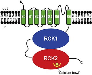
BK channels (big potassium), are large conductance calcium-activated potassium channels, also known as Maxi-K, slo1, or Kca1.1. BK channels are voltage-gated potassium channels that conduct large amounts of potassium ions (K+) across the cell membrane, hence their name, big potassium. These channels can be activated (opened) by either electrical means, or by increasing Ca2+ concentrations in the cell. BK channels help regulate physiological processes, such as circadian behavioral rhythms and neuronal excitability. BK channels are also involved in many processes in the body, as it is a ubiquitous channel. They have a tetrameric structure that is composed of a transmembrane domain, voltage sensing domain, potassium channel domain, and a cytoplasmic C-terminal domain, with many X-ray structures for reference. Their function is to repolarize the membrane potential by allowing for potassium to flow outward, in response to a depolarization or increase in calcium levels.

<span class="mw-page-title-main">Potassium channel</span> Ion channel that selectively passes K+

Potassium channels are the most widely distributed type of ion channel found in virtually all organisms. They form potassium-selective pores that span cell membranes. Potassium channels are found in most cell types and control a wide variety of cell functions.

Hyperkalemic periodic paralysis is an inherited autosomal dominant disorder that affects sodium channels in muscle cells and the ability to regulate potassium levels in the blood. It is characterized by muscle hyperexcitability or weakness which, exacerbated by potassium, heat or cold, can lead to uncontrolled shaking followed by paralysis. Onset usually occurs in early childhood, but it still occurs with adults.

<span class="mw-page-title-main">Voltage-gated ion channel</span> Type of ion channel transmembrane protein

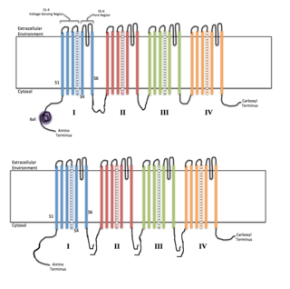
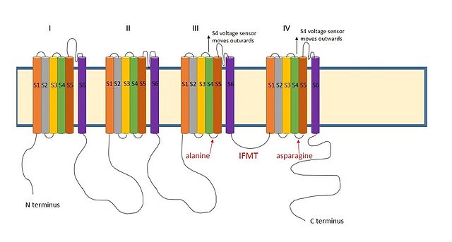
Voltage-gated ion channels are a class of transmembrane proteins that form ion channels that are activated by changes in the electrical membrane potential near the channel. The membrane potential alters the conformation of the channel proteins, regulating their opening and closing. Cell membranes are generally impermeable to ions, thus they must diffuse through the membrane through transmembrane protein channels. They have a crucial role in excitable cells such as neuronal and muscle tissues, allowing a rapid and co-ordinated depolarization in response to triggering voltage change. Found along the axon and at the synapse, voltage-gated ion channels directionally propagate electrical signals. Voltage-gated ion-channels are usually ion-specific, and channels specific to sodium (Na+), potassium (K+), calcium (Ca2+), and chloride (Cl) ions have been identified. The opening and closing of the channels are triggered by changing ion concentration, and hence charge gradient, between the sides of the cell membrane.

Sodium channels are integral membrane proteins that form ion channels, conducting sodium ions (Na+) through a cell's membrane. They belong to the superfamily of cation channels.

<span class="mw-page-title-main">Kv1.1</span>

Potassium voltage-gated channel subfamily A member 1 also known as Kv1.1 is a shaker related voltage-gated potassium channel that in humans is encoded by the KCNA1 gene. Isaacs syndrome is a result of an autoimmune reaction against the Kv1.1 ion channel.

<span class="mw-page-title-main">Voltage-gated potassium channel</span> Class of transport proteins

Voltage-gated potassium channels (VGKCs) are transmembrane channels specific for potassium and sensitive to voltage changes in the cell's membrane potential. During action potentials, they play a crucial role in returning the depolarized cell to a resting state.

Na<sub>v</sub>1.4 Protein-coding gene in the species Homo sapiens

Sodium channel protein type 4 subunit alpha is a protein that in humans is encoded by the SCN4A gene.

<span class="mw-page-title-main">SK channel</span> Protein subfamily of calcium-activated potassium channels

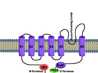
SK channels are a subfamily of calcium-activated potassium channels. They are so called because of their small single channel conductance in the order of 10 pS. SK channels are a type of ion channel allowing potassium cations to cross the cell membrane and are activated (opened) by an increase in the concentration of intracellular calcium through N-type calcium channels. Their activation limits the firing frequency of action potentials and is important for regulating afterhyperpolarization in the neurons of the central nervous system as well as many other types of electrically excitable cells. This is accomplished through the hyperpolarizing leak of positively charged potassium ions along their concentration gradient into the extracellular space. This hyperpolarization causes the membrane potential to become more negative. SK channels are thought to be involved in synaptic plasticity and therefore play important roles in learning and memory.

<span class="mw-page-title-main">Scorpion toxin</span>

Scorpion toxins are proteins found in the venom of scorpions. Their toxic effect may be mammal- or insect-specific and acts by binding with varying degrees of specificity to members of the Voltage-gated ion channel superfamily; specifically, voltage-gated sodium channels, voltage-gated potassium channels, and Transient Receptor Potential (TRP) channels. The result of this action is to activate or inhibit the action of these channels in the nervous and cardiac organ systems. For instance, α-scorpion toxins MeuNaTxα-12 and MeuNaTxα-13 from Mesobuthus eupeus are neurotoxins that target voltage-gated Na+ channels (Navs), inhibiting fast inactivation. In vivo assays of MeuNaTxα-12 and MeuNaTxα-13 effects on mammalian and insect Navs show differential potency. These recombinants exhibit their preferential affinity for mammalian and insect Na+ channels at the α-like toxins' active site, site 3, in order to inactivate the cell membrane depolarization faster[6]. The varying sensitivity of different Navs to MeuNaTxα-12 and MeuNaTxα-13 may be dependent on the substitution of a conserved Valine residue for a Phenylalanine residue at position 1630 of the LD4:S3-S4 subunit or due to various changes in residues in the LD4:S5-S6 subunit of the Navs. Ultimately, these actions can serve the purpose of warding off predators by causing pain or to subdue predators.

<span class="mw-page-title-main">KCNMB2</span> Protein-coding gene in the species Homo sapiens

Calcium-activated potassium channel subunit beta-2 is a protein that in humans is encoded by the KCNMB2 gene.

<span class="mw-page-title-main">Gating (electrophysiology)</span>

In electrophysiology, the term gating refers to the opening (activation) or closing of ion channels. This change in conformation is a response to changes in transmembrane voltage.

<span class="mw-page-title-main">Phrixotoxin</span>

Phrixotoxins are peptide toxins derived from the venom of the Chilean copper tarantula Phrixotrichus auratus, also named Paraphysa scrofa. Phrixotoxin-1 and -2 block A-type voltage-gated potassium channels; phrixotoxin-3 blocks voltage-gated sodium channels. Similar toxins are found in other species, for instance the Chilean rose tarantula.

<span class="mw-page-title-main">Heteroscodratoxin-1</span>

Heteroscodratoxin-1 is a neurotoxin produced by the venom glands of Heteroscodra maculata that shifts the activation threshold of voltage-gated potassium channels and the inactivation of Nav1.1 sodium channels to more positive potentials.

<span class="mw-page-title-main">KcsA potassium channel</span> Prokaryotic potassium ion channel

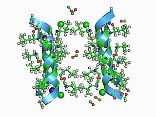
KcsA (Kchannel of streptomyces A) is a prokaryotic potassium channel from the soil bacterium Streptomyces lividans that has been studied extensively in ion channel research. The pH activated protein possesses two transmembrane segments and a highly selective pore region, responsible for the gating and shuttling of K+ ions out of the cell. The amino acid sequence found in the selectivity filter of KcsA is highly conserved among both prokaryotic and eukaryotic K+ voltage channels; as a result, research on KcsA has provided important structural and mechanistic insight on the molecular basis for K+ ion selection and conduction. As one of the most studied ion channels to this day, KcsA is a template for research on K+ channel function and its elucidated structure underlies computational modeling of channel dynamics for both prokaryotic and eukaryotic species.

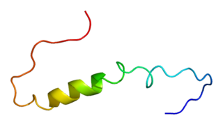
Halcurin is a polypeptide neurotoxin from the sea anemone Halcurias sp. Based on sequence homology to type 1 and type 2 sea anemone toxins it is thought to delay channel inactivation by binding to the extracellular site 3 on the voltage gated sodium channels in a membrane potential-dependent manner.

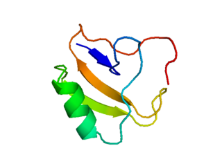
Blood-depressing substance-1 (BDS-1), also known as kappa-actitoxin-Avd4a, is a polypeptide found in the venom of the snakelocks anemone Anemonia sulcata. BDS-1 is a neurotoxin that modulates voltage-dependent potassium channels, in particular Kv3-family channels, as well as certain sodium channels. This polypeptide belongs to the sea anemone type 3 toxin peptide family.

HsTx1 is a toxin from the venom of the scorpion Heterometrus spinifer. HsTx1 is a very potent inhibitor of the rat Kv1.3 voltage-gated potassium channel.

LmαTX3 is an α-scorpion toxin from Lychas mucronatus. that inhibits fast inactivation of voltage gated sodium-channels (VGSCs).

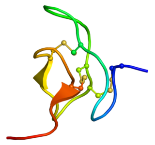
Protoxin-I, also known as ProTx-I, or Beta/omega-theraphotoxin-Tp1a, is a 35-amino-acid peptide neurotoxin extracted from the venom of the tarantula Thrixopelma pruriens. Protoxin-I belongs to the inhibitory cystine knot (ICK) family of peptide toxins, which have been known to potently inhibit voltage-gated ion channels. Protoxin-I selectively blocks low voltage threshold T-type calcium channels, voltage gated sodium channels and the nociceptor cation channel TRPA1. Due to its unique ability to bind to TRPA1, Protoxin-I has been implicated as a valuable pharmacological reagent with potential applications in clinical contexts with regards to pain and inflammation

References

  1. Nicholls JG, Martin AR, Wallace BG, Fuchs PA (2011). From neuron to brain (8th ed.). Sunderland, Mas.: Sinauer Associates. pp. 123–124. ISBN   978-0878936090.
  2. Brady S, Siegel G, Albers RW, Price D (2012). Basic neurochemistry: molecular, cellular and medical aspects (8th ed.). Amsterdam; London: Academic Press. pp.  106–107. ISBN   978-0080959016.
  3. Aldrich RW (2001). "Fifty years of inactivation". Nature . Nature Publishing Group. 411 (6838): 643–644. Bibcode:2001Natur.411..643A. doi:10.1038/35079705. PMID   11395746. S2CID   4373704.
  4. Armstrong CM & Bezanilla, F (1977). "Inactivation of the sodium channel. II. Gating current experiments". The Journal of General Physiology . Rockefeller University Press. 70 (5): 567–590. doi:10.1085/jgp.70.5.567. PMC   2228472 . PMID   591912.
  5. 1 2 Zagotta WN, Hoshi T, Aldrich RW (1990). "Restoration of inactivation in mutants of Shaker potassium channels by a peptide derived from ShB". Science . American Association for the Advancement of Science. 250 (4980): 568–571. Bibcode:1990Sci...250..568Z. doi:10.1126/science.2122520. PMID   2122520. S2CID   15260935.
  6. Choi KL, Aldrich RW, Yellen G (1991). "Tetraethylammonium blockade distinguishes two inactivation mechanisms in voltage-activated K+ channels". Proceedings of the National Academy of Sciences . National Academy of Science. 88 (12): 5092–5095. Bibcode:1991PNAS...88.5092C. doi: 10.1073/pnas.88.12.5092 . PMC   51817 . PMID   2052588.
  7. Xia XM, Ding JP, Lingle CJ (2003). "Inactivation of BK Channels by the NH2 Terminus of the beta Auxiliary Subunit An Essential Role of a Terminal Peptide Segment of Three Hydrophobic Residues". The Journal of General Physiology . Rockefeller University Press. 121 (2): 125–148. doi:10.1085/jgp.20028667. PMC   2217327 . PMID   12566540.
  8. Bentrop D, Beyermann M, Wissmann R, Fakler B (2001). "NMR structure of the "ball-and-chain" domain of KCNMB2, the beta2-subunit of large conductance Ca2+-and voltage-activated potassium channels". Journal of Biological Chemistry . American Society for Biochemistry and Molecular Biology. 276 (45): 42116–42121. doi: 10.1074/jbc.M107118200 . PMID   11517232.
  9. 1 2 Hall ZW (1992). An introduction to molecular neurobiology (1st ed.). Sunderland, Mas.: Sinauer Associates. pp.  113. ISBN   978-0878933075.
  10. Holmgren M, Jurman ME, Yellen G (1996). "N-type inactivation and the S4-S5 region of the Shaker K+ channel". The Journal of General Physiology . Rockefeller University Press. 108 (3): 195–206. doi:10.1085/jgp.108.3.195. PMC   2229322 . PMID   8882863.
  11. Sokolova O, Kolmakova-Partensky L, Grigorieff N (2001). "Three-dimensional structure of a voltage-gated potassium channel at 2.5 nm resolution". Structure . Elsevier. 9 (3): 215–220. doi: 10.1016/s0969-2126(01)00578-0 . PMID   11286888.
  12. Zhou M, Morais-Cabral JH, Mann S, MacKinnon R (2002). "Potassium channel receptor site for the inactivation gate and quaternary amine inhibitors". Nature . Nature Publishing Group. 411 (6838): 657–661. doi:10.1038/35079500. PMID   11395760. S2CID   4415329.
  13. 1 2 3 Goldin AL (2003). "Mechanisms of sodium channel inactivation". Current Opinion in Neurobiology . Elsevier. 13 (3): 284–290. doi:10.1016/S0959-4388(03)00065-5. PMID   12850212. S2CID   17568128.
  14. Miyamoto K, Nakagawa T, Kuroda Y (2001). "Solution structure of the cytoplasmic linker between domain III-S6 and domain IV-S1 (III--IV linker) of the rat brain sodium channel in SDS micelles". Biopolymers . Wiley Online Library. 59 (5): 380–393. doi:10.1002/1097-0282(20011015)59:5<380::AID-BIP1035>3.0.CO;2-T. PMID   11514941.
  15. Miyamoto K, Nakagawa T, Kuroda Y (2001). "Solution structures of the cytoplasmic linkers between segments S4 and S5 (S4- S5) in domains III and IV of human brain sodium channels in SDS micelles". The Journal of Peptide Research. Wiley Online Library. 58 (3): 193–203. doi:10.1034/j.1399-3011.2001.00912.x. PMID   11576325.
  16. Payandeh J, Scheuer T, Zheng N, Catterall WA (2011). "The crystal structure of a voltage-gated sodium channel". Nature . Nature Publishing Group. 475 (7356): 353–358. doi:10.1038/nature10238. PMC   3266868 . PMID   21743477.
  17. Gonzalez-Perez V, Zeng XH, Henzler-Wildman K, Lingle CJ (2012). "Stereospecific binding of a disordered peptide segment mediates BK channel inactivation". Nature . Nature Publishing Group. 485 (7396): 133–136. Bibcode:2012Natur.485..133G. doi:10.1038/nature10994. PMC   3348258 . PMID   22522931.
  18. Lewis AH, Raman IM (2014). "Resurgent current of voltage-gated Na(+) channels". J Physiol. 592 (22): 4825–38. doi:10.1113/jphysiol.2014.277582. PMC   4259529 . PMID   25172941.
  19. Bant JS, Raman IM (2010). "Control of transient, resurgent, and persistent current by open-channel block by Na channel beta4 in cultured cerebellar granule neurons". Proceedings of the National Academy of Sciences . National Academy of Sciences. 107 (27): 12357–12362. Bibcode:2010PNAS..10712357B. doi: 10.1073/pnas.1005633107 . PMC   2901465 . PMID   20566860.
  20. Roeper J, Sewing S, Zhang Y, Sommer T, Wanner SG, Pongs O (1998). "NIP domain prevents N-type inactivation in voltage-gated potassium channels". Nature . Nature Publishing Group. 391 (6665): 390–393. Bibcode:1998Natur.391..390R. doi:10.1038/34916. PMID   9450755. S2CID   205003887.
  21. Yellen G (1998). "The moving parts of voltage-gated ion channels". Quarterly Reviews of Biophysics. Cambridge University Press. 31 (3): 239–295. doi:10.1017/s0033583598003448. PMID   10384687. S2CID   2605660.
  22. Zimmer T, Benndorf K (2002). "The human heart and rat brain IIA Na+ channels interact with different molecular regions of the beta subunit". The Journal of General Physiology . Rockefeller University Press. 120 (6): 887–895. doi:10.1085/jgp.20028703. PMC   2229568 . PMID   12451056.
  23. McCormick KA, Isom LL, Ragsdale D, Smith D, Scheuer T, Catterall WA (1998). "Molecular determinants of Na+ channel function in the extracellular domain of the beta1 subunit". Journal of Biological Chemistry . American Society for Biochemistry and Molecular Biology. 273 (7): 3954–3962. doi: 10.1074/jbc.273.7.3954 . PMID   9461582.
  24. Alekov AK, Rahman MM, Mitrovic N, Lehmann-Horn F, Lerche H (2000). "A sodium channel mutation causing epilepsy in man exhibits subtle defects in fast inactivation and activation in vitro". The Journal of Physiology . Wiley Online Library. 529 (3): 533–540. doi:10.1111/j.1469-7793.2000.00533.x. PMC   2270215 . PMID   11118488.
  25. Stafstrom CE (2007). "Persistent sodium current and its role in epilepsy". Epilepsy Currents . Wiley Online Library. 7 (1): 15–22. doi:10.1111/j.1535-7511.2007.00156.x. PMC   1797888 . PMID   17304346.
  26. Rivolta I, Abriel H, Tateyama M, Liu H, Memmi M, Vardas P, Napolitano C, Priori SG, Kass RS (2001). "Inherited Brugada and long QT-3 syndrome mutations of a single residue of the cardiac sodium channel confer distinct channel and clinical phenotypes". Journal of Biological Chemistry . American Society for Biochemistry and Molecular Biology. 276 (33): 30623–30630. doi: 10.1074/jbc.M104471200 . PMID   11410597.
  27. Lerche H, Heine R, Pika U, George AL, Mitrovic N, Browatzki M, Weiss T, Rivet-Bastide M, Franke C, Lomonaco M (1993). "Human sodium channel myotonia: slowed channel inactivation due to substitutions for a glycine within the III-IV linker". The Journal of Physiology . Wiley Online Library. 470 (1): 113–120. doi:10.1113/jphysiol.1993.sp019843. PMC   1143902 . PMID   8308722.