Chlamydia psittaci

Last updated

Chlamydia psittaci
Chlamydophila psittaci FA stain.jpg
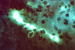
Direct fluorescent antibody stain of a mouse brain impression smear showing C. psittaci.
Scientific classification
Domain:
Phylum:
Class:
Order:
Family:
Genus:
Species:
C. psittaci
Binomial name
Chlamydia psittaci [1]
Synonyms

Chlamydophila psittaci

Chlamydia psittaci is a lethal intracellular bacterial species that may cause endemic avian chlamydiosis, epizootic outbreaks in other mammals, and respiratory psittacosis in humans. Potential hosts include feral birds and domesticated poultry, as well as cattle, pigs, sheep, and horses. C. psittaci is transmitted by inhalation, contact, or ingestion among birds and to mammals. Psittacosis in birds and in humans often starts with flu-like symptoms and becomes a life-threatening pneumonia. Many strains remain quiescent in birds until activated by stress. Birds are excellent, highly mobile vectors for the distribution of chlamydia infection, because they feed on, and have access to, the detritus of infected animals of all sorts.

Contents

C. psittaci in birds is often systemic, and infections can be inapparent, severe, acute, or chronic with intermittent shedding. [2] [3] [4] C. psittaci strains in birds infect mucosal epithelial cells and macrophages of the respiratory tract. Septicaemia eventually develops and the bacteria become localized in epithelial cells and macrophages of most organs, conjunctiva, and gastrointestinal tracts. It can also be passed in the eggs. Stress will commonly trigger onset of severe symptoms, resulting in rapid deterioration and death. C. psittaci strains are similar in virulence, grow readily in cell culture, have 16S rRNA genes that differ by <0.8%, and belong to eight known serotypes. All should be considered to be readily transmissible to humans.

C. psittaci serovar A is endemic among psittacine birds and has caused sporadic zoonotic disease in humans, other mammals, and tortoises. Serovar B is endemic among pigeons, has been isolated from turkeys, and has also been identified as the cause of abortion in herds of dairy cattle. Serovars C and D are occupational hazards for slaughterhouse workers and for people in contact with birds. Serovar E isolates (known as Cal-10, MP or MN) have been obtained from a variety of avian hosts worldwide and, although they were associated with the 1920s–1930s outbreak in humans, a specific reservoir for serovar E has not been identified. The M56 and WC serovars were isolated during outbreaks in mammals. Many C. psittaci strains are susceptible to bacteriophages.

Life cycle and method of infection

Lifecycle of C. psittaci Chlamydophila pneumoniae.jpg
Lifecycle of C. psittaci

Chlamydia psittaci is a small bacterium (0.5μm) that undergoes several transformations during its lifecycle. It exists as an elementary body (EB) between hosts. The EB is not biologically active, but is resistant to environmental stresses and can survive outside a host. The EB travels from an infected bird to the lungs of an uninfected bird or person in small droplets, and is responsible for infection. Once in the lungs, the EB is taken up by cells in a pouch called an endosome by phagocytosis. However, the EB is not destroyed by fusion with lysosomes, as is typical for phagocytosed material. Instead, it transforms into a reticulate body and begins to replicate within the endosome. The reticulate bodies must use some of the host's cellular machinery to complete their replication. The reticulate bodies then convert back to elementary bodies, and are released back into the lung, often after causing the death of the host cell. The EBs are thereafter able to infect new cells, either in the same organism or in a new host. Thus, the lifecycle of C. psittaci is divided between the elementary body which is able to infect new hosts, but can not replicate, and the reticulate body, which replicates, but is not able to cause new infection.

History

The disease caused by C. psittaci, psittacosis, was first characterized in 1879 when seven individuals in Switzerland were found to experience pneumonia after exposure to tropical pet birds. The causative pathogen was not known. The related bacterial species Chlamydia trachomatis was described in 1907, but was assumed to be a virus, as it could not be grown on artificial media. In the winter of 1929–1930, a psittacosis pandemic spread across the United States and Europe. Its mortality rate was 20% and as high as 80% for pregnant women. The disease's spread was eventually attributed to exposure to Amazon parrots imported from Argentina. Though C. psittaci was identified in 1930 as the agent responsible for psittacosis, it was not found to be a bacterium until examination by electron microscopy in the 1960s. [5]

Taxonomy

For several decades, the family Chlamydiaceae contained a sole genus, Chlamydia. C. psittaci was originally classified from the 1960s to 1999 as a species of this sole genus. In 1999, the order Chlamydiales was assigned two new families (Parachlamydiaceae and Simkaniaceae), and within the family Chlamydiaceae, the genus Chlamydia was divided into two genera, Chlamydia and the newly designated genus Chlamydophila , with C. psittaci becoming Chlamydophila psittaci. [1] However, this reclassification "was not wholly accepted or adopted" [6] among microbiologists, which "resulted in a reversion to the single, original genus Chlamydia, which now encompasses all 9 species including C. psittaci." [6] A new species was added to the reunited genus Chlamydia in 2013, [7] two more were added in 2014. [8]

What were once classified as the mammal-endemic strains C. psittaci abortion, 'C. psittaci feline, and C. psittaci guinea pig are today three separate species, C. abortus , C. felis , and C. caviae .

Diseases

C. psittaci infection is also associated with schizophrenia. Many other kinds of infections have been associated with schizophrenia. [9]

Genomics

Like other Chlamydia, C. psittaci is an intracellular pathogen and has thus undergone significant genome reduction. Most C. psittaci genomes encode between 1,000 and 1,400 proteins. A total of 911 core genes were found to be present in all 20 strains sequenced by Read et al., corresponding to 90% of the genes present in each genome. [10]

Confirmation of diagnosis

In addition to symptoms and CHX, complement fixation, microimmunofluorescence, and polymerase chain reaction tests can be used to confirm the diagnosis.

Treatment

Tetracycline or macrolides can be used to treat this condition. The drugs are given intravenously or orally, depending on drug choice. Treatment should continue for 10–14 days after the fever subsides. In children or pregnant women, though, tetracycline should not be used. Ibuprofen or acetominophen, and fluids are also administered. Cannabis or tobacco smoke should be avoided. While taking tetracycline, dairy products should be avoided.

See also

Related Research Articles

<span class="mw-page-title-main">Psittacosis</span> Infectious disease in humans

Psittacosis—also known as parrot fever, and ornithosis—is a zoonotic infectious disease in humans caused by a bacterium called Chlamydia psittaci and contracted from infected parrots, such as macaws, cockatiels, and budgerigars, and from pigeons, sparrows, ducks, hens, gulls and many other species of birds. The incidence of infection in canaries and finches is believed to be lower than in psittacine birds.

<i>Plasmodium</i> Genus of parasitic protists that can cause malaria

Plasmodium is a genus of unicellular eukaryotes that are obligate parasites of vertebrates and insects. The life cycles of Plasmodium species involve development in a blood-feeding insect host which then injects parasites into a vertebrate host during a blood meal. Parasites grow within a vertebrate body tissue before entering the bloodstream to infect red blood cells. The ensuing destruction of host red blood cells can result in malaria. During this infection, some parasites are picked up by a blood-feeding insect, continuing the life cycle.

<i>Chlamydia trachomatis</i> Species of bacterium

Chlamydia trachomatis, commonly known as chlamydia, is a bacterium that causes chlamydia, which can manifest in various ways, including: trachoma, lymphogranuloma venereum, nongonococcal urethritis, cervicitis, salpingitis, pelvic inflammatory disease. C. trachomatis is the most common infectious cause of blindness and the most common sexually transmitted bacterium.

<i>Orthomyxoviridae</i> Family of RNA viruses including the influenza viruses

Orthomyxoviridae is a family of negative-sense RNA viruses. It includes seven genera: Alphainfluenzavirus, Betainfluenzavirus, Gammainfluenzavirus, Deltainfluenzavirus, Isavirus, Thogotovirus, and Quaranjavirus. The first four genera contain viruses that cause influenza in birds and mammals, including humans. Isaviruses infect salmon; the thogotoviruses are arboviruses, infecting vertebrates and invertebrates. The Quaranjaviruses are also arboviruses, infecting vertebrates (birds) and invertebrates (arthropods).

<span class="mw-page-title-main">Chlamydiota</span> Phylum of bacteria

The Chlamydiota are a bacterial phylum and class whose members are remarkably diverse, including pathogens of humans and animals, symbionts of ubiquitous protozoa, and marine sediment forms not yet well understood. All of the Chlamydiota that humans have known about for many decades are obligate intracellular bacteria; in 2020 many additional Chlamydiota were discovered in ocean-floor environments, and it is not yet known whether they all have hosts. Historically it was believed that all Chlamydiota had a peptidoglycan-free cell wall, but studies in the 2010s demonstrated a detectable presence of peptidoglycan, as well as other important proteins.

<i>Chlamydia pneumoniae</i> Species of bacterium

Chlamydia pneumoniae is a species of Chlamydia, an obligate intracellular bacterium that infects humans and is a major cause of pneumonia. It was known as the Taiwan acute respiratory agent (TWAR) from the names of the two original isolates – Taiwan (TW-183) and an acute respiratory isolate designated AR-39. Briefly, it was known as Chlamydophila pneumoniae, and that name is used as an alternate in some sources. In some cases, to avoid confusion, both names are given.

<span class="mw-page-title-main">H5N1 genetic structure</span>

H5N1 genetic structure is the molecular structure of the H5N1 virus's RNA.

<i>Chlamydophila</i> Genus of bacteria

Chlamydophila is a controversial bacterial genus belonging to the family Chlamydiaceae.

<span class="mw-page-title-main">Chlamydiaceae</span> Family of bacteria

The Chlamydiaceae are a family of gram-negative bacteria that belongs to the phylum Chlamydiota, order Chlamydiales. Chlamydiaceae species express the family-specific lipopolysaccharide epitope αKdo-(2→8)-αKdo-(2→4)-αKdo. Chlamydiaceae ribosomal RNA genes all have at least 90% DNA sequence identity. Chlamydiaceae species have varying inclusion morphology, varying extrachromosomal plasmid content, and varying sulfadiazine resistance.

Chlamydia muridarum is an intracellular bacterial species that at one time belonged to Chlamydia trachomatis. However, C. trachomatis naturally only infects humans and C. muridarum naturally infects only members of the family Muridae.

Chlamydia suis is a member of the genus Chlamydia. C. suis has only been isolated from swine, in which it may be endemic. Glycogen has been detected in Chlamydia suis inclusions in infected swine tissues and in cell culture. C. suis is associated with conjunctivitis, enteritis and pneumonia in swine.

<i>Chlamydia abortus</i> Species of bacterium

Chlamydia abortus is a species in Chlamydiota that causes abortion and fetal death in mammals, including humans. Chlamydia abortus was renamed in 1999 as Chlamydophila psittaci along with all Chlamydiota except Chlamydia trachomatis. This was based on a lack of evident glycogen production and on resistance to the antibiotic sulfadiazine. In 1999 C. psittaci and C. abortus were recognized as distinct species based on differences of pathogenicity and DNA–DNA hybridization. In 2015, this new name was reverted to Chlamydia.

Chlamydia felis is a Gram-negative, obligate intracellular bacterial pathogen that infects cats. It is endemic among domestic cats worldwide, primarily causing inflammation of feline conjunctiva, rhinitis and respiratory problems. C. felis can be recovered from the stomach and reproductive tract. Zoonotic infection of humans with C. felis has been reported. Strains FP Pring and FP Cello have an extrachromosomal plasmid, whereas the FP Baker strain does not. FP Cello produces lethal disease in mice, whereas the FP Baker does not. An attenuated FP Baker strain, and an attenuated 905 strain, are used as live vaccines for cats.

Chlamydia caviae is a bacterium that can be recovered from the conjunctiva of Guinea pigs suffering from ocular inflammation and eye discharge. It is also possible to infect the genital tract of Guinea pigs with C. caviae and elicit a disease that is very similar to human Chlamydia trachomatis infection. C. caviae infects primarily the mucosal epithelium and is not invasive.

Chlamydia pecorum, also known as Chlamydophila pecorum is a species of Chlamydiaceae that originated from ruminants, such as cattle, sheep and goats. It has also infected koalas and swine. C. pecorum strains are serologically and pathogenically diverse.

<i>Pasteurella multocida</i> Species of bacterium

Pasteurella multocida is a Gram-negative, nonmotile, penicillin-sensitive coccobacillus of the family Pasteurellaceae. Strains of the species are currently classified into five serogroups based on capsular composition and 16 somatic serovars (1–16). P. multocida is the cause of a range of diseases in mammals and birds, including fowl cholera in poultry, atrophic rhinitis in pigs, and bovine hemorrhagic septicemia in cattle and buffalo. It can also cause a zoonotic infection in humans, which typically is a result of bites or scratches from domestic pets. Many mammals and birds harbor it as part of their normal respiratory microbiota.

<i>Chlamydia</i> (genus) Genus of bacteria

Chlamydia is a genus of pathogenic Gram-negative bacteria that are obligate intracellular parasites. Chlamydia infections are the most common bacterial sexually transmitted diseases in humans and are the leading cause of infectious blindness worldwide.

The transmission of hepadnaviruses between their natural hosts, humans, non-human primates, and birds, including intra-species host transmission and cross-species transmission, is a topic of study in virology.

Parachlamydia acanthamoebae are bacterium that fall into the category of host-associated microorganisms. This bacterium lives within free-living amoebae that are an intricate part of their reproduction. Originally named Candidatus Parachlamydia acanthamoebae, its current scientific name was introduced shortly after. This species has shown to have over eighty percent 16S rRNA gene sequencing identity with the class Chlamydiia. Parachlamydia acanthamoebae has the same family as the genus Neochlamydia with which it shares many similarities.

<i>Avian metaavulavirus 2</i> Species of virus

Avian metaavulavirus 2, formerly Avian paramyxovirus 2, is a species of virus belonging to the family Paramyxoviridae and genus Metaavulavirus. The virus is a negative strand RNA virus containing a monopartite genome. Avian metaavulavirus 2 is one of nine species belonging to the genus Metaavulavirus. The most common serotype of Avulavirinae is serotype 1, the cause of Newcastle disease (ND). Avian metaavulavirus 2 has been known to cause disease, specifically mild respiratory infections in domestic poultry, including turkeys and chickens, and has many economic effects on egg production and poultry industries. The virus was first isolated from a strain in Yucaipa, California in 1956. Since then, other isolates of the virus have been isolated worldwide.

References

  1. 1 2 Everett KD, Bush RM, Andersen AA (April 1999). "Emended description of the order Chlamydiales, proposal of Parachlamydiaceae fam. nov. and Simkaniaceae fam. nov., each containing one monotypic genus, revised taxonomy of the family Chlamydiaceae, including a new genus and five new species, and standards for the identification of organisms". International Journal of Systematic Bacteriology. 49 Pt 2 (2): 415–40. doi: 10.1099/00207713-49-2-415 . PMID   10319462.
  2. Andersen AA (September 2005). "Serotyping of US isolates of Chlamydophila psittaci from domestic and wild birds". J. Vet. Diagn. Invest. 17 (5): 479–82. doi: 10.1177/104063870501700514 . PMID   16312243.
  3. Dorrestein GM, Wiegman LJ (December 1989). "[Inventory of the shedding of Chlamydia psittaci by parakeets in the Utrecht area using ELISA]". Tijdschr Diergeneeskd (in Dutch). 114 (24): 1227–36. PMID   2617495.
  4. Sareyyupoglu B, Cantekin Z, Bas B (2007). "Chlamydophila psittaci DNA detection in the faeces of cage birds". Zoonoses Public Health. 54 (6–7): 237–42. doi:10.1111/j.1863-2378.2007.01060.x. PMID   17803512. S2CID   9087999.
  5. Harkinezhad, Taher; Geens, Tom; Vanrompay, Daisy (1 March 2009). "Chlamydophila psittaci infections in birds: A review with emphasis on zoonotic consequences". Veterinary Microbiology. 135 (1–2): 68–77. doi:10.1016/j.vetmic.2008.09.046. PMID   19054633.
  6. 1 2 Balsamo G, Maxted AM, Midla JW, Murphy JM, Wohrle R, Edling TM, et al. (September 2017). "Compendium of Measures to Control Chlamydia psittaci Infection Among Humans (Psittacosis) and Pet Birds (Avian Chlamydiosis), 2017" (PDF). Journal of Avian Medicine and Surgery. 31 (3): 262–282. doi: 10.1647/217-265 . PMID   28891690.
  7. Vorimore F, Hsia RC, Huot-Creasy H, Bastian S, Deruyter L, Passet A, Sachse K, Bavoil P, Myers G, Laroucau K (20 September 2013). "Isolation of a New Chlamydia species from the Feral Sacred Ibis (Threskiornis aethiopicus): Chlamydia ibidis". PLOS ONE. 8 (9): e74823. Bibcode:2013PLoSO...874823V. doi: 10.1371/journal.pone.0074823 . PMC   3779242 . PMID   24073223.
  8. Joseph SJ, Marti H, Didelot X, Castillo-Ramirez S, Read TD, Dean D (October 2015). "Chlamydiaceae Genomics Reveals Interspecies Admixture and the Recent Evolution of Chlamydia abortus Infecting Lower Mammalian Species and Humans". Genome Biology and Evolution. 7 (11): 3070–84. doi:10.1093/gbe/evv201. PMC   4994753 . PMID   26507799.
  9. Arias I (April 2012). "Infectious agents associated with schizophrenia: a meta-analysis". Schizophr. Res. 136 (1–3): 128–36. doi:10.1016/j.schres.2011.10.026. PMID   22104141. S2CID   2687441.
  10. Read TD, Joseph SJ, Didelot X, Liang B, Patel L, Dean D (March 2013). "Comparative analysis of Chlamydia psittaci genomes reveals the recent emergence of a pathogenic lineage with a broad host range". mBio. 4 (2): e00604–12. doi:10.1128/mBio.00604-12. PMC   3622922 . PMID   23532978.

Further reading