Clostridium difficile (bacteria)

Last updated

Clostridium difficile
Clostridium difficile 01.png
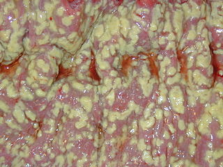
C. difficile colonies on a blood agar plate
Clostridium difficile 01.jpg
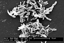
Micrograph of Clostridium difficile
Scientific classification
Kingdom: Bacteria
Phylum: Firmicutes
Class: Clostridia
Order: Clostridiales
Family: Peptostreptococcaceae
Genus: Clostridioides
Species:C. difficile
Binomial name
Clostridioides difficile
Hall & O'Toole, 1935; Lawson & Rainey, 2016

Clostridium difficile (etymology and pronunciation), also known as C. difficile, C. diff ( /sdɪf/ ), or sometimes CDF/cdf, is a species of Gram-positive spore-forming bacterium. [1]

In biology, a species ( ) is the basic unit of classification and a taxonomic rank of an organism, as well as a unit of biodiversity. A species is often defined as the largest group of organisms in which any two individuals of the appropriate sexes or mating types can produce fertile offspring, typically by sexual reproduction. Other ways of defining species include their karyotype, DNA sequence, morphology, behaviour or ecological niche. In addition, paleontologists use the concept of the chronospecies since fossil reproduction cannot be examined. While these definitions may seem adequate, when looked at more closely they represent problematic species concepts. For example, the boundaries between closely related species become unclear with hybridisation, in a species complex of hundreds of similar microspecies, and in a ring species. Also, among organisms that reproduce only asexually, the concept of a reproductive species breaks down, and each clone is potentially a microspecies.

Contents

Clostridia (members of the genus Clostridium and of the Clostridiaceae family) are anaerobic, motile bacteria, ubiquitous in nature, and especially prevalent in soil. Its vegetative cells are rod shaped, pleomorphic, and occur in pairs or short chains. Under the microscope, they appear as long, irregular (often drumstick- or spindle-shaped) cells with a bulge at their terminal ends (forms subterminal spores). Under Gram staining, C. difficile cells are Gram-positive and show optimum growth on blood agar at human body temperatures in the absence of oxygen. C. difficile is catalase and superoxide dismutase negative, and produces two types of toxins: enterotoxin A and cytotoxin B, which disrupts cytoskeleton signal transductions in the host. When stressed, the bacteria produce spores that are able to tolerate extreme conditions that the active bacteria cannot tolerate. [2]

<i>Clostridium</i> genus of bacteria

Clostridium is a genus of Gram-positive bacteria, which includes several significant human pathogens, including the causative agent of botulism. The genus formerly included an important cause of diarrhea, Clostridium difficile, which was separated after 16S rRNA analysis. They are obligate anaerobes capable of producing endospores. The normal, reproducing cells of Clostridium, called the vegetative form, are rod-shaped, which gives them their name, from the Greek κλωστήρ or spindle. Clostridium endospores have a distinct bowling pin or bottle shape, distinguishing them from other bacterial endospores, which are usually ovoid in shape. Clostridium species inhabit soils and the intestinal tract of animals, including humans. Clostridium is a normal inhabitant of the healthy lower reproductive tract of women.

An anaerobic organism or anaerobe is any organism that does not require oxygen for growth. It may react negatively or even die if free oxygen is present.

Motility Ability to move spontaneously and actively, consuming energy in the process

Motility is the ability of an organism to move independently, using metabolic energy. This is in contrast to mobility, which describes the ability of an object to be moved. Motility is genetically determined, but may be affected by environmental factors. For instance, muscles give animals motility but the consumption of hydrogen cyanide would adversely affect muscle physiology, causing them to stiffen, leading to rigor mortis. In addition to animal locomotion, most animals are motile – the term applies to bacteria and other microorganisms, and to some multicellular organisms, as well as to some mechanisms of fluid flow in multicellular organs and tissue. Motile marine animals are commonly called free-swimming, and motile non-parasitic organisms are called free-living.

C. difficile may become established in the human colon; it is present in 2–5% of the adult population. [2] Sometimes antibiotic therapy for various infections has the adverse effect of disrupting the normal balance of the gut microbiota, in which case C. difficile may opportunistically dominate, causing Clostridium difficile infection.

Large intestine last part of the digestive system in vertebrate animals, including the cecum, colon, rectum, and anal canal

The large intestine, also known as the large bowel, is the last part of the gastrointestinal tract and of the digestive system in vertebrates. Water is absorbed here and the remaining waste material is stored as feces before being removed by defecation.

Infection invasion of a host by disease-causing organisms

Infection is the invasion of an organism's body tissues by disease-causing agents, their multiplication, and the reaction of host tissues to the infectious agents and the toxins they produce. Infectious disease, also known as transmissible disease or communicable disease, is illness resulting from an infection.

In medicine, an adverse effect is an undesired harmful effect resulting from a medication or other intervention such as surgery.


Renaming and scientific reclassification

This species has been officially renamed, in 2016, to Clostridioides difficile. [3] [4] This new name reflects the taxonomic differences between this species and other members of the Clostridium genus, while maintaining the common name as C. diff. [5] Currently, the only other species in this new Clostridioides genus is Clostridioides mangenotii (formerly known as Clostridium mangenotii). [6]

Human pathogen

Pathogenic C. difficile strains produce multiple toxins. [7] The best-characterized are enterotoxin (Clostridium difficile toxin A) and cytotoxin (Clostridium difficile toxin B), both of which may produce diarrhea and inflammation in infected patients (Clostridium difficile colitis), although their relative contributions have been debated. The diarrhea may range from a few days of intestinal fluid loss to life-threatening pseudomembranous colitis. Pseudomembranous colitis is associated with intense inflammation of the colon and formation of pseudomembranes on the intestinal mucosal surface. [2] Toxins A and B are glucosyltransferases that target and inactivate the Rho family of GTPases. Toxin B (cytotoxin) induces actin depolymerization by a mechanism correlated with a decrease in the ADP-ribosylation of the low molecular mass GTP-binding Rho proteins. [8] Another toxin, binary toxin, also has been described, but its role in disease is not fully understood. [9]

A toxin is a poisonous substance produced within living cells or organisms; synthetic toxicants created by artificial processes are thus excluded. The term was first used by organic chemist Ludwig Brieger (1849–1919), derived from the word toxic.

Enterotoxin

An enterotoxin is a protein exotoxin released by a microorganism that targets the intestines.

Clostridium difficile toxin A

Clostridium difficile toxin A (TcdA) is a toxin generated by Clostridium difficile. It is similar to Clostridium difficile Toxin B. The toxins are the main virulence factors produced by the gram positive, anaerobic, Clostridium difficile bacteria. The toxins function by damaging the intestinal mucosa and cause the symptoms of C. difficile infection, including pseudomembranous colitis.

Additional virulence factors include an adhesin factor which mediate the binding to human colonic cells and a hyaluronidase. [10]

Hyaluronidase pharmaceutical drug

Hyaluronidases are a family of enzymes that catalyse the degradation of hyaluronic acid (HA). Karl Meyer classified these enzymes in 1971 into three distinct groups, a scheme based on the enzyme reaction products. The three main types of hyaluronidases are two classes of eukaryotic endoglycosidase hydrolases and a prokaryotic lyase-type of glycosidase.

Antibiotic treatment of C. diff infections may be difficult, due both to antibiotic resistance and physiological factors of the bacterium (spore formation, protective effects of the pseudomembrane). [2] The emergence of a new, highly toxic strain of C. difficile, resistant to fluoroquinolone antibiotics, such as ciprofloxacin and levofloxacin, said to be causing geographically dispersed outbreaks in North America, was reported in 2005. [11] The U.S. Centers for Disease Control (CDC) in Atlanta warned of the emergence of an epidemic strain with increased virulence, antibiotic resistance, or both. [12]

Transmission

C. difficile is transmitted from person to person by the fecal-oral route. Clostridium difficile is shed in faeces. Any surface, device, or material (e.g., toilets, bathing tubs, and electronic rectal thermometers) that becomes contaminated with faeces may serve as a reservoir for the Clostridium difficile spores. Clostridium difficile spores are transferred to patients mainly via the hands of healthcare personnel who have touched a contaminated surface or item. Clostridium difficile can live for long periods of time on surfaces. [13] The organism forms heat-resistant spores that are not killed by alcohol-based hand cleansers or routine surface cleaning, thus, these spores survive in clinical environments for long periods. Because of this, the bacterium may be cultured from almost any surface. Once spores are ingested, their acid-resistance allows them to pass through the stomach unscathed. They germinate and multiply into vegetative cells in the colon upon exposure to bile acids. Consequently, the World Health Organization advocates the use of soap in addition to alcohol solutions in order to limit the spread of the spores. [14]

Host range

C. difficile infects pigs, calves, and humans, and inhabits a natural reservoir of soil, faeces of domestic animals and humans, sewage, the human intestinal tract, and retail meat. [15]

A 2015 CDC study estimated that C. diff afflicted almost half a million Americans and caused 29,000 deaths in 2011. The study estimated that 40 percent of cases began in nursing homes or community health care settings, while 24 percent occurred in hospitals. [16]

C. difficile is common in the human digestive system. However, C. difficile is a poor competitor, and is often out competed by other bacteria for nutrients in the digestive system. As a result, C. difficile is kept to a manageable amount. If the sudden introduction of antibiotic disrupts the microbiome, C. difficile may be able to grow as a result of many of its competitors being killed off. The incubation period is 5–10 days, with a range of 1 day to weeks following antibiotic treatment for AAD (antibiotic associated diarrhea). Additionally, carriage of C. difficile with high levels of toxins are common in young children while disease is rare. The production of one or even both toxins is not always sufficient for producing symptoms. [17]

Signs and symptoms

Symptoms of C. difficile include: Diarrhea [18] —watery, at least a minimum of three times a day and up to 15 times a day. Abdominal cramping and pain that can be severe. Loss of appetite and dehydration. Weight loss and nausea. [19]

Treatment

Patients being treated with antibiotics when symptoms begin should stop taking them, if possible. This break in antibiotic therapy can sometimes lead to spontaneous resolution of symptoms. Patients that don't respond to the cessation of broad-spectrum antibiotics will need to be treated with antibiotics capable of killing C. difficile spores. Primary infections are typically treated with metronidazole. This medication is normally given 3 times a day orally and should be taken for a minimum of 10 days. Some patients can't tolerate the side effects of metronidazole, which include severe nausea and vomiting, which can lead to dehydration. In this case, patients should be given oral vancomycin, usually at a dosage of 125 milligrams every 6 hours. [20]

About 20% of patients who successfully complete therapy of primary infection with metronidazole or vancomycin will experience a relapse. A fraction of those patients will experience continuous reoccurrences of the infection. The first relapse of a C. difficile is usually treated with the same antibiotic used to treat the primary infection. Any subsequent infections should not be treated with metronidazole. Occasionally, a standard 10 day course of oral vancomycin will not work. In these cases, a vancomycin taper is the preferred treatment. Patients will take decreasing doses of vancomycin over a period of up to 3 months, depending on the severity of the infection. [19]

Each subsequent relapse of C. difficile tends to be more severe than previous infections. Long term treatment with a vancomycin taper supplemented with probiotics, especially Saccharomyces boulardii, is associated with a higher rate of success. [21]

After three relapses, patients may be treated with oral fidaxomicin, a narrow spectrum antibiotic. The usual dosage is 200 mg twice a day orally for 10 days. Fidaxomicin is considered to be superior to vancomycin for severe CDI (C. difficile infection). [20] The major downside of treatment with fidaxomicin is the cost of medication. A 10-day course may cost up to 3500 USD.

Patients that do not respond to traditional antibiotic therapy may be eligible for a fecal transplant. Healthcare providers can transfer stool from a healthy person to the colon of a patient with repeated CDI. This process is the most successful treatment for severe CDI with a cure rate of around 93%. Long term effects of fecal transplantation are unknown, as the procedure has only been FDA approved since 2011 and relatively few procedures have been performed. If transplantation is not an option, removal of the infected part of the colon can cure C. difficile. [20] [19]

Strains

In 2005, molecular analysis led to the identification of the C. difficile strain type characterized as group BI by restriction endonuclease analysis, as North American pulse-field-type NAP1 by pulsed-field gel electrophoresis and as ribotype 027; the differing terminology reflects the predominant techniques used for epidemiological typing. This strain is referred to as C. difficile BI/NAP1/027. [22]

Two strains, ribotypes RT078 and RT027, can live on low concentrations of the sugar trehalose; both strains became more common after trehalose was introduced as a food additive in the early 2000s and thus increasing dietary trehalose intake. [23]

Genome

Genomic information
NCBI genome ID 535?
Ploidy haploid
Genome size 4.3 Mb
Number of chromosomes 1
Year of completion 2005

The first complete genome sequence of a C. difficile strain was first published in 2005 by Sanger Institute in the UK. This was of the strain 630, a virulent and multiple drug-resistant strain isolated in Switzerland in 1982. Scientists at Sanger Institute have sequenced genomes of about 30 C. difficile isolates using next-generation sequencing technologies from 454 Life Sciences and Illumina. [24]

Researchers at McGill University in Montreal sequenced the genome of the highly virulent Quebec strain of C. difficile in 2005 using ultra-high-throughput sequencing technology. The tests involved doing 400,000 DNA parallel-sequencing reactions of the bacterium's genome, which had been fragmented for sequencing. These sequences were assembled computationally to form a complete genome sequence. [11] [25]

In 2012, scientists at University of Oxford sequenced C. difficile genomes from 486 cases arising over four years in Oxfordshire using next-generation sequencing technologies from Illumina. [26]

Bacteriophage

At least eight mainly temperate bacteriophages have been isolated from C. difficile, ranging in genome size from about 30 to about 60 kb. [27] Both environmentally and clinically derived C. difficile strains carry a diverse and prevalent set of prophages. [27]

Naming and pronunciation

The species name is New Latin, from the Greek kloster (κλωστήρ), "spindle", [28] and Latin difficile, "difficult, obstinate". [29] It is pronounced /klɔːˈstrɪdiəmdɪˈfɪsɪli/ in medical English [30] (compare classical Latin /difˈfi.ki.leː/), although spelling pronunciations such as /ˈdɪfɪsil/ and /ˌdɪfɪˈsl/ are commonly heard. The latter two are approximately comparable to the pronunciations of the obsolete English adjective difficile and the French adjective difficile.

It has been proposed, in a July 2013 paper from Environmental Microbiology , one of the journals of the Society for Applied Microbiology, to rename the species Peptoclostridium difficile. [31] [32]

Notes

  1. Moreno MA, Furtner F, Rivara FP (June 2013). "Clostridium difficile: A Cause of Diarrhea in Children". JAMA Pediatrics. 167 (6): 592. doi:10.1001/jamapediatrics.2013.2551. PMID   23733223.
  2. 1 2 3 4 Ryan KJ, Ray CG, eds. (2004). Sherris Medical Microbiology (4th ed.). McGraw Hill. pp. 322–4. ISBN   0-8385-8529-9.
  3. Oren, Aharon; Garrity, George M. (2017). "List of new names and new combinations previously effectively, but not validly, published". International Journal of Systematic and Evolutionary Microbiology. 67 (9): 3140–3143. doi:10.1099/ijsem.0.002278. PMC   5817221 Lock-green.svg. PMID   28891789.
  4. Zhu, Duolong; Sorg, Joseph A.; Sun, Xingmin (2018). "Clostridioides difficile Biology: Sporulation, Germination, and Corresponding Therapies for C. difficile Infection". Frontiers in Cellular and Infection Microbiology. 8. doi:10.3389/fcimb.2018.00029. ISSN   2235-2988. PMC   5809512 Lock-green.svg. PMID   29473021.
  5. Lawson, Paul A.; Citron, Diane M.; Tyrrell, Kerin L.; Finegold, Sydney M. (2016-08). "Reclassification of Clostridium difficile as Clostridioides difficile (Hall and O'Toole 1935) Prévot 1938". Anaerobe. 40: 95–99. doi:10.1016/j.anaerobe.2016.06.008. ISSN   1075-9964.Check date values in: |date= (help)
  6. Galperin, Michael Y.; Brover, Vyacheslav; Tolstoy, Igor; Yutin, Natalya (2016). "Phylogenomic analysis of the family Peptostreptococcaceae (Clostridium cluster XI) and proposal for reclassification of Clostridium litorale (Fendrich et al. 1991) and Eubacterium acidaminophilum (Zindel et al. 1989) as Peptoclostridium litorale gen. nov. comb. nov. and Peptoclostridium acidaminophilum comb. nov". International Journal of Systematic and Evolutionary Microbiology. 66 (12): 5506–5513. doi:10.1099/ijsem.0.001548. PMC   5244501 Lock-green.svg. PMID   27902180.
  7. Di Bella, Stefano; Ascenzi, Paolo; Siarakas, Steven; Petrosillo, Nicola; di Masi, Alessandra (2016-01-01). "Clostridium difficile Toxins A and B: Insights into Pathogenic Properties and Extraintestinal Effects". Toxins. 8 (5): 134. doi:10.3390/toxins8050134. ISSN   2072-6651. PMC   4885049 Lock-green.svg. PMID   27153087.
  8. Just I, Selzer J, von Eichel-Streiber C, Aktories K (1995). "The low molecular mass GTP-binding protein Rh is affected by toxin a from Clostridium difficile". The Journal of Clinical Investigation. 95 (3): 1026–31. doi:10.1172/JCI117747. PMC   441436 Lock-green.svg. PMID   7883950.
  9. Barth H, Aktories K, Popoff MR, Stiles BG (2004). "Binary Bacterial Toxins: Biochemistry, Biology, and Applications of Common Clostridium and Bacillus Proteins". Microbiology and Molecular Biology Reviews : MMBR. 68 (3): 373–402, table of contents. doi:10.1128/MMBR.68.3.373-402.2004. PMC   515256 Lock-green.svg. PMID   15353562.
  10. [Medical Micriobiology, Fifth Edition, Patrick Murray, Elsevier Mosby, 2005, page 412]
  11. 1 2 Loo VG, Poirier L, Miller MA, Oughton M, Libman MD, Michaud S, Bourgault AM, Nguyen T, Frenette C, Kelly M, Vibien A, Brassard P, Fenn S, Dewar K, Hudson TJ, Horn R, René P, Monczak Y, Dascal A (December 2005). "A predominantly clonal multi-institutional outbreak of Clostridium difficile-associated diarrhea with high morbidity and mortality". The New England Journal of Medicine. 353 (23): 2442–9. doi:10.1056/NEJMoa051639. PMID   16322602.
  12. McDonald LC (August 2005). "Clostridium difficile: responding to a new threat from an old enemy" (PDF). Infection Control and Hospital Epidemiology. 26 (8): 672–5. doi:10.1086/502600. PMID   16156321.
  13. "Clostridium difficile Infection Information for Patients | HAI | CDC". www.cdc.gov. Retrieved 22 May 2017.
  14. "WHO Guidelines on Hand Hygiene in Health Care: a Summary" (PDF). World Health Organization. 2009. p. 31. Retrieved June 18, 2018.
  15. Gould, L. Hannah; Limbago, Brandi (2010). "Clostridium difficile in Food and Domestic Animals: A New Foodborne Pathogen?". Clinical Infectious Diseases. 51 (5): 577. doi:10.1086/655692. PMID   20642351.
  16. Belluck, Pam (February 25, 2015). "Death Toll From C. Difficile Is Raised". The New York Times . Retrieved 25 February 2015.
  17. [Medical Microbiology, Fifth Edition, Patrick Murray, Elsevier Mosby, 2005, page 412]
  18. https://www.jmscleaningservices.co.uk/news/7-germs-commonly-found-office-theyre-dangerous/
  19. 1 2 3 https://www.cdc.gov/hai/organisms/cdiff/cdiff-patient.html
  20. 1 2 3 Surawicz, Christina M; Brandt, Lawrence J; Binion, David G; Ananthakrishnan, Ashwin N; Curry, Scott R; Gilligan, Peter H; McFarland, Lynne V; Mellow, Mark; Zuckerbraun, Brian S (2013-02-26). "Guidelines for Diagnosis, Treatment and Prevention of Clostridium difficile Infections". The American Journal of Gastroenterology. 108 (4): 478–498. doi:10.1038/ajg.2013.4. ISSN   0002-9270.
  21. "Treatment of Recurrent Clostridium difficile Colitis with Vancomycin and Saccharomyces boulardii" (PDF). The American Journal of Gastroenterology.
  22. Rupnik M, Wilcox MH, Gerding DN (July 2009). "Clostridium difficile infection: New developments in epidemiology and pathogenesis". Nature Reviews. Microbiology. 7 (7): 526–36. doi:10.1038/nrmicro2164. PMID   19528959.
  23. Collins, J.; Robinson, C.; Danhof, H.; Knetsch, C. W.; van Leeuwen, H. C.; Lawley, T. D.; Auchtung, J. M.; Britton, R. A. (2018). "Dietary trehalose enhances virulence of epidemic Clostridium difficile". Nature. doi:10.1038/nature25178. ISSN   0028-0836.
  24. He M, Sebaihia M, Lawley TD, Stabler RA, Dawson LF, Martin MJ, Holt KE, Seth-Smith HM, Quail MA, Rance R, Brooks K, Churcher C, Harris D, Bentley SD, Burrows C, Clark L, Corton C, Murray V, Rose G, Thurston S, van Tonder A, Walker D, Wren BW, Dougan G, Parkhill J (April 2010). "Evolutionary dynamics of Clostridium difficile over short and long time scales" (PDF). Proceedings of the National Academy of Sciences of the United States of America. 107 (16): 7527–32. Bibcode:2010PNAS..107.7527H. doi:10.1073/pnas.0914322107. PMC   2867753 Lock-green.svg. PMID   20368420.
  25. Scientists map C. difficile strain – Institute of Public Affairs, Montreal
  26. Didelot X, Eyre DW, Cule M, Ip CL, Ansari MA, Griffiths D, Vaughan A, O'Connor L, Golubchik T, Batty EM, Piazza P, Wilson DJ, Bowden R, Donnelly PJ, Dingle KE, Wilcox M, Walker AS, Crook DW, A Peto TE, Harding RM (December 2012). "Microevolutionary analysis of Clostridium difficile genomes to investigate transmission" (PDF). Genome Biology. 13 (12): R118. doi:10.1186/gb-2012-13-12-r118. PMC   4056369 Lock-green.svg. PMID   23259504.
  27. 1 2 Hargreaves KR, Clokie MR (2014). "Clostridium difficile phages: Still difficult?". Frontiers in Microbiology. 5: 184. doi:10.3389/fmicb.2014.00184. PMC   4009436 Lock-green.svg. PMID   24808893.
  28. Liddell-Scott. "κλωστήρ". Greek-English Lexicon. Oxford{{inconsistent citations}}
  29. Cawley, Kevin. "Difficilis". Latin Dictionary and Grammar Aid. University of Notre Dame . Retrieved 2013-03-16{{inconsistent citations}}
  30. Wolters Kluwer, Stedman's Medical Dictionary, Wolters Kluwer.
  31. Yutin N, Galperin MY (2013). "A genomic update on clostridial phylogeny: Gram-negative spore formers and other misplaced clostridia". Environ. Microbiol. 15 (10): 2631–41. doi:10.1111/1462-2920.12173. PMC   4056668 Lock-green.svg. PMID   23834245.
  32. Gerard J. Tortora; Berdell R. Funke; Christine L. Case (2015-01-13). Microbiology: An Introduction. Pearson Education. ISBN   978-0-13-392339-1.

Related Research Articles

<i>Escherichia coli</i> O157:H7 serotype of the bacterial species Escherichia coli and is one of the Shiga toxin–producing types of E. coli

Escherichia coli O157:H7 is a serotype of the bacterial species Escherichia coli and is one of the Shiga toxin–producing types of E. coli. It is a cause of disease, typically foodborne illness, through consumption of contaminated and raw food, including raw milk and undercooked ground beef. Infection with this type of pathogenic bacteria may lead to hemorrhagic diarrhea, and to kidney failure; these have been reported to cause the deaths of children younger than five years of age, of elderly patients, and of patients whose immune systems are otherwise compromised.

Vancomycin pharmaceutical drug

Vancomycin is an antibiotic used to treat a number of bacterial infections. It is recommended intravenously as a treatment for complicated skin infections, bloodstream infections, endocarditis, bone and joint infections, and meningitis caused by methicillin-resistant Staphylococcus aureus. Blood levels may be measured to determine the correct dose. Vancomycin is also recommended by mouth as a treatment for severe Clostridium difficile colitis. When taken by mouth it is very poorly absorbed.

Metronidazole chemical compound

Metronidazole, marketed under the brand name Flagyl among others, is an antibiotic and antiprotozoal medication. It is used either alone or with other antibiotics to treat pelvic inflammatory disease, endocarditis, and bacterial vaginosis. It is effective for dracunculiasis, giardiasis, trichomoniasis, and amebiasis. It is an option for a first episode of mild-to-moderate Clostridium difficile colitis if vancomycin or fidaxomicin is unavailable. Metronidazole is available by mouth, as a cream, and intravenously.

<i>Clostridioides difficile</i> infection

Clostridioides difficile infection, also known as Clostridium difficile infection, is a symptomatic infection due to the spore-forming bacterium, Clostridioides difficile. Symptoms include watery diarrhea, fever, nausea, and abdominal pain. It makes up about 20% of cases of antibiotic-associated diarrhea. Complications may include pseudomembranous colitis, toxic megacolon, perforation of the colon, and sepsis.

Fecal microbiota transplant process of transplantation of fecal bacteria from a healthy individual into a recipient

Fecal microbiota transplant (FMT), also known as a stool transplant, is the process of transplantation of fecal bacteria from a healthy individual into a recipient. FMT involves restoration of the colonic microflora by introducing healthy bacterial flora through infusion of stool, e.g. by colonoscopy, enema, orogastric tube or by mouth in the form of a capsule containing freeze-dried material, obtained from a healthy donor. The effectiveness of FMT has been established in clinical trials for the treatment of Clostridium difficile infection (CDI), whose effects can range from diarrhea to pseudomembranous colitis.

Antibiotic-associated diarrhea (AAD) results from an imbalance in the colonic microbiota caused by antibiotic therapy. Microbiota alteration changes carbohydrate metabolism with decreased short-chain fatty acid absorption and an osmotic diarrhea as a result. Another consequence of antibiotic therapy leading to diarrhea is overgrowth of potentially pathogenic organisms such as Clostridium difficile. It is defined as frequent loose and watery stools with no other complications.

Saccharomyces boulardii is a tropical species of yeast first isolated from lychee and mangosteen fruit in 1923 by French scientist Henri Boulard. Although early reports described distinct taxonomic, metabolic, and genetic properties, S. boulardii is a strain of S. cerevisiae, sharing >99% genomic relatedness—(synonym: S. cerevisiae var boulardii and Saccharomyces cerevisiae Hansen CBS 5926). S. boulardii is sometimes used as a probiotic with the purpose of introducing beneficial active cultures into the large and small intestines, as well as conferring protection against pathogenic microorganisms in the host. It grows at 37 °C (98.6 °F). In addition, the popular genome-editing tool CRISPR-Cas9 was proven to be effective in S. boulardii. Boulard first isolated this yeast after he observed natives of Southeast Asia chewing on the skin of lychee and mangosteen in an attempt to control the symptoms of cholera. In healthy patients, S. boulardii has been shown to be nonpathogenic and nonsystemic.

Oritavancin pharmaceutical drug

Oritavancin is a novel semisynthetic glycopeptide antibiotic for the treatment of serious Gram-positive bacterial infections. Its chemical structure as a lipoglycopeptide is similar to vancomycin.

<i>Clostridium tetani</i> species of bacterium

Clostridium tetani is a common soil bacterium and the causative agent of tetanus. When growing in soil, C. tetani are rod-shaped and up to 2.5 micrometres long. However, when forming spores C. tetani becomes substantially enlarged at one end, resembling tennis rackets or drumsticks. C. tetani spores are extremely hardy and can be found globally in soil or in the gastrointestinal tract of animals. If inoculated into a wound, C. tetani can grow and produce a potent toxin, tetanospasmin, which interferes with motor neurons, causing tetanus. All mammals are susceptible to the disease. The toxin's action can be prevented with tetanus toxoid vaccines, which are often administered to children worldwide.

Clostridium difficile toxin B

Clostridium difficile toxin B is a toxin produced by the bacteria Clostridium difficile. C. difficile produces two major kinds of toxins that are very potent and lethal; an enterotoxin and a cytotoxin.

<i>Clostridium butyricum</i> species of bacterium

Clostridium butyricum is a strictly anaerobic endospore-forming Gram-positive butyric acid producing bacillus subsisting by means of fermentation using an intracellularly accumulated amylopectin-like α-polyglucan (granulose) as a substrate. It is uncommonly reported as a human pathogen and is widely used as a probiotic in Asia. C. butyricum is a soil inhabitant in various parts of the world, has been cultured from the stool of healthy children and adults, and is common in soured milk and cheeses.

Fidaxomicin pharmaceutical drug

Fidaxomicin is the first member of a class of narrow spectrum macrocyclic antibiotic drugs. It is a fermentation product obtained from the actinomycete Dactylosporangium aurantiacum subspecies hamdenesis. Fidaxomicin is minimally absorbed into the bloodstream when taken orally, is bactericidal, and selectively eradicates pathogenic Clostridium difficile with relatively little disruption to the multiple species of bacteria that make up the normal, healthy intestinal flora. The maintenance of normal physiological conditions in the colon may reduce the probability of recurrence of Clostridium difficile infection.

Colitis X, equine colitis X or peracute toxemic colitis is a catchall term for various fatal forms of acute or peracute colitis found in horses, but particularly a fulminant colitis where clinical signs include sudden onset of severe diarrhea, abdominal pain, shock, and dehydration. Death is common, with 90% to 100% mortality, usually in less than 24 hours. The causative factor may be Clostridium difficile, but it also may be caused by other intestinal pathogens. Horses under stress appear to be more susceptible to developing colitis X, and like the condition pseudomembranous colitis in humans, an association with prior antibiotic use also exists. Immediate and aggressive treatment can sometimes save the horse, but even in such cases, 75% mortality is considered a best-case scenario.

Clostridium innocuum is an anaerobic, non-motile, gram-positive bacterium that reproduces by sporulation. While there are over 130 species of Clostridium, C. innocuum is the third most commonly isolated. Although it is not normally considered an aggressive human pathogen, it has been isolated in some disease processes. C. innocuum and other Clostridium line the oropharynx and gastrointestinal tract, and are considered normal gut flora.

Clostridium cadaveris is an enteric, gas-forming, motile, strictly anaerobic gram-positive bacterium of the genus Clostridium. First described by Klein in 1899, it was noted to be the most prominent bacteria during human decomposition; historically it was described as "putrefying flora".

Rifalazil chemical compound

Rifalazil is an antibiotic. Rifalazil kills bacterial cells by blocking off the β-subunit in RNA polymerase. Rifalazil is used as treatments for many different diseases. Of the most common are Chlamydia infection, Clostridium difficile associated diarrhea (CDAD), and tuberculosis (TB). Using rifalazil and the effects that coincide with taking rifalazil for treating a bacterial disease vary from person to person, as does any drug put into the human body. Food interactions and genetic variation are a few causes for the variation in side effects from the use of rifalazil. Its development was terminated in 2013 due to severe side effects.

Cadazolid chemical compound

Cadazolid is an experimental antibiotic of the oxazolidinone class made by Actelion Pharmaceuticals Ltd. which is effective against Clostridium difficile, a major cause of drug resistant diarrhea in the elderly. Current drug treatments for this infection involve orally delivered antibiotics, principally fidaxomicin, metronidazole and vancomycin; the last two drugs are the principal therapeutic agents in use, but fail in approximately 20 to 45% of the cases. The drug is in Phase III trials. The drug works by inhibiting synthesis of proteins in the bacteria, thus inhibiting the production of toxins and the formation of spores. In its financial results for Q1 2018, Idorsia mentions that Actelion informed them that "following completion of Phase 3 data analysis of cadazolid - it has decided to discontinue the development of the compound."

Ridinilazole chemical compound

Ridinilazole is a non-absorbable small molecule antibiotic for oral administration to treat Clostridium difficile infection (CDI). The Centers for Disease Control and Prevention (CDC) estimates 500,000 cases of CDI per year and on average 29,000 deaths per year attributed to CDI in the United States (USA). The CDC estimates a similar number of cases in the European Union (EU) each year.

References