Colon cancer staging

Last updated

Colon cancer staging is an estimate of the amount of penetration of a particular cancer. It is performed for diagnostic and research purposes, and to determine the best method of treatment. The systems for staging colorectal cancers depend on the extent of local invasion, the degree of lymph node involvement and whether there is distant metastasis.

Contents

Definitive staging can only be done after surgery and histopathology of colorectal carcinoma. An exception to this principle would be after a colonoscopic polypectomy of a malignant pedunculated polyp with minimal invasion. Preoperative staging of rectal cancers may be done with endoscopic ultrasound. Adjunct staging of metastasis include abdominal ultrasound, MRI, CT, PET scanning, and other imaging studies.

TNM staging system

The most common staging system is the TNM (for tumors/nodes/metastases) system, from the American Joint Committee on Cancer. [1] This system assigns a number based on three categories. "T" denotes the degree of invasion of the intestinal wall, "N" the degree of lymphatic node involvement, and "M" the degree of metastasis. Possibly, the overall Joint Committee stage is a shorter format of the TNM stage, and is usually quoted as a number I, II, III, IV derived from the TNM value grouped by prognosis; a higher number indicates a more advanced cancer and likely a worse outcome. Following is the eighth edition from 2017:

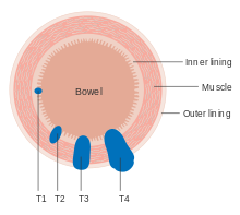
Tumor (T)

The T stages of bowel cancer. Diagram showing T stages of bowel cancer CRUK 276.svg
The T stages of bowel cancer.

Numbers 0 to 4, with subgroups, are used to describe deepest tumor depth: [2]

Node (N)

Numbers 0 to 2, and subgroups, are used to describe lymph node involvement: [2]

Metastasis (M)

Numbers 0 and 1, with subgroups, describe the metastasis status: [2]

Overall AJCC stage

AJCC stage [2] TNM stage [2] TNM stage criteria [2]
Stage 0Tis N0 M0Tis: Tumor confined to mucosa; cancer-in-situ
Stage IT1 N0 M0T1: Tumor invades submucosa
T2 N0 M0T2: Tumor invades muscularis propria
Stage II-AT3 N0 M0T3: Tumor invades subserosa or beyond (without other organs involved)
Stage II-BT4a N0 M0T4a: Tumor perforates the visceral peritoneum
Stage II-CT4b N0 M0T4b: Tumor invades adjacent organs
Stage III-A
  • T1-2 N1 M0 or
  • T1, N2a, M0
  • N1: Tumor cells in 1 to 3 regional lymph nodes. T1 or T2.
  • N2a: Tumor cells in 4 to 6 regional lymph nodes. T1
Stage III-B
  • T3-4a, N1 M0 or
  • T2-3, N2a, M0 or
  • T1-2 N2b M0
  • N1: Tumor cells in 1 to 3 regional lymph nodes. T3 or T4
  • N2a: Tumor cells in 4 to 6 regional lymph nodes. T2 or T3
  • N2b: Tumor cells in 7 or more regional lymph nodes. T1 or 2
Stage III-C
  • T4a N2a M0 or
  • T3-4a N2b M0 or
  • T4b N1-2, M0
  • N2a: Tumor cells in 4 to 6 regional lymph nodes. T4a
  • N2b: Tumor cells in 7 or more regional lymph nodes. T3-4a
  • N1-2: Tumor cells in at least one regional lymph node. T4b
Stage IVaany T, any N, M1aM1a: Metastasis to 1 other part of the body beyond the colon, rectum or regional lymph nodes. Any T, any N.
Stage IVbany T, any N, M1bM1b: Metastasis to more than 1 other part of the body beyond the colon, rectum or regional lymph nodes. Any T, any N.
Stage IVcany T, any N, M1cM1c: Metastasis to the peritoneal surface. Any T, any N.

Dukes classification

Micrograph of a colorectal adenocarcinoma metastasis to a lymph node. The cancerous cells are at the top center-left of the image, in glands (circular/ovoid structures) and eosinophilic (bright pink). H&E stain. Crc met to node1.jpg
Micrograph of a colo rectal adenocarcinoma metastasis to a lymph node. The cancerous cells are at the top center-left of the image, in glands (circular/ovoid structures) and eosinophilic (bright pink). H&E stain.

In 1932 the British pathologist Cuthbert Dukes (1890–1977) devised a classification system for colorectal cancer. [3] Several different forms of the Dukes classification were developed. [4] [5] However, this system has largely been replaced by the more detailed TNM staging system and is no longer recommended for use in clinical practice. [6]

Astler-Coller classification

An adaptation by the Americans Astler and Coller in 1954 further divided stages B and C [8] [9]

The stage gives valuable information for the prognosis and management of the particular cancer. [ citation needed ]

Full Dukes classification

Another modification of the original Dukes classification was made in 1935 by Gabriel, Dukes and Bussey. [10] This subdivided stage C. This staging system was noted to be prognostically relevant to rectal and colonic adenocarcinoma. [11] Stage D was added by Turnbull to denote the presence of liver and other distant metastases [12]

Related Research Articles

<span class="mw-page-title-main">Metastasis</span> Spread of a disease inside a body

Metastasis is a pathogenic agent's spread from an initial or primary site to a different or secondary site within the host's body; the term is typically used when referring to metastasis by a cancerous tumor. The newly pathological sites, then, are metastases (mets). It is generally distinguished from cancer invasion, which is the direct extension and penetration by cancer cells into neighboring tissues.

<span class="mw-page-title-main">Colorectal cancer</span> Cancer of the colon or rectum

Colorectal cancer (CRC), also known as bowel cancer, colon cancer, or rectal cancer, is the development of cancer from the colon or rectum. Signs and symptoms may include blood in the stool, a change in bowel movements, weight loss, and fatigue. Most colorectal cancers are due to old age and lifestyle factors, with only a small number of cases due to underlying genetic disorders. Risk factors include diet, obesity, smoking, and lack of physical activity. Dietary factors that increase the risk include red meat, processed meat, and alcohol. Another risk factor is inflammatory bowel disease, which includes Crohn's disease and ulcerative colitis. Some of the inherited genetic disorders that can cause colorectal cancer include familial adenomatous polyposis and hereditary non-polyposis colon cancer; however, these represent less than 5% of cases. It typically starts as a benign tumor, often in the form of a polyp, which over time becomes cancerous.

<span class="mw-page-title-main">Carcinoma</span> Malignancy that develops from epithelial cells

Carcinoma is a malignancy that develops from epithelial cells. Specifically, a carcinoma is a cancer that begins in a tissue that lines the inner or outer surfaces of the body, and that arises from cells originating in the endodermal, mesodermal or ectodermal germ layer during embryogenesis.

The TNM Classification of Malignant Tumors (TNM) is a globally recognised standard for classifying the anatomical extent of the spread of malignant tumours (cancer). It has gained wide international acceptance for many solid tumor cancers, but is not applicable to leukaemia or tumors of the central nervous system. Most common tumors have their own TNM classification. The TNM staging system is sometimes referred to as the AJCC/UICC staging system or the UICC/AJCC staging system.

<span class="mw-page-title-main">Anal cancer</span> Medical condition

Anal cancer is a cancer which arises from the anus, the distal opening of the gastrointestinal tract. Symptoms may include bleeding from the anus or a lump near the anus. Other symptoms may include pain, itchiness, or discharge from the anus. A change in bowel movements may also occur.

Cancer staging is the process of determining the extent to which a cancer has grown and spread. A number from I to IV is assigned, with I being an isolated cancer and IV being a cancer that has metastasized and spread from its origin. The stage generally takes into account the size of a tumor, whether it has invaded adjacent organs, how many regional (nearby) lymph nodes it has spread to, and whether it has appeared in more distant locations (metastasized).

<span class="mw-page-title-main">Invasive carcinoma of no special type</span> Medical condition

Invasive carcinoma of no special type, invasive breast carcinoma of no special type (IBC-NST), invasive ductal carcinoma (IDC), infiltrating ductal carcinoma (IDC) or invasive ductal carcinoma, not otherwise specified (NOS) is a disease. For international audiences this article will use "invasive carcinoma NST" because it is the preferred term of the World Health Organization (WHO).

Prostate cancer staging is the process by which physicians categorize the risk of cancer having spread beyond the prostate, or equivalently, the probability of being cured with local therapies such as surgery or radiation. Once patients are placed in prognostic categories, this information can contribute to the selection of an optimal approach to treatment. Prostate cancer stage can be assessed by either clinical or pathological staging methods. Clinical staging usually occurs before the first treatment and tumour presence is determined through imaging and rectal examination, while pathological staging is done after treatment once a biopsy is performed or the prostate is removed by looking at the cell types within the sample.

<span class="mw-page-title-main">Papillary thyroid cancer</span> Medical condition

Papillary thyroid cancer is the most common type of thyroid cancer, representing 75 percent to 85 percent of all thyroid cancer cases. It occurs more frequently in women and presents in the 20–55 year age group. It is also the predominant cancer type in children with thyroid cancer, and in patients with thyroid cancer who have had previous radiation to the head and neck. It is often well-differentiated, slow-growing, and localized, although it can metastasize.

The uterine sarcomas form a group of malignant tumors that arises from the smooth muscle or connective tissue of the uterus.

<span class="mw-page-title-main">Vulvar cancer</span> Cancer involving the vulva

Vulvar cancer is a cancer of the vulva, the outer portion of the female genitals. It most commonly affects the labia majora. Less often, the labia minora, clitoris, or vaginal glands are affected. Symptoms include a lump, itchiness, changes in the skin, or bleeding from the vulva.

<span class="mw-page-title-main">Colorectal polyp</span> Growth found in bowel wall

A colorectal polyp is a polyp occurring on the lining of the colon or rectum. Untreated colorectal polyps can develop into colorectal cancer.

Cancer of unknown primary origin (CUP) is a cancer that is determined to be at the metastatic stage at the time of diagnosis, but a primary tumor cannot be identified. A diagnosis of CUP requires a clinical picture consistent with metastatic disease and one or more biopsy results inconsistent with a tumor cancer

Lung cancer staging is the assessment of the extent to which a lung cancer has spread from its original source. As with most cancers, staging is an important determinant of treatment and prognosis. In general, more advanced stages of cancer are less amenable to treatment and have a worse prognosis.

Uterine clear-cell carcinoma (CC) is a rare form of endometrial cancer with distinct morphological features on pathology; it is aggressive and has high recurrence rate. Like uterine papillary serous carcinoma CC does not develop from endometrial hyperplasia and is not hormone sensitive, rather it arises from an atrophic endometrium. Such lesions belong to the type II endometrial cancers.

Lymph node metastasis is the spread (metastasis) of cancer cells into a lymph node.

Cervical cancer staging is the assessment of cervical cancer to determine the extent of the disease. This is important for determining disease prognosis and treatment. Cancer staging generally runs from stage 0, which is pre-cancerous or non-invasive, to stage IV, in which the cancer has spread throughout a significant part of the body.

<span class="mw-page-title-main">Lymphovascular invasion</span>

Lymphovascular invasion is the invasion of a cancer to the blood vessels and/or lymphatics.

Carcinoma of the tonsil is a type of squamous cell carcinoma. The tonsil is the most common site of squamous cell carcinoma in the oropharynx. It comprises 23.1% of all malignancies of the oropharynx. The tumors frequently present at advanced stages, and around 70% of patients present with metastasis to the cervical lymph nodes. . The most reported complaints include sore throat, otalgia or dysphagia. Some patients may complain of feeling the presence of a lump in the throat. Approximately 20% patients present with a node in the neck as the only symptom.

<span class="mw-page-title-main">Histopathology of colorectal adenocarcinoma</span> Analysis of tissue from a biopsy or surgery to identify colorectal cancer characteristics

The histopathology of colorectal cancer of the adenocarcinoma type involves analysis of tissue taken from a biopsy or surgery. A pathology report contains a description of the microscopical characteristics of the tumor tissue, including both tumor cells and how the tumor invades into healthy tissues and finally if the tumor appears to be completely removed. The most common form of colon cancer is adenocarcinoma, constituting between 95% and 98% of all cases of colorectal cancer. Other, rarer types include lymphoma, adenosquamous and squamous cell carcinoma. Some subtypes have been found to be more aggressive.
References

  1. "Cancer Staging". American Cancer Society. Retrieved 12 Sep 2020.
  2. 1 2 3 4 5 6 "Colorectal Cancer: Stages". Cancer.net (American Society of Clinical Oncology). Retrieved 2019-09-26. Approved by the Cancer.Net Editorial Board, 11/2018. In turn citing:
    Amin, Mahul B.; Greene, Frederick L.; Edge, Stephen B.; Compton, Carolyn C.; Gershenwald, Jeffrey E.; Brookland, Robert K.; Meyer, Laura; Gress, Donna M.; Byrd, David R.; Winchester, David P. (2017). "The Eighth Edition AJCC Cancer Staging Manual: Continuing to build a bridge from a population-based to a more "personalized" approach to cancer staging". CA: A Cancer Journal for Clinicians. 67 (2): 93–99. doi: 10.3322/caac.21388 . ISSN   0007-9235.
  3. Who Named It, showing correct grammatical usage
  4. Kyriakos M: The President cancer, the Dukes classification, and confusion, Arch Pathol Lab Med 109:1063, 1985
  5. Dukes CE. The classification of cancer of the rectum. Journal of Pathological Bacteriology 1932;35:323
  6. AJCC (American Joint Committee on Cancer) Cancer Staging Manual, 7th ed, Edge, SB, Byrd, DR, Compton, CC, et al (Eds), Springer, New York 2010. p 143.
  7. Single Best Answers in Surgery, Patten DK et al. Hodder Education 2009. p.107 ( ISBN   9780340972359)
  8. Astler VB, Coller FA: The prognostic significance of direct extension of carcinoma of the colon and rectum. Ann Surg 139:846, 1954
  9. Banias, Laura; Jung, Ioan; Chiciudean, Rebeca; Gurzu, Simona (August 2022). "From Dukes-MAC Staging System to Molecular Classification: Evolving Concepts in Colorectal Cancer". International Journal of Molecular Sciences. 23 (16): 1–14. doi: 10.3390/ijms23169455 . Retrieved 19 November 2022.
  10. Gabriel WB, Dukes C, Busset HJR: Lymphatic spread in cancer of the rectum. Br J Surg 23:395–413, 1935
  11. Grinnell RS: The grading and prognosis of carcinoma of the colon and rectum. Ann Surg 109:500-33, 1939
  12. Turnbull RB Jr, Kyle K, Watson FR, et al: Cancer of the colon: the influence of the no touch isolation technique on survival rates. Ann Surg 166:420-7, 1967