Coniochaeta hoffmannii

Last updated

Coniochaeta hoffmannii
Scientific classification OOjs UI icon edit-ltr.svg
Domain: Eukaryota
Kingdom: Fungi
Division: Ascomycota
Class: Sordariomycetes
Order: Coniochaetales
Family: Coniochaetaceae
Genus: Coniochaeta
Species:
C. hoffmannii
Binomial name
Coniochaeta hoffmannii
Synonyms [1]

Coniochaeta hoffmannii, also known as Lecythophora hoffmannii, is an ascomycete fungus that grows commonly in soil. [2] It has also been categorized as a soft-rot fungus capable of bringing the surface layer of timber into a state of decay, even when safeguarded with preservatives. [3] Additionally, it has pathogenic properties, although it causes serious infection only in rare cases. [4] A plant pathogen lacking a known sexual state, [5] C. hoffmannii has been classified as a "dematiaceous fungus" despite its contradictory lack of pigmentation; both in vivo and in vitro, there is no correlation between its appearance and its classification. [4]

Contents

History

Coniochaeta hoffmannii was originally described by J.F.H. van Beyma in 1939 as Margarinomyces hoffmannii. [6] The genus Lecythophora, originally described by John Axel Nannfeldt in 1934, was reintroduced by Konrad Walter Gams and Michael R. McGinnis to accommodate this species in 1983. [7] In 2013, Lecythophora was recombined with Coniochaeta , with all species transferred to the latter genus. [1]

Ecology

Occurrence

Coniochaeta hoffmannii is a plant pathogen that commonly inhabits fertile soil. [3] It is also known as an agent of soft-rot wood in terrestrial and aquatic environments, it is known to colonize wood surfaces through the use of soft rot hyphae. [8]

Environmental Interactions

In soil, [2] C. hoffmannii is thought primarily to be a decomposer of wood. To this end, C. hoffmannii is one of, if not the most important fungal agents of soft rot in preservative-treated wood. It, along with soft rot species of other genera, is known to metabolize aromatic compounds of low molecular mass; this includes nine phenolic compounds metabolized by C. hoffmannii itself, such as p-hydroxybenzoic acid, vanillic acid, and vanillin, to name a few. This allows for the utilization of part of the amorphous granular material, found in said phenolic compounds, which contains the lignin breakdown products produced in soft-rot activities. [3]

Decay of wood by soft rot fungi such as C. hoffmannii occurs in one of two ways: either the enzymes released from the hyphae on the lumen surface of the wood cell wall erode it, or the cavities around the hyphae in the S2region of the cell wall get excavated. The cavity itself is created by the enzymatic activity along the length of the hyphae after its growth. [8]

Morphology

As a former member of the genus Lecythophora, it was considered to be poorly differentiated morphologically. [9] Coniochaeta hoffmannii colonies range in colour from a pale salmon to a pallid orange, with degenerate strains presenting a creamy white colour. [10] Colonies are also flat, smooth and moist. [11] Collarettes are distinct, [10] yet unpigmented, while conidia are also hyaline, smooth, and thin-walled. [11] The production of slimy, orange- to salmon-colored colonies on short adelophialides (phialides lacking a basal septum) with cylindrical collarettes can be quite distinct, with ventricose phialide formations less frequent. Narrow and hyaline, hyphae, through the use of small collarettes, [11] produce conidia directly upon them laterally, directly upon the vegetative hyphae. [9] Alternatively, flask-shaped lateral cells, which are practically cylindrical, can serve as the medium through which conidia are produced; they are sometimes found to be arranged in densely packed groups. [11] Conidia are slightly curved, appearing broadly ellipsoidal to cylindrical to bean-shaped; [9] measurements vary from 3.5–7.0 µm in length by 1.0–2.5 µm in width, [10] to 3.0–3.5 µm by 1.5–2.5 µm. [11]

Ultrastructure

There are multiple types of hyphae within C. hoffmannii. Ones to note are fine soft rot hyphae, T-branch hyphae and proboscis hyphae. T-branch hyphae, which penetrate the wood cell wall, arise from lumen-colonizing hyphae. During early stages of their development, T-branch hyphae contain few cellular organelles; later on in that development, the T-branch arms elongate, complexifying the internal organization of the hyphae. The tip of the T-branch is lacking in hyphal distortion, as well. [8] Proboscis hyphae, when young, exhibit a similar ultrastructural organization and overall diameter to young T-branch hyphae upon emergence from the cone region of parent cavities. They are also fine (only 0.24 µm in diameter (0.34 µm diam including the fungal cell wall) with the tips showing an electron-dense zone. [8] The occurrence of membrane configurations at the base of fine hyphae within proboscis hyphae has importance when it comes to the transport and release of extracellular lytic enzymes. [12] Hyphal diameter is measured to be 0.87 to 1.82 µm. [8]

Physiology

Intolerant to benomyl, [13] C. hoffmannii is capable of proteolytic activity (hydrolysis, in particular), with proteolytic enzymes detected in cultures of C. hoffmannii with various media, including Loeffler blood serum, and skim milk and Litmus milk. [14] Coniochaeta hoffmannii is also quite wikt:thermotolerant, easily growing at 30 and 35 °C, with two out of four isolates (both environmental) growing at 40 °C. [14]

Disease

Despite being one of only two species of medical interest within the genus Coniochaeta, [11] Coniochaeta hoffmannii is rarely encountered as a pathogen primarily due to its related taxa. It is one of the more predominant species of its genus concerning the number of clinical cases reporting infection, having been associated with cases of subcutaneous infections, keratitis, sinusitis, and peritonitis. [9] Coniochaeta hoffmannii rarely causes disease, but when it does, it is only seen in immunocompromised patients, thus marking it as an opportunistic fungal pathogen. [4] Other identifying factors for opportunistic pathogens like this include an association with other underlying diseases capable of compromising the host's immune system, an association with new therapeutic modalities, and cases of invasive fungal sinusitis with no determinable predisposing factors. While great advances in medical technology have decreased previously high rates of morbidity and mortality, the amount and variation of opportunistic pathogens has also increased. [15]

Coniochaeta hoffmannii has been inculpated in human phaeohyphomycosis, leading to the aforementioned abscesses, sinusitis, and mastoiditis, to name a few. [16] It has also been implicated as a food contaminant, which could explain how people or animals interact with it in their respective environments. [17]

Human

In rare cases, C. hoffmannii has demonstrated its ability to cause serious infection; in one case, it actually induced chronic sinusitis in an AIDS patient. The patient in question was immunocompromised and thus severely immunodeficient due to contracting the human immunodeficiency virus; this allowed for the fungal pathogen to not only infect the patient (where an intact immune system would have made an infection by C. hoffmannii a non-issue), but inflict a recurrent, chronic disease such as sinusitis. [4]

Coniochaeta hoffmannii has also been isolated as a pure growth fungal aggregate from a human gluteal abscess; it actually develops an aggregate not only after treatment is attempted (in this case, for a respiratory tract infection, where various antibacterial drug treatments were implemented), but at the exact point of inoculation, [6] once again demonstrating its immunity to antifungal agents and treatment methods.[ citation needed ]

Non-human

There are parallels in both humans and animals when it comes to infections caused by C. hoffmannii. In humans, C. hoffmannii is not only the causative agent when it comes to original infection, but it also exacerbates the symptoms upon administration of antifungal treatments. In the case of a household dog suffering from claudication in the jaw, rigorous treatment with ketoconazole and itraconazole (for 3 total times per day over 3 months) had no effect, since this fungus is resistant to such treatments. [17]

Treatment

Coniochaeta hoffmannii has proven to be resistant to multiple antifungal agents, including amphotericin B, flucytosine, ketoconazole, and fluconazole. [4] Because of this, precautions have been taken, such as antifungal susceptibility testing, in order to circumvent such drawbacks. Through this method, polyhexamethylene biguanide (PHMB) has been identified and utilized in conjunction with invasive surgical procedures to successfully treat one of the only cases of infection at the hands of this fungus. This method of treatment is employed in order to safeguard against fungal infection even in an immunocompetent host. [15]

Related Research Articles

<i>Aspergillus fumigatus</i> Species of fungus

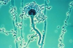
Aspergillus fumigatus is a species of fungus in the genus Aspergillus, and is one of the most common Aspergillus species to cause disease in individuals with an immunodeficiency.

<i>Sporothrix schenckii</i> Species of fungus

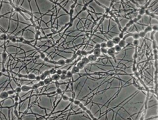
Sporothrix schenckii, a fungus that can be found worldwide in the environment, is named for medical student Benjamin Schenck, who in 1896 was the first to isolate it from a human specimen. The species is present in soil as well as in and on living and decomposing plant material such as peat moss. It can infect humans as well as animals and is the causative agent of sporotrichosis, commonly known as "rose handler's disease." The most common route of infection is the introduction of spores to the body through a cut or puncture wound in the skin. Infection commonly occurs in otherwise healthy individuals but is rarely life-threatening and can be treated with antifungals. In the environment it is found growing as filamentous hyphae. In host tissue it is found as a yeast. The transition between the hyphal and yeast forms is temperature dependent making S. schenckii a thermally dimorphic fungus.

<i>Cochliobolus lunatus</i> Fungal plant pathogen

Cochliobolus lunatus is a fungal plant pathogen that can cause disease in humans and other animals. The anamorph of this fungus is known as Curvularia lunata, while C. lunatus denotes the teleomorph or sexual stage. They are, however, the same biological entity. C. lunatus is the most commonly reported species in clinical cases of reported Cochliobolus infection.

<i>Setosphaeria rostrata</i> Pathogenic fungus

Setosphaeria rostrata is a heat tolerant fungus with an asexual reproductive form (anamorph) known as Exserohilum rostratum. This fungus is a common plant pathogen, causing leaf spots as well as crown rot and root rot in grasses. It is also found in soils and on textiles in subtropical and tropical regions. Exserohilum rostratum is one of the 35 Exserohilum species implicated uncommonly as opportunistic pathogens of humans where it is an etiologic agent of sinusitis, keratitis, skin lesions and an often fatal meningoencephalitis. Infections caused by this species are most often seen in regions with hot climates like Israel, India and the southern USA.

Acremonium strictum is an environmentally widespread saprotroph species found in soil, plant debris, and rotting mushrooms. Isolates have been collected in North and Central America, Asia, Europe and Egypt. A. strictum is an agent of hyalohyphomycosis and has been identified as an increasingly frequent human pathogen in immunosuppressed individuals, causing localized, disseminated and invasive infections. Although extremely rare, A. strictum can infect immunocompetent individuals, as well as neonates. Due to the growing number of infections caused by A. strictum in the past few years, the need for new medical techniques in the identification of the fungus as well as for the treatment of human infections has risen considerably.

<i>Acrophialophora fusispora</i> Species of ascomycete fungus found in soil, air and various plants

Acrophialophora fusispora is a poorly studied ascomycete fungus found in soil, air and various plants. A. fusispora is morphologically similar to the genera Paecilomyces and Masonia, but differ in the presence of pigmented conidiophores, verticillate phialides, and frequent sympodial proliferation. Moreover, A. fusispora is distinguished by its pigmented spindle-shaped conidia, covered with spiral bands. The fungus is naturally found in soils of tropical to temperate regions. The fungus has been identified as a plant and animal pathogen, and has recently been recognized as an emerging opportunistic human pathogen. A. fusispora infection in human is rare and has few documented clinical cases, but due to the rarity of the fungus and potential misidentification, the infections may be underdiagnosed. Clinical cases of A. fusispora include cases of keratitis, pulmonary colonization and infection, and cerebral infections. The fungus also has two documented cases of infection in dogs.

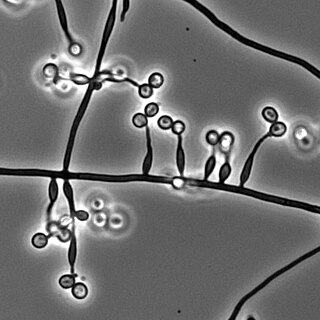
Exophiala jeanselmei is a saprotrophic fungus in the family Herpotrichiellaceae. Four varieties have been discovered: Exophiala jeanselmei var. heteromorpha, E. jeanselmei var. lecanii-corni, E. jeanselmei var. jeanselmei, and E. jeanselmei var. castellanii. Other species in the genus Exophiala such as E. dermatitidis and E. spinifera have been reported to have similar annellidic conidiogenesis and may therefore be difficult to differentiate.

<i>Pseudallescheria boydii</i> Species of fungus

Pseudallescheria boydii is a species of fungus classified in the Ascomycota. It is associated with some forms of eumycetoma/maduromycosis and is the causative agent of pseudallescheriasis. Typically found in stagnant and polluted water, it has been implicated in the infection of immunocompromised and near-drowned pneumonia patients. Treatment of infections with P. boydii is complicated by resistance to many of the standard antifungal agents normally used to treat infections by filamentous fungi.

<i>Lomentospora prolificans</i> Species of fungus

Lomentospora prolificans is an emerging opportunistic fungal pathogen that causes a wide variety of infections in immunologically normal and immunosuppressed people and animals. It is resistant to most antifungal drugs and infections are often fatal. Drugs targeting the Class II dihydroorotate dehydrogenase (DHODH) proteins of L. prolificans, Scedosporium, Aspergillus and other deadly moulds are the basis for at least one new therapy, Olorofim, which is currently in phase 2b clinical trials and has received breakthrough status by FDA. For information on all DHODH proteins, please see Dihydroorotate dehydrogenase.

Phialemonium curvatum is a pathogenic fungus in the phylum Ascomycota. The genus was created to accommodate taxa intermediate to Acremonium and Phialophora. This genus is characterized by its abundance of adelophialides and few discrete phialides with no signs of collarettes. Specifically, P. curvatum is characterized by its grayish white colonies and its allantoid conidia. Phialemonium curvatum is typically found in a variety of environments including air, soil, industrial water and sewage. Furthermore, P. curvatum affects mainly immunocompromised and is rarely seen in immunocompetent people. The species has been known to cause peritonitis, endocarditis, endovascular infections, osteomyelitis as well as cutaneous infections of wounds and burns.

<i>Madurella mycetomatis</i> Species of fungus

Madurella mycetomatis is a fungus primarily reported in Central Africa as a cause of mycetoma in humans. It has been misclassified for many years, but with improvement of molecular techniques, its phylogenetic classification has been established. Many methods exist to identify M. mycetomatis, both in lesions and in culture. Histological examination is especially useful, as it has many unique morphological features. Strain-level differences in response to antifungal agents is informative for treatment and laboratory isolation of cultures.

<i>Phialemonium obovatum</i> Species of fungus

Phialemonium obovatum is a saprotrophic filamentous fungus able to cause opportunistic infections in humans with weakened immune systems. P. obovatum is widespread throughout the environment, occurring commonly in sewage, soil, air and water. Walter Gams and Michael McGinnis described the genus Phialemonium to accommodate species intermediate between the genera Acremonium and Phialophora. Currently, three species of Phialemonium are recognized of which P. obovatum is the only one to produce greenish colonies and obovate conidia. It has been investigated as one of several microfungi with potential use in the accelerated aging of wine.

<i>Rhinocladiella mackenziei</i> Species of fungus

Rhinocladiella mackenziei is a deeply pigmented mold that is a common cause of human cerebral phaeohyphomycosis. Rhinocladiella mackenziei was believed to be endemic solely to the Middle East, due to the first cases of infection being limited to the region. However, cases of R. mackenziei infection are increasingly reported from regions outside the Middle East. This pathogen is unique in that the majority of cases have been reported from immunologically normal people.

<i>Cladophialophora carrionii</i> Species of fungus

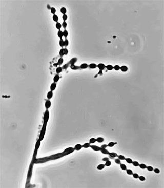
Cladophialophora carrionii is a melanized fungus in the genus Cladophialophora that is associated with decaying plant material like cacti and wood. It is one of the most frequent species of Cladophialophora implicated in human disease. Cladophialophora carrionii is a causative agent of chromoblastomycosis, a subcutaneous infection that occurs in sub-tropical areas such as Madagascar, Australia and northwestern Venezuela. Transmission occurs through traumatic implantation of plant material colonized by C. carrionii, mainly infecting rural workers. When C. carrionii infects its host, it transforms from a mycelial state to a muriform state to better tolerate the extreme conditions in the host's body.

<i>Phialophora verrucosa</i> Species of fungus

Phialophora verrucosa is a pathogenic, dematiaceous fungus that is a common cause of chromoblastomycosis. It has also been reported to cause subcutaneous phaeohyphomycosis and mycetoma in very rare cases. In the natural environment, it can be found in rotting wood, soil, wasp nests, and plant debris. P. verrucosa is sometimes referred to as Phialophora americana, a closely related environmental species which, along with P. verrucosa, is also categorized in the P. carrionii clade.

<i>Metarhizium granulomatis</i> Species of fungus

Metarhizium granulomatis is a fungus in the family Clavicipitaceae associated with systemic mycosis in veiled chameleons. The genus Metarhizium is known to infect arthropods, and collectively are referred to green-spored asexual pathogenic fungi. This species grows near the roots of plants and has been reported as an agent of disease in captive veiled chameleons. The etymology of the species epithet, "granulomatis" refers to the ability of the fungus to cause granulomatous disease in susceptible reptiles.

Sarocladium kiliense is a saprobic fungus that is occasionally encountered as a opportunistic pathogen of humans, particularly immunocompromised and individuals. The fungus is frequently found in soil and has been linked with skin and systemic infections. This species is also known to cause disease in the green alga, Cladophora glomerata as well as various fruit and vegetable crops grown in warmer climates.

Microascus manginii is a filamentous fungal species in the genus Microascus. It produces both sexual (teleomorph) and asexual (anamorph) reproductive stages known as M. manginii and Scopulariopsis candida, respectively. Several synonyms appear in the literature because of taxonomic revisions and re-isolation of the species by different researchers. M. manginii is saprotrophic and commonly inhabits soil, indoor environments and decaying plant material. It is distinguishable from closely related species by its light colored and heart-shaped ascospores used for sexual reproduction. Scopulariopsis candida has been identified as the cause of some invasive infections, often in immunocompromised hosts, but is not considered a common human pathogen. There is concern about amphotericin B resistance in S. candida.

Arthrographis kalrae is an ascomycetous fungus responsible for human nail infections described in 1938 by Cochet as A. langeronii. A. kalrae is considered a weak pathogen of animals including human restricted to the outermost keratinized layers of tissue. Infections caused by this species are normally responsive to commonly used antifungal drugs with only very rare exceptions.

Aspergillus giganteus is a species of fungus in the genus Aspergillus that grows as a mold. It was first described in 1901 by Wehmer, and is one of six Aspergillus species from the Clavati section of the subgenus Fumigati. Its closest taxonomic relatives are Aspergillus rhizopodus and Aspergillus longivescia.

References

  1. 1 2 3 Khan, Ziauddin; Gené, Josepa; Ahmad, Suhail; Cano, Josep; Al-Sweih, Noura; Joseph, Leena; Chandy, Rachel; Guarro, Josep (August 2013). "Coniochaeta polymorpha, a new species from endotracheal aspirate of a preterm neonate, and transfer of Lecythophora species to Coniochaeta". Antonie van Leeuwenhoek. 104 (2): 243–252. doi:10.1007/s10482-013-9943-z. ISSN   1572-9699. PMID   23748934. S2CID   15404078.
  2. 1 2 Domsch, K.H; W. Gams; T.H. Anderson (1993). Compendium of Soil Fungi (Reprint [der Ausg. London] 1980. ed.). Eching: IHW-Verl. ISBN   978-3980308380.{{cite book}}: CS1 maint: multiple names: authors list (link)
  3. 1 2 3 Bugos, RC; Sutherland, JB; Adler, JH (July 1988). "Phenolic Compound Utilization by the Soft Rot Fungus Lecythophora hoffmannii". Applied and Environmental Microbiology. 54 (7): 1882–5. Bibcode:1988ApEnM..54.1882B. doi:10.1128/AEM.54.7.1882-1885.1988. PMC   202766 . PMID   16347701.
  4. 1 2 3 4 5 Marriott, DJ; Wong, KH; Aznar, E; Harkness, JL; Cooper, DA; Muir, D (November 1997). "Scytalidium dimidiatum and Lecythophora hoffmannii: unusual causes of fungal infections in a patient with AIDS". Journal of Clinical Microbiology. 35 (11): 2949–52. doi:10.1128/JCM.35.11.2949-2952.1997. PMC   230093 . PMID   9350765.
  5. "Synonym and Classification Data for Lecythophora". Doctor Fungus. Archived from the original on 19 October 2013. Retrieved 18 October 2013.
  6. 1 2 Rinaldi, MG; McCoy, EL; Winn, DF (July 1982). "Gluteal abscess caused by Phialophora hoffmannii and review of the role of this organism in human mycoses". Journal of Clinical Microbiology. 16 (1): 181–5. doi:10.1128/JCM.16.1.181-185.1982. PMC   272316 . PMID   7107854.
  7. Damm, U.; Fourie, P.H.; Crous, P.W. (18 June 2010). "Coniochaeta (Lecythophora), Collophora gen. nov. and Phaeomoniella species associated with wood necroses of Prunus trees". Persoonia. 24 (1): 60–80. doi:10.3767/003158510X500705. PMC   2890157 . PMID   20664761.
  8. 1 2 3 4 5 Hale, Michael D.; Eaton, Rodney A. (May–June 1985). "The Ultrastructure of Soft Rot Fungi. I. Fine Hyphae in Wood Cell Walls". Mycologia. 77 (3): 447–463. doi:10.2307/3793202. JSTOR   3793202.
  9. 1 2 3 4 Perdomo, H.; Sutton, DA; García, D; Fothergill, AW; Gené, J; Cano, J; Summerbell, RC; Rinaldi, MG; Guarro, J (April 2011). "Molecular and phenotypic characterization of Phialemonium and Lecythophora isolates from clinical samples". Journal of Clinical Microbiology. 49 (4): 1209–16. doi:10.1128/jcm.01979-10. PMC   3122869 . PMID   21270235.
  10. 1 2 3 Howard, Dexter H. (2003). Pathogenic fungi in humans and animals (2. ed.). New York [u.a.]: Dekker. ISBN   978-0824706838.
  11. 1 2 3 4 5 6 "Lecythophora hoffmannii". Mycology Online. University of Adelaide. Archived from the original on 22 September 2013. Retrieved 18 October 2013.
  12. Hale, Michael D.; Eaton, Rodney A. (Jul–Aug 1985). "The Ultrastructure of Soft Rot Fungi. II. Cavity-Forming Hyphae in Wood Cell Walls". Mycologia. 77 (4): 594–605. doi:10.2307/3793358. JSTOR   3793358.
  13. "Lecythophora hoffmannii". MycoBank. Retrieved 18 October 2013.
  14. 1 2 Espinel-Ingroff, A; Goldson, PR; McGinnis, MR; Kerkering, TM (February 1988). "Evaluation of proteolytic activity to differentiate some dematiaceous fungi". Journal of Clinical Microbiology. 26 (2): 301–7. doi:10.1128/JCM.26.2.301-307.1988. PMC   266272 . PMID   3343325.
  15. 1 2 Chang, Christopher Y.; Schell, Wiley A.; Perfect, John R.; Hulka, Gregory F. (1 June 2005). "Novel Use of a Swimming Pool Biocide in the Treatment of a Rare Fungal Mastoiditis". The Laryngoscope. 115 (6): 1065–1069. doi:10.1097/01.MLG.0000163338.45700.FE. PMID   15933522. S2CID   36450444.
  16. Liu, Dongyou (2011-06-17). Molecular detection of human fungal pathogens. Boca Raton, Fla.: CRC Press. ISBN   978-1439812402.
  17. 1 2 Sakaeyama, S; Sano, A; Murata, Y; Kamei, K; Nishimura, K; Hatai, K (May 2007). "Lecythophora hoffmannii isolated from a case of canine osteomyelitis in Japan". Medical Mycology. 45 (3): 267–72. doi: 10.1080/13693780601188602 . PMC   7537612 . PMID   17464847.