Cryptogenic organizing pneumonia

Last updated
Cryptogenic organizing pneumonia
Other namesBronchiolitis obliterans with organizing pneumonia, idiopathic interstitial pneumonia [1]
Masson body - high mag.jpg
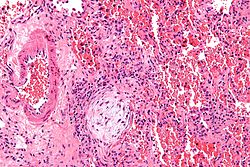
Micrograph showing a Masson body (off center left/bottom of the image – pale circular and paucicellular), as may be seen in cryptogenic organizing pneumonia. The Masson body plugs the airway. The artery associated with the obliterated airway is also seen (far left of the image). H&E stain.
Specialty Pulmonology   OOjs UI icon edit-ltr-progressive.svg
Symptoms cough, labored breathing, fever, fatigue, unexpected weight loss [1]

Cryptogenic organizing pneumonia (COP), formerly known as bronchiolitis obliterans organizing pneumonia (BOOP), is an inflammation of the bronchioles (bronchiolitis) and surrounding tissue in the lungs. [2] [3] It is a form of idiopathic interstitial pneumonia. [4]

Contents

It is often a complication of an existing chronic inflammatory disease such as rheumatoid arthritis, dermatomyositis, or it can be a side effect of certain medications such as amiodarone. COP was first described by Gary Epler in 1985. [5]

The clinical features and radiological imaging resemble infectious pneumonia. However, diagnosis is suspected after there is no response to multiple antibiotics, and blood and sputum cultures are negative for organisms.

Terminology

"Organizing" refers to unresolved pneumonia (in which the alveolar exudate persists and eventually undergoes fibrosis) in which fibrous tissue forms in the alveoli. The phase of resolution and/or remodeling following bacterial infections is commonly referred to as organizing pneumonia, both clinically and pathologically.

The American Thoracic Society and the European Respiratory Society hold that "cryptogenic organizing pneumonia" is the preferred clinical term for this disease for multiple reasons: [6] [7]

Signs and symptoms

The classic presentation of COP is the development of nonspecific systemic (e.g., fevers, chills, night sweats, fatigue, weight loss) and respiratory (e.g. difficulty breathing, cough) symptoms in association with filling of the lung alveoli that is visible on chest x-ray. [8] This presentation is usually so suggestive of an infection that the majority of patients with COP have been treated with at least one failed course of antibiotics by the time the true diagnosis is made. [8] Symptoms are usually subacute, occurring over weeks to months with dry cough (seen in 71% of people), dyspnea (shortness of breath)(62%) and fever (44%) being the most common symptoms. [9]

Causes

It was identified in 1985, although its symptoms had been noted before but not recognised as a separate lung disease. The risk of COP is higher for people with inflammatory diseases like lupus, dermatomyositis, rheumatoid arthritis, and scleroderma. [16] It most commonly presents in the 5th or 6th decade of life and it is exceedingly rare in children. [9] [17]

Pathophysiology

Organizing pneumonia is usually preceded by some type of lung injury that causes a localized denudation or disruption in continuity of the epithelial basal laminae of the type 1 alveolar pneumocytes that line the alveoli. [9] This injury to the epithelial basal lamina results in inflammatory cells and plasma proteins leaking into the alveolar space and forming fibrin, resulting in an initial fibroblast driven intra-alveolar fibroproliferation. [9] The fibroblasts differentiate into myofibroblasts and continue to form fibrosis resulting in intra-alveolar fibroinflammatory buds (Masson's Bodies) that are characteristic of organizing pneumonia. [9] These Masson's bodies consist of inflammatory cells contained in an extracellular matrix consisting of type I collagen, fibronectin, procollagen type III, tenascin C and proteoglycans. [9] Angiogenesis , or the formation of blood vessels, occurs in the Masson's bodies and this is driven by vascular endothelial growth factor. [9] Remodeling occurs, resulting in the intra-alveolar fibroinflammatory buds (Masson's Bodies) moving into the interstitial space and forming collagen globules that are then covered by type 1 alveolar epithelial cells with well developed basement membranes. These type 1 alveolar epithelial cells (pneumocytes) then proliferate, restoring the continuity and function of the alveolar unit. [9] This process is in contrast to the histopathologic changes seen in usual interstitial pneumonia where extensive fibrosis and inflammation occur leading to fibroblastic foci to form in the alveolar spaces resulting in obliteration of the alveolar space, scarring and significant damage to lung architecture (the alveoli). [9]

Tissue inhibitors of metalloproteinases (which inhibit breakdown of the extracellular matrix connective tissue) are more active in usual interstitial pneumonia as compared to organizing pneumonia, this is thought to lead to a greater deposition of connective tissue in the alveolar space in interstitial pneumonia as compared to organizing pneumonia and may explain the progressive, irreversible fibrosis seen in usual interstitial pneumonia. [9] Gelatinolytic activity (resulting in the breakdown of extracellular matrix connective tissue) is greater in organizing pneumonia as compared to usual interstitial pneumonia, and this is thought to contribute to the reversible fibroproliferation characteristic of organizing pneumonia. [9]

Diagnosis

On clinical examination, crackles are common, and more rarely, patients may have clubbing (<5% of cases). Laboratory findings are nonspecific but inflammatory markers such as the erythrocyte sedimentation rate or C-reactive protein and the lymphocyte count are frequently elevated. [9] If the organizing pneumonia is secondary to a connective tissue disorder, then the associated laboratory values such as the anti-nuclear antibody, rheumatoid factor, anti-citrullinated protein antibodies, anti-dsDNA antibodies and other similar connective tissue associated antibodies are elevated. [9]

Pulmonary function testing in people with organizing pneumonia, either cryptogenic or due to secondary causes, shows a restrictive defect with a decrease in the gas absorptive capacity of the lungs (seen as a decrease in the diffusion capacity of carbon monoxide). [9] Airflow obstruction is usually not seen on pulmonary function testing. [9]

Bronchoscopy with bronchoalveolar lavage is recommended in possible cases of organizing pneumonia to rule out infection and other causes of alveolar infiltrates. [9] The bronchoalveolar lavage in organizing pneumonia shows a lymphocytic predominant inflammation of the alveoli with increases in neutrophils and eosinophils. [9] Resolution of inflammatory cells in the bronchoalveolar lavage is usually delayed in organizing pneumonia, lagging behind clinical and radiographic improvement. [9]

Biopsy findings in patients with organizing pneumonia consist of loose connective tissue plugs involving the alveoli, alveolar ducts and bronchioles. The loose connective tissue plugs occupying the alveolar spaces often connect to other connective tissue plugs in nearby alveoli via the pores of Kohn creating a characteristic butterfly pattern on histology. [9] There is usually minimal to no interstitial inflammatory changes in biopsies of organizing pneumonia. [9]

Imaging

CT scan showing cryptogenic organizing pneumonia (biopsy-proven) CT BOOP.jpg
CT scan showing cryptogenic organizing pneumonia (biopsy-proven)
The reversed halo sign is seen in about 20% of individuals with COP. Chest CT with reversed halo sign.jpg
The reversed halo sign is seen in about 20% of individuals with COP.

The chest x-ray is distinctive with features that appear similar to an extensive pneumonia, with both lungs showing widespread white patches. The white patches may seem to migrate from one area of the lung to another as the disease persists or progresses. Computed tomography (CT) may be used to confirm the diagnosis. Often the findings are typical enough to allow the doctor to make a diagnosis without ordering additional tests. [19] To confirm the diagnosis, a doctor may perform a lung biopsy using a bronchoscope. Many times, a larger specimen is needed and must be removed surgically.

Plain chest radiography shows normal lung volumes, with characteristic patchy unilateral or bilateral consolidation. Small nodular opacities occur in up to 50% of patients and large nodules in 15%. On high resolution computed tomography, airspace consolidation with air bronchograms is present in more than 90% of patients, often with a lower zone predominance. A subpleural or peribronchiolar distribution is noted in up to 50% of patients. Ground glass appearance or hazy opacities associated with the consolidation are detected in most patients.

Histologically, cryptogenic organizing pneumonia is characterized by the presence of polypoid plugs of loose organizing connective tissue (Masson bodies) within alveolar ducts, alveoli, and bronchioles.

Unusual presentations of organizing pneumonia

While patchy bilateral disease is typical, there are unusual variants of organizing pneumonia where it may appear as multiple nodules or masses. One rare presentation, focal organizing pneumonia, may be indistinguishable from lung cancer based on imaging alone, requiring biopsy or surgical resection to make the diagnosis. [20]

Complications

Rare cases of COP have induced with lobar cicatricial atelectasis. [21]

Treatment

Systemic steroids are considered the first line treatment for organizing pneumonia, with patient's often having clinical improvement within 72 hours of steroid initiation and most patient's achieving recovery. [22] [9] A prolonged treatment course is indicated, with patients usually requiring at least 4-6 months of treatment. [9] Patient's who are treated with larger doses of steroids require prophylaxis against pneumocystis jirovecii. [9] Relapses may occur and are more likely to occur in severe disease or when steroids are tapered too soon or too quickly. [9] Alternative or adjunct treatment options include macrolide antibiotics (due to anti-inflammatory properties), azathioprine and cyclophosphamide. [9]

Related Research Articles

<span class="mw-page-title-main">Pulmonary alveolar proteinosis</span> Medical condition

Pulmonary alveolar proteinosis (PAP) is a rare lung disorder characterized by an abnormal accumulation of surfactant-derived lipoprotein compounds within the alveoli of the lung. The accumulated substances interfere with the normal gas exchange and expansion of the lungs, ultimately leading to difficulty breathing and a predisposition to developing lung infections. The causes of PAP may be grouped into primary, secondary, and congenital causes, although the most common cause is a primary autoimmune condition in an individual.

<span class="mw-page-title-main">Interstitial lung disease</span> Group of diseases

Interstitial lung disease (ILD), or diffuse parenchymal lung disease (DPLD), is a group of respiratory diseases affecting the interstitium and space around the alveoli of the lungs. It concerns alveolar epithelium, pulmonary capillary endothelium, basement membrane, and perivascular and perilymphatic tissues. It may occur when an injury to the lungs triggers an abnormal healing response. Ordinarily, the body generates just the right amount of tissue to repair damage, but in interstitial lung disease, the repair process is disrupted, and the tissue around the air sacs (alveoli) becomes scarred and thickened. This makes it more difficult for oxygen to pass into the bloodstream. The disease presents itself with the following symptoms: shortness of breath, nonproductive coughing, fatigue, and weight loss, which tend to develop slowly, over several months. The average rate of survival for someone with this disease is between three and five years. The term ILD is used to distinguish these diseases from obstructive airways diseases.

<span class="mw-page-title-main">Chest radiograph</span> Projection X-ray of the chest

A chest radiograph, called a chest X-ray (CXR), or chest film, is a projection radiograph of the chest used to diagnose conditions affecting the chest, its contents, and nearby structures. Chest radiographs are the most common film taken in medicine.

<span class="mw-page-title-main">Pulmonary fibrosis</span> Disease that causes scarring of the lungs

Pulmonary fibrosis is a condition in which the lungs become scarred over time. Symptoms include shortness of breath, a dry cough, feeling tired, weight loss, and nail clubbing. Complications may include pulmonary hypertension, respiratory failure, pneumothorax, and lung cancer.

<span class="mw-page-title-main">Hypersensitivity pneumonitis</span> Medical condition

Hypersensitivity pneumonitis (HP) or extrinsic allergic alveolitis (EAA) is a syndrome caused by the repetitive inhalation of antigens from the environment in susceptible or sensitized people. Common antigens include molds, bacteria, bird droppings, bird feathers, agricultural dusts, bioaerosols and chemicals from paints or plastics. People affected by this type of lung inflammation (pneumonitis) are commonly exposed to the antigens by their occupations, hobbies, the environment and animals. The inhaled antigens produce a hypersensitivity immune reaction causing inflammation of the airspaces (alveoli) and small airways (bronchioles) within the lung. Hypersensitivity pneumonitis may eventually lead to interstitial lung disease.

<span class="mw-page-title-main">Pneumonitis</span> General inflammation of lung tissue

Pneumonitis describes general inflammation of lung tissue. Possible causative agents include radiation therapy of the chest, exposure to medications used during chemo-therapy, the inhalation of debris, aspiration, herbicides or fluorocarbons and some systemic diseases. If unresolved, continued inflammation can result in irreparable damage such as pulmonary fibrosis.

<span class="mw-page-title-main">Acute interstitial pneumonitis</span> Medical condition

Acute interstitial pneumonitis (also known as acute interstitial pneumoniais a rare, severe lung disease that usually affects otherwise healthy individuals. There is no known cause or cure.

Bronchoalveolar lavage (BAL), also known as bronchoalveolar washing, is a diagnostic method of the lower respiratory system in which a bronchoscope is passed through the mouth or nose into an appropriate airway in the lungs, with a measured amount of fluid introduced and then collected for examination. This method is typically performed to diagnose pathogenic infections of the lower respiratory airways, though it also has been shown to have utility in diagnosing interstitial lung disease. Bronchoalveolar lavage can be a more sensitive method of detection than nasal swabs in respiratory molecular diagnostics, as has been the case with SARS-CoV-2 where bronchoalveolar lavage samples detect copies of viral RNA after negative nasal swab testing.

<span class="mw-page-title-main">Bronchiolitis obliterans</span> Medical condition

Bronchiolitis obliterans (BO), also known as obliterative bronchiolitis, constrictive bronchiolitis and popcorn lung, is a disease that results in obstruction of the smallest airways of the lungs (bronchioles) due to inflammation. Symptoms include a dry cough, shortness of breath, wheezing and feeling tired. These symptoms generally get worse over weeks to months. It is not related to cryptogenic organizing pneumonia, previously known as bronchiolitis obliterans organizing pneumonia.

<span class="mw-page-title-main">Alveolar lung disease</span> Medical condition

Alveolar lung diseases, are a group of diseases that mainly affect the alveoli of the lungs.

Air trapping, also called gas trapping, is an abnormal retention of air in the lungs where it is difficult to exhale completely. It is observed in obstructive lung diseases such as asthma, bronchiolitis obliterans syndrome and chronic obstructive pulmonary diseases such as emphysema and chronic bronchitis.

<span class="mw-page-title-main">Idiopathic pulmonary fibrosis</span> Medical condition

Idiopathic pulmonary fibrosis (IPF), or (formerly) fibrosing alveolitis, is a rare, progressive illness of the respiratory system, characterized by the thickening and stiffening of lung tissue, associated with the formation of scar tissue. It is a type of chronic scarring lung disease characterized by a progressive and irreversible decline in lung function. The tissue in the lungs becomes thick and stiff, which affects the tissue that surrounds the air sacs in the lungs. Symptoms typically include gradual onset of shortness of breath and a dry cough. Other changes may include feeling tired, and abnormally large and dome shaped finger and toenails. Complications may include pulmonary hypertension, heart failure, pneumonia or pulmonary embolism.

<span class="mw-page-title-main">Usual interstitial pneumonia</span> Medical condition

Usual interstitial pneumonia (UIP) is a form of lung disease characterized by progressive scarring of both lungs. The scarring (fibrosis) involves the pulmonary interstitium. UIP is thus classified as a form of interstitial lung disease.

<span class="mw-page-title-main">High-resolution computed tomography</span> Diagnostic imaging test

High-resolution computed tomography (HRCT) is a type of computed tomography (CT) with specific techniques to enhance image resolution. It is used in the diagnosis of various health problems, though most commonly for lung disease, by assessing the lung parenchyma. On the other hand, HRCT of the temporal bone is used to diagnose various middle ear diseases such as otitis media, cholesteatoma, and evaluations after ear operations.

<span class="mw-page-title-main">Idiopathic interstitial pneumonia</span> Medical condition

Idiopathic interstitial pneumonia (IIP), or noninfectious pneumonia are a class of diffuse lung diseases. These diseases typically affect the pulmonary interstitium, although some also have a component affecting the airways. There are seven recognized distinct subtypes of IIP.

<span class="mw-page-title-main">Desquamative interstitial pneumonia</span> Medical condition

Desquamative interstitial pneumonia (DIP) is a form of idiopathic interstitial pneumonia featuring elevated numbers of macrophages within the alveoli of the lung. The alveolar macrophages have a characteristic light brown pigmentation and accumulate in the alveolar lumen and septa regions of the lower lobes of the lungs. The typical effects of the macrophage accumulation are inflammation and later fibrosis of the lung tissue.

<span class="mw-page-title-main">Respiratory bronchiolitis</span> Medical condition

Respiratory bronchiolitis is a lung disease associated with tobacco smoking. In pathology, it is defined by the presence of "smoker's macrophages". When manifesting significant clinical symptoms it is referred to as respiratory bronchiolitis interstitial lung disease (RB-ILD).

<span class="mw-page-title-main">Diffuse alveolar damage</span> Medical condition

Diffuse alveolar damage (DAD) is a histologic term used to describe specific changes that occur to the structure of the lungs during injury or disease. Most often DAD is described in association with the early stages of acute respiratory distress syndrome (ARDS). It is important to note that DAD can be seen in situations other than ARDS (such as acute interstitial pneumonia) and that ARDS can occur without DAD.

Rheumatoid lung disease is a disease of the lung associated with RA, rheumatoid arthritis. Rheumatoid lung disease is characterized by pleural effusion, pulmonary fibrosis, lung nodules and pulmonary hypertension. Common symptoms associated with the disease include shortness of breath, cough, chest pain and fever. It is estimated that about one quarter of people with rheumatoid arthritis develop this disease, which are more likely to develop among elderly men with a history of smoking.

<span class="mw-page-title-main">Ground-glass opacity</span> Radiologic sign on radiographs and computed tomography scans

Ground-glass opacity (GGO) is a finding seen on chest x-ray (radiograph) or computed tomography (CT) imaging of the lungs. It is typically defined as an area of hazy opacification (x-ray) or increased attenuation (CT) due to air displacement by fluid, airway collapse, fibrosis, or a neoplastic process. When a substance other than air fills an area of the lung it increases that area's density. On both x-ray and CT, this appears more grey or hazy as opposed to the normally dark-appearing lungs. Although it can sometimes be seen in normal lungs, common pathologic causes include infections, interstitial lung disease, and pulmonary edema.

References

  1. 1 2 "Cryptogenic organizing pneumonia". Autoimmune Registry Inc. Retrieved 15 June 2022.
  2. " bronchiolitis obliterans with organizing pneumonia " at Dorland's Medical Dictionary
  3. White, Eric J. Stern, Charles S. (1999). Chest radiology companion. Philadelphia: Lippincott Williams & Wilkins. p. 76. ISBN   978-0-397-51732-9.{{cite book}}: CS1 maint: multiple names: authors list (link)
  4. "Cryptogenic Organizing Pneumonia - Pulmonary Disorders".
  5. Epler GR (June 2011). "Bronchiolitis obliterans organizing pneumonia, 25 years: a variety of causes, but what are the treatment options?". Expert Rev Respir Med. 5 (3): 353–61. doi:10.1586/ers.11.19. PMID   21702658. S2CID   207222916.
  6. Joseph F. Tomashefski; Carol Farver; Armando E. Fraire (2009). Dail and Hammar's Pulmonary Pathology: Volume I: Nonneoplastic Lung Disease (3rd ed.). Springer Science & Business Media. p.  64. ISBN   9780387687926.
  7. Geddes DM (August 1991). "BOOP and COP". Thorax. 46 (8): 545–7. doi:10.1136/thx.46.8.545. PMC   463266 . PMID   1926020.
  8. 1 2 "Pulmonary Question 27: Diagnose cryptogenic organizing pneumonia". MKSAP 5 For Students Online. American College of Physicians. Retrieved 23 November 2012.
  9. 1 2 3 4 5 6 7 8 9 10 11 12 13 14 15 16 17 18 19 20 21 22 23 24 25 King, Talmadge E.; Lee, Joyce S. (17 March 2022). "Cryptogenic Organizing Pneumonia". New England Journal of Medicine. 386 (11): 1058–1069. doi:10.1056/NEJMra2116777. PMID   35294814. S2CID   247498540.
  10. Levy, Barry S.; Wegman, David H.; Baron, Sherry L.; Sokas, Rosemary K., eds. (2011). Occupational and environmental health recognizing and preventing disease and injury (6th ed.). New York: Oxford University Press. p. 414. ISBN   9780199750061 . Retrieved June 23, 2015.
  11. Mukhopadhyay, Sanjay; Mehrad, Mitra; Dammert, Pedro; Arrossi, Andrea V; Sarda, Rakesh; Brenner, David S; Maldonado, Fabien; Choi, Humberto; Ghobrial, Michael (2019). "Lung Biopsy Findings in Severe Pulmonary Illness Associated With E-Cigarette Use (Vaping): A Report of Eight Cases". American Journal of Clinical Pathology. 153 (1): 30–39. doi:10.1093/ajcp/aqz182. ISSN   0002-9173. PMID   31621873.
  12. Nogi, S; Nakayama, H; Tajima, Y; Okubo, M; Mikami, R; Sugahara, S; Akata, S; Tokuuye, K (2014). "Cryptogenic organizing pneumonia associated with radiation: A report of two cases". Oncology Letters. 7 (2): 321–324. doi:10.3892/ol.2013.1716. PMC   3881924 . PMID   24396439.
  13. Oie, Y; Saito, Y; Kato, M; Ito, F; Hattori, H; Toyama, H; Kobayashi, H; Katada, K (2013). "Relationship between radiation pneumonitis and organizing pneumonia after radiotherapy for breast cancer". Radiation Oncology. 8: 56. doi: 10.1186/1748-717X-8-56 . PMC   3605133 . PMID   23497657.
  14. Radzikowska, E; Nowicka, U; Wiatr, E; Jakubowska, L; Langfort, R; Chabowski, M; Roszkowski, K (2007). "Organising pneumonia and lung cancer - case report and review of the literature". Pneumonologia I Alergologia Polska. 75 (4): 394–7. PMID   18080991.
  15. Kory, Pierre; Kanne, Jeffrey P (2020-09-22). "SARS-CoV-2 organizing pneumonia:'Has there been a widespread failure to identify and treat this prevalent condition in COVID-19?'". BMJ. 7 (1): e000724. doi: 10.1136/bmjresp-2020-000724 . PMC   7509945 . PMID   32963028.
  16. Al-Ghanem Sara; Al-Jahdali Hamdan; Bamefleh Hanaa; Khan Ali Nawaz (Apr–Jun 2008). "Bronchiolitis obliterans organizing pneumonia: Pathogenesis, clinical features, imaging and therapy review". Ann Thorac Med. 3 (2): 67–75. doi: 10.4103/1817-1737.39641 . PMC   2700454 . PMID   19561910.
  17. Drakopanagiotakis, Fotios; Paschalaki, Koralia; Abu-Hijleh, Muhanned; Aswad, Bassam; Karagianidis, Napoleon; Kastanakis, Emmanouil; Braman, Sidney S.; Polychronopoulos, Vlasis (April 2011). "Cryptogenic and Secondary Organizing Pneumonia". Chest. 139 (4): 893–900. doi:10.1378/chest.10-0883. PMID   20724743.
  18. Radswiki; et al. "Reversed halo sign (lungs)". Radiopaedia . Retrieved 2018-01-02.
  19. Zare Mehrjardi, Mohammad; Kahkouee, Shahram; Pourabdollah, Mihan (March 2017). "Radio-pathological correlation of organizing pneumonia (OP): a pictorial review". The British Journal of Radiology. 90 (1071): 20160723. doi:10.1259/bjr.20160723. ISSN   1748-880X. PMC   5601538 . PMID   28106480.
  20. Oikonomou, A; Hansell, DM (2001). "Organizing pneumonia: the many morphological faces". European Radiology. 12 (6): 1486–96. doi:10.1007/s00330-001-1211-3. PMID   12042959. S2CID   10180778.
  21. Yoshida, K; Nakajima, M; Niki, Y; Matsushima, T (2001). "Atelectasis of the right lower lobe in association with bronchiolitis obliterans organizing pneumonia". Nihon Kokyuki Gakkai Zasshi = the Journal of the Japanese Respiratory Society. 39 (4): 260–5. PMID   11481825.
  22. Oymak FS, Demirbaş HM, Mavili E, et al. (2005). "Bronchiolitis obliterans organizing pneumonia. Clinical and roentgenological features in 26 cases". Respiration. 72 (3): 254–62. doi:10.1159/000085366. PMID   15942294. S2CID   71769382.