Cuboid bone

Last updated
Cuboid bone
Gray274.png
The left cuboid. Antero-medial view.
Gray275.png
The left cuboid. Postero-lateral view
Details
Articulations Calcaneocuboid, cuboideonavicular and cuneocuboid articulation
Identifiers
Latin os cuboideum
TA98 A02.5.16.001
TA2 1489
FMA 24527
Anatomical terms of bone

In the human body, the cuboid bone is one of the seven tarsal bones of the foot.

Contents

Structure

The cuboid bone is the most lateral of the bones in the distal row of the tarsus. It is roughly cubical in shape, and presents a prominence in its inferior (or plantar) surface, the tuberosity of the cuboid. The bone provides a groove where the tendon of the peroneus longus muscle passes to reach its insertion in the first metatarsal and medial cuneiform bones. [1]

Surfaces

The dorsal surface, directed upward and lateralward, is rough, for the attachment of ligaments.

The plantar surface presents in front a deep groove, the peroneal sulcus, which runs obliquely forward and medialward; it lodges the tendon of the peroneus longus, and is bounded behind by a prominent ridge, to which the long plantar ligament is attached.

The ridge ends laterally in an eminence, the tuberosity, the surface of which presents an oval facet; on this facet glides the sesamoid bone or cartilage frequently found in the tendon of the peroneus longus. The surface of bone behind the groove is rough, for the attachment of the plantar calcaneocuboid ligament, a few fibers of the flexor hallucis brevis, and a fasciculus from the tendon of the tibialis posterior.

The lateral surface presents a deep notch formed by the commencement of the peroneal sulcus.

The posterior surface is smooth, triangular, and concavo-convex, for articulation with the anterior surface of the calcaneus (the calcaneocuboid joint); its infero-medial angle projects backward as a process which underlies and supports the anterior end of the calcaneus.

The anterior surface, of smaller size, but also irregularly triangular, is divided by a vertical ridge into two facets, forming the fourth and fifth tarsometatarsal joints: the medial facet, quadrilateral in form, articulates with the fourth metatarsal; the lateral, larger and more triangular, articulates with the fifth.

The medial surface is broad, irregularly quadrilateral, and presents at its middle and upper part a smooth oval facet, for articulation with the third cuneiform; and behind this (occasionally) a smaller facet, for articulation with the navicular bone; it is rough in the rest of its extent, for the attachment of strong interosseous ligaments.

Muscle attachments

Only one muscle is attached to the cuboid bone; the tibialis posterior. The tibialis posterior inserts to the under surface of the cuboid bone. [2] While the flexor hallucis brevis arises, by a pointed tendinous process, from the medial part of the under surface of the cuboid bone, from the contiguous portion of the lateral cuneiform bone, and from the prolongation of the tendon of the tibialis posterior.[ citation needed ]

Clinical significance

In a condition known as cuboid syndrome, the cuboid can be subluxated downward causing a swollen kind of ache along the central portion of the lateral border of the foot.

See also

Related Research Articles

<span class="mw-page-title-main">Foot</span> Anatomical structure found in vertebrates

The foot is an anatomical structure found in many vertebrates. It is the terminal portion of a limb which bears weight and allows locomotion. In many animals with feet, the foot is a separate organ at the terminal part of the leg made up of one or more segments or bones, generally including claws and/or nails.

<span class="mw-page-title-main">Human leg</span> Lower extremity or limb of the human body (foot, lower leg, thigh and hip)

The leg is the entire lower limb of the human body, including the foot, thigh or sometimes even the hip or buttock region. The major bones of the leg are the femur, tibia, and adjacent fibula. The thigh is between the hip and knee, while the calf (rear) and shin (front) are between the knee and foot.

<span class="mw-page-title-main">Humerus</span> Long bone of the upper arm

The humerus is a long bone in the arm that runs from the shoulder to the elbow. It connects the scapula and the two bones of the lower arm, the radius and ulna, and consists of three sections. The humeral upper extremity consists of a rounded head, a narrow neck, and two short processes. The body is cylindrical in its upper portion, and more prismatic below. The lower extremity consists of 2 epicondyles, 2 processes, and 3 fossae. As well as its true anatomical neck, the constriction below the greater and lesser tubercles of the humerus is referred to as its surgical neck due to its tendency to fracture, thus often becoming the focus of surgeons.

<span class="mw-page-title-main">Fibularis longus</span> Superficial muscle in the lateral compartment of the leg

In human anatomy, the fibularis longus is a superficial muscle in the lateral compartment of the leg. It acts to tilt the sole of the foot away from the midline of the body (eversion) and to extend the foot downward away from the body at the ankle.

<span class="mw-page-title-main">Tibia</span> Leg bone in vertebrates

The tibia, also known as the shinbone or shankbone, is the larger, stronger, and anterior (frontal) of the two bones in the leg below the knee in vertebrates ; it connects the knee with the ankle. The tibia is found on the medial side of the leg next to the fibula and closer to the median plane. The tibia is connected to the fibula by the interosseous membrane of leg, forming a type of fibrous joint called a syndesmosis with very little movement. The tibia is named for the flute tibia. It is the second largest bone in the human body, after the femur. The leg bones are the strongest long bones as they support the rest of the body.

<span class="mw-page-title-main">Fibula</span> Leg bone in vertebrates

The fibula or calf bone is a leg bone on the lateral side of the tibia, to which it is connected above and below. It is the smaller of the two bones and, in proportion to its length, the most slender of all the long bones. Its upper extremity is small, placed toward the back of the head of the tibia, below the knee joint and excluded from the formation of this joint. Its lower extremity inclines a little forward, so as to be on a plane anterior to that of the upper end; it projects below the tibia and forms the lateral part of the ankle joint.

<span class="mw-page-title-main">Cuneiform bones</span> Three bones in the human foot

There are three cuneiform ("wedge-shaped") bones in the human foot:

<span class="mw-page-title-main">Calcaneus</span> Bone of the tarsus of the foot

In humans and many other primates, the calcaneus or heel bone is a bone of the tarsus of the foot which constitutes the heel. In some other animals, it is the point of the hock.

<span class="mw-page-title-main">Talus bone</span> One of the foot bones that forms the tarsus

The talus, talus bone, astragalus, or ankle bone is one of the group of foot bones known as the tarsus. The tarsus forms the lower part of the ankle joint. It transmits the entire weight of the body from the lower legs to the foot.

<span class="mw-page-title-main">Flexor digitorum longus muscle</span> Muscle located on the tibial side of the leg

The flexor digitorum longus muscle is situated on the tibial side of the leg. At its origin it is thin and pointed, but it gradually increases in size as it descends. It serves to flex the second, third, fourth, and fifth toes.

<span class="mw-page-title-main">Fibularis brevis</span> Shorter and smaller of the fibularis (peroneus) muscles

In human anatomy, the fibularis brevis is a muscle that lies underneath the fibularis longus within the lateral compartment of the leg. It acts to tilt the sole of the foot away from the midline of the body (eversion) and to extend the foot downward away from the body at the ankle.

<span class="mw-page-title-main">Flexor hallucis brevis muscle</span> Muscle in sole of the foot that leads to the big toe

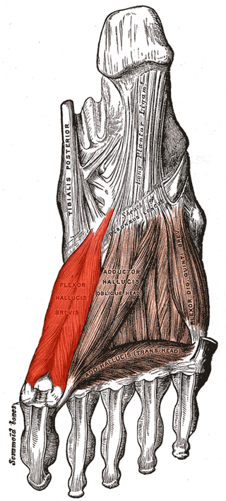
Flexor hallucis brevis muscle is a muscle of the foot that flexes the big toe.

<span class="mw-page-title-main">Quadratus plantae muscle</span> Muscles of the sole of the foot

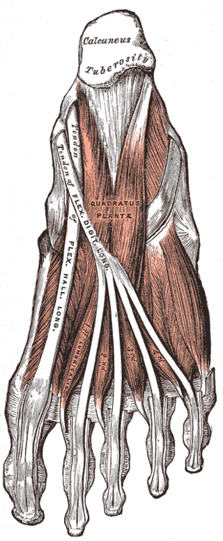
The quadratus plantae is separated from the muscles of the first layer by the lateral plantar vessels and nerve. It acts to aid in flexing the 2nd to 5th toes and is one of the few muscles in the foot with no homolog in the hand.

<span class="mw-page-title-main">Inferior extensor retinaculum of foot</span> Y-shaped band placed in front of the ankle-joint

The inferior extensor retinaculum of the foot is a Y-shaped band placed in front of the ankle-joint, the stem of the Y being attached laterally to the upper surface of the calcaneus, in front of the depression for the interosseous talocalcaneal ligament; it is directed medialward as a double layer, one lamina passing in front of, and the other behind, the tendons of the peroneus tertius and extensor digitorum longus.

<span class="mw-page-title-main">Arches of the foot</span> Load-bearing curves in the tarsal and metatarsal bones of the feet

The arches of the foot, formed by the tarsal and metatarsal bones, strengthened by ligaments and tendons, allow the foot to support the weight of the body in the erect posture with the least weight.

<span class="mw-page-title-main">Plantar calcaneocuboid ligament</span>

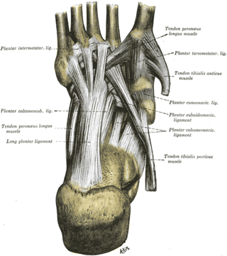
The plantar calcaneocuboid ligament is a ligament on the bottom of the foot that connects the calcaneus to the cuboid bone. It lies deep to the long plantar ligament.

<span class="mw-page-title-main">Malleolus</span> Ankle bone protrusion

A malleolus is the bony prominence on each side of the human ankle.

<span class="mw-page-title-main">Fourth metatarsal bone</span>

The fourth metatarsal bone is a long bone in the foot. It is smaller in size than the third metatarsal bone and is the third longest of the five metatarsal bones. The fourth metatarsal is analogous to the fourth metacarpal bone in the hand

<span class="mw-page-title-main">First metatarsal bone</span>

The first metatarsal bone is the bone in the foot just behind the big toe. The first metatarsal bone is the shortest of the metatarsal bones and by far the thickest and strongest of them.

<span class="mw-page-title-main">Deep fascia of leg</span> Forms a complete investment to the muscles

The deep fascia of leg, or crural fascia forms a complete investment to the muscles, and is fused with the periosteum over the subcutaneous surfaces of the bones.

References

PD-icon.svgThis article incorporates text in the public domain from page 269 of the 20th edition of Gray's Anatomy (1918)

  1. Moore, KL.; Dalley, AF.; Agur, AM. (2013). Clinically Oriented Anatomy, 7th ed. Lippincott Williams & Wilkins. p. 524. ISBN   978-1-4511-8447-1.
  2. Bojsen-Møller, Finn; Simonsen, Erik B.; Tranum-Jensen, Jørgen (2001). Bevægeapparatets anatomi[Anatomy of the Locomotive Apparatus] (in Danish) (12th ed.). p. 293. ISBN   978-87-628-0307-7.