The foot is an anatomical structure found in many vertebrates. It is the terminal portion of a limb which bears weight and allows locomotion. In many animals with feet, the foot is a separate organ at the terminal part of the leg made up of one or more segments or bones, generally including claws and/or nails.

The femur, or thigh bone is the only bone in the thigh. The thigh is the region of the lower limb between the hip and the knee. In many four-legged animals the femur is the upper bone of the hindleg.

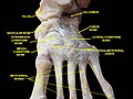
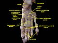
In the human body, the cuboid bone is one of the seven tarsal bones of the foot.

The tibia, also known as the shinbone or shankbone, is the larger, stronger, and anterior (frontal) of the two bones in the leg below the knee in vertebrates ; it connects the knee with the ankle. The tibia is found on the medial side of the leg next to the fibula and closer to the median plane. The tibia is connected to the fibula by the interosseous membrane of leg, forming a type of fibrous joint called a syndesmosis with very little movement. The tibia is named for the flute tibia. It is the second largest bone in the human body, after the femur. The leg bones are the strongest long bones as they support the rest of the body.

The fibula or calf bone is a leg bone on the lateral side of the tibia, to which it is connected above and below. It is the smaller of the two bones and, in proportion to its length, the most slender of all the long bones. Its upper extremity is small, placed toward the back of the head of the tibia, below the knee joint and excluded from the formation of this joint. Its lower extremity inclines a little forward, so as to be on a plane anterior to that of the upper end; it projects below the tibia and forms the lateral part of the ankle joint.

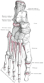
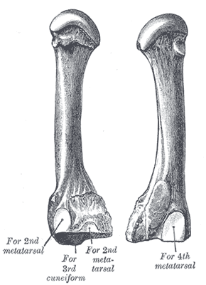
The metatarsal bones, or metatarsus, are a group of five long bones in the midfoot, located between the tarsal bones and the phalanges (toes). Lacking individual names, the metatarsal bones are numbered from the medial side : the first, second, third, fourth, and fifth metatarsal. The metatarsals are analogous to the metacarpal bones of the hand. The lengths of the metatarsal bones in humans are, in descending order, second, third, fourth, fifth, and first. A bovine hind leg has two metatarsals.

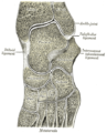
In humans and many other primates, the calcaneus or heel bone is a bone of the tarsus of the foot which constitutes the heel. In some other animals, it is the point of the hock.

The navicular bone is a small bone found in the feet of most mammals.
In the human body, the tarsus is a cluster of seven articulating bones in each foot situated between the lower end of the tibia and the fibula of the lower leg and the metatarsus. It is made up of the midfoot and hindfoot.

The tibialis posterior muscle is the most central of all the leg muscles, and is located in the deep posterior compartment of the leg. It is the key stabilizing muscle of the lower leg.
The talus, talus bone, astragalus, or ankle bone is one of the group of foot bones known as the tarsus. The tarsus forms the lower part of the ankle joint. It transmits the entire weight of the body from the lower legs to the foot.

The adductor muscles of the hip are a group of muscles in the medial compartment of the thigh mostly used for bringing the thighs together.

The lateral rotator group is a group of six small muscles of the hip which all externally (laterally) rotate the femur in the hip joint. It consists of the following muscles: piriformis, gemellus superior, obturator internus, gemellus inferior, quadratus femoris and the obturator externus.
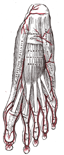
The abductor digiti minimi is a muscle which lies along the lateral (outer) border of the foot, and is in relation by its medial margin with the lateral plantar artery, vein and nerves.

The upper extremity, proximal extremity or superior epiphysis of the femur is the part of the femur closest to the pelvic bone and the trunk. It contains the following structures:
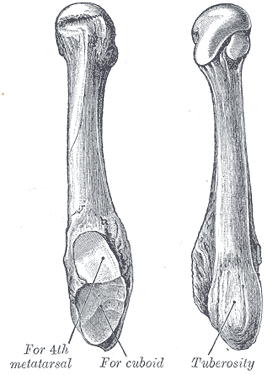
The fifth metatarsal bone is a long bone in the foot, and is palpable along the distal outer edges of the feet. It is the second smallest of the five metatarsal bones. The fifth metatarsal is analogous to the fifth metacarpal bone in the hand.

The fourth metatarsal bone is a long bone in the foot. It is smaller in size than the third metatarsal bone and is the third longest of the five metatarsal bones. The fourth metatarsal is analogous to the fourth metacarpal bone in the hand
The third metatarsal bone is a long bone in the foot. It is the second longest metatarsal. The longest being the second metatarsal. The third metatarsal is analogous to the third metacarpal bone in the hand

The second metatarsal bone is a long bone in the foot. It is the longest of the metatarsal bones, being prolonged backward and held firmly into the recess formed by the three cuneiform bones. The second metatarsal forms joints with the second proximal phalanx through the metatarsophalangeal joint, the cuneiform bones, third metatarsal and occasionally the first metatarsal bone.

The first metatarsal bone is the bone in the foot just behind the big toe. The first metatarsal bone is the shortest of the metatarsal bones and by far the thickest and strongest of them.