Demodex

Last updated

Contents

Demodex
Demodex folliculorum SEM crop.jpg
Scientific classification OOjs UI icon edit-ltr.svg
Domain: Eukaryota
Kingdom: Animalia
Phylum: Arthropoda
Subphylum: Chelicerata
Class: Arachnida
Order: Trombidiformes
Family: Demodecidae
Genus: Demodex
Owen, 1843 [1]
Type species
Acarus folliculorum
Simon, 1842
Species

Demodex /ˈdɛmədɛks/ is a genus of tiny mites that live in or near hair follicles of mammals. Around 65 species of Demodex are known. [2] Two species live on humans: Demodex folliculorum and Demodex brevis , both frequently referred to as eyelash mites, alternatively face mites or skin mites. [3]

Different species of animals host different species of Demodex. Demodex canis lives on the domestic dog. The presence of Demodex species on mammals is common and usually does not cause any symptoms. Demodex is derived from Greek δημός (dēmos) 'fat' and δήξ, δηκός (dēx, dēkós) 'woodworm'. [4] [5] [6]

Notable species

D. folliculorum and D. brevis

D. folliculorum and D. brevis are typically found on humans. D. folliculorum was first described in 1842 by German physician and dermatologist Gustav Simon, [7] with English biologist Richard Owen naming the genus Demodex the following year. [8]

D. brevis was identified as separate in 1963 by Akbulatova. D. folliculorum is found in hair follicles, while D. brevis lives in sebaceous glands connected to hair follicles. Both species are primarily found in the face near the nose, the eyelashes, and eyebrows but also occur elsewhere on the body. D. folliculorum is occasionally found as a cause of folliculitis, although most people with D. folliculorum mites have no obvious ill effects.

The adult mites are 0.3–0.4 mm (3256164 in) long, with D. brevis slightly shorter than D. folliculorum. [9] Each has a semitransparent, elongated body that consists of two fused parts. Eight short, segmented legs are attached to the first body segment. The body is covered with scales for anchoring itself in the hair follicle, and the mite has pin-like mouthparts for eating skin cells and oils that accumulate in the hair follicles. The mites can leave the follicles and slowly walk around on the skin, at a speed of 8–16 mm (3858 in) per hour, especially at night, as they try to avoid light. [9] :2 The mites are transferred between hosts through contact with hair, eyebrows, and the sebaceous glands of the face.

Females of D. folliculorum are larger and rounder than males. Both male and female Demodex mites have a genital opening, and fertilization is internal. [10] Mating takes place in the follicle opening, and eggs are laid inside the hair follicles or sebaceous glands. The six-legged larvae hatch after 3–4 days, and the larvae develop into adults in about 7 days. The total lifespan of a Demodex mite is several weeks. [11]

Prevalence

Older people are much more likely to carry face mites; about a third of children and young adults, half of adults, and two-thirds of elderly people carry them. [12] The lower rate in children may be because children produce less sebum, or simply have had less time to acquire the mite. A 2014 study of 29 people in North Carolina, USA, found that all of the adults (19) carried mites, and that 70% of those under 18 years of age carried mites. [13] This study (using a DNA-detection method, more sensitive than traditional sampling and observation by microscope), along with several studies of cadavers, suggests that previous work might have underestimated the mites' prevalence. The small sample size and small geographical area involved prevent drawing broad conclusions from these data.

Research

Research about human infestation by Demodex mites is ongoing: [14] [15] [16] [17]

  • Evidence of a correlation between Demodex infestation and acne vulgaris exists, suggesting it may play a role in promoting acne, including in immunocompetent infants displaying pityriasis and erythema toxicum neonatorum, or simply that Demodex mites thrive in the same oily conditions where acne bacteria thrive. [16] [18]
  • Several preliminary studies suggest an association between mite infestation and rosacea. [19] [20]
  • Demodex mites can cause blepharitis, which can be treated with solutions of tea tree oil. [21] [22] [23]
  • Demodex mites causing a reaction in healthy individuals depends on genealogy. Mites may evolve differently with different bloodlines. [24] [25]
  • New studies suggest demodex mites are involved in psoriasis, allergic rhinitis, and seborrheic dermatitis in immunosuppressed individuals. [26] [27] [28]
  • Atopic triad is widely known as atopic dermatitis, allergic rhinitis and asthma." [29]

Consequently, it has been suggested that effective management of atopic dermatitis could deter the progression of the atopic march, therefore preventing or at least reducing the subsequent development of asthma and allergic rhinitis. [30]

D. canis

D. canis Demodex mite 1.JPG
D. canis

The natural host of D. canis is the domestic dog. D. canis mites can survive on immunosuppressed human skin and human mites can infect immunosuppressed dogs, although reported cases are rare. Ivermectin is used for demodex mites requiring up to four treatments to eradicate in humans; only one treatment is usually given to dogs to reduce mite count. Naturally, the D. canis mite has a commensal relationship with the dog, and under normal conditions does not produce any clinical signs or disease. The escalation of a commensal D. canis infestation into one requiring clinical attention usually involves complex immune factors.

Under normal health conditions, the mite can live within the dermis of the dog without causing any harm to the animal. However, whenever an immunosuppressive condition is present and the dog's immune system (which normally ensures that the mite population cannot escalate to an infestation that can damage the dermis of the host) is compromised, it allows the mites to proliferate. As they continue to infest the host, clinical signs begin to become apparent and demodicosis/demodectic mange/red mange is diagnosed.

Since "D. canis" is a part of the natural fauna on a canine's skin, the mite is not considered to be contagious. Many dogs receive an initial exposure from their mothers while nursing, during the first few days of life. [31] The immune system of the healthy animal keeps the population of the mite in check, so subsequent exposure to dogs possessing clinical demodectic mange does not increase an animal's chance of developing demodicosis. Subsequent infestations after treatment can occur.

D. canis was first described by Franz Leydig in 1859. [32]

Related Research Articles

<span class="mw-page-title-main">Scabies</span> Human disease

Scabies is a contagious human skin infestation by the tiny (0.2–0.45 mm) mite Sarcoptes scabiei, variety hominis. The word is from Latin: scabere, lit. 'to scratch'. The most common symptoms are severe itchiness and a pimple-like rash. Occasionally, tiny burrows may appear on the skin. In a first-ever infection, the infected person usually develops symptoms within two to six weeks. During a second infection, symptoms may begin within 24 hours. These symptoms can be present across most of the body or just certain areas such as the wrists, between fingers, or along the waistline. The head may be affected, but this is typically only in young children. The itch is often worse at night. Scratching may cause skin breakdown and an additional bacterial infection in the skin.

<span class="mw-page-title-main">Mite</span> Small eight-legged arthropod

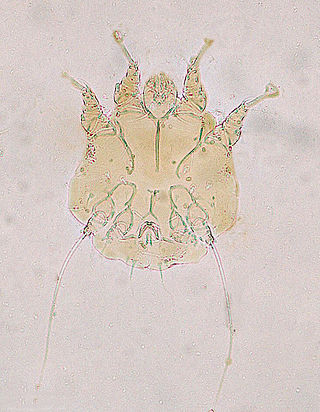
Mites are small arachnids. Mites span two large orders of arachnids, the Acariformes and the Parasitiformes, which were historically grouped together in the subclass Acari. However, most recent genetic analyses do not recover the two as each other's closest relative within Arachnida, rendering the group non-monophyletic. Most mites are tiny, less than 1 mm (0.04 in) in length, and have a simple, unsegmented body plan. The small size of most species makes them easily overlooked; some species live in water, many live in soil as decomposers, others live on plants, sometimes creating galls, while others are predators or parasites. This last type includes the commercially destructive Varroa parasite of honey bees, as well as scabies mites of humans. Most species are harmless to humans, but a few are associated with allergies or may transmit diseases.

<span class="mw-page-title-main">Sebaceous gland</span> Gland to lubricate the hair and skin

A sebaceous gland or oil gland is a microscopic exocrine gland in the skin that opens into a hair follicle to secrete an oily or waxy matter, called sebum, which lubricates the hair and skin of mammals. In humans, sebaceous glands occur in the greatest number on the face and scalp, but also on all parts of the skin except the palms of the hands and soles of the feet. In the eyelids, meibomian glands, also called tarsal glands, are a type of sebaceous gland that secrete a special type of sebum into tears. Surrounding the female nipple, areolar glands are specialized sebaceous glands for lubricating the nipple. Fordyce spots are benign, visible, sebaceous glands found usually on the lips, gums and inner cheeks, and genitals.

<span class="mw-page-title-main">Mange</span> Type of skin disease caused by parasitic mites

Mange is a type of skin disease caused by parasitic mites. Because various species of mites also infect plants, birds and reptiles, the term "mange", or colloquially "the mange", suggesting poor condition of the skin and fur due to the infection, is sometimes reserved for pathological mite-infestation of nonhuman mammals. Thus, mange includes mite-associated skin disease in domestic mammals, in livestock, and in wild mammals. Severe mange caused by mites has been observed in wild bears. Since mites belong to the arachnid subclass Acari, another term for mite infestation is acariasis.

<span class="mw-page-title-main">Demodicosis</span> Medical condition

Demodicosis, also called Demodex folliculitis in humans and demodectic mange or red mange in animals, is caused by a sensitivity to and overpopulation of Demodex spp. as the host's immune system is unable to keep the mites under control.

<span class="mw-page-title-main">Blepharitis</span> Medical condition of the eyelid

Blepharitis, sometimes known as granulated eyelids, is one of the most common ocular conditions characterized by inflammation, scaling, reddening, and crusting of the eyelid. This condition may also cause swelling, burning, itching, or a grainy sensation when introducing foreign objects or substances to the eye. Although blepharitis by itself is not sight-threatening, it can lead to permanent alterations of the eyelid margin. The primary cause is bacteria and inflammation from congested meibomian oil glands at the base of each eyelash. Other conditions may give rise to blepharitis, whether they be infectious or noninfectious, including, but not limited to, bacterial infections or allergies.

<span class="mw-page-title-main">Keratosis pilaris</span> Skin condition characterized by small bumps caused by overproduction of keratin

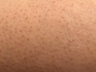
Keratosis pilaris is a common, autosomal-dominant, genetic condition of the skin's hair follicles characterized by the appearance of possibly itchy, small, gooseflesh-like bumps, with varying degrees of reddening or inflammation. It most often appears on the outer sides of the upper arms, thighs, face, back, and buttocks; KP can also occur on the hands, and tops of legs, sides, or any body part except glabrous (hairless) skin. Often the lesions can appear on the face, which may be mistaken for acne or folliculitis.

Skin disorders are among the most common health problems in dogs, and have many causes. The condition of a dog's skin and coat is also an important indicator of its general health. Skin disorders of dogs vary from acute, self-limiting problems to chronic or long-lasting problems requiring life-time treatment. Skin disorders may be primary or secondary in nature, making diagnosis complicated.

Acariasis is an infestation with mites.

<span class="mw-page-title-main">Perioral dermatitis</span> Medical condition

Perioral dermatitis, also known as periorificial dermatitis, is a common type of skin rash. Symptoms include multiple small (1–2 mm) bumps and blisters sometimes with background redness and scale, localized to the skin around the mouth and nostrils. Less commonly the eyes and genitalia may be involved. It can be persistent or recurring and resembles particularly rosacea and to some extent acne and allergic dermatitis. The term "dermatitis" is a misnomer because this is not an eczematous process.

<i>Demodex folliculorum</i> Species of mite

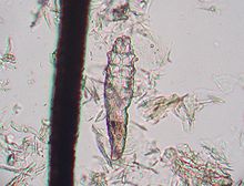
Demodex folliculorum is a microscopic mite that can only survive on the skin of humans. Most people have D. folliculorum on their skin. Usually, the mites do not cause any harm, so are considered an example of commensalism rather than parasitism; but they can cause disease, known as demodicosis.

<span class="mw-page-title-main">Skin flora</span> Microbiota that reside on the skin

Skin flora, also called skin microbiota, refers to microbiota that reside on the skin, typically human skin.

<span class="mw-page-title-main">Feline acne</span> Medical condition

Feline acne is a problem seen in cats primarily involving the formation of blackheads accompanied by inflammation on the cat's chin and surrounding areas that can cause lesions, alopecia, and crusty sores. In many cases symptoms are mild and the disease does not require treatment. Mild cases will resemble dirt on the cat's chin, but the "dirt" will not brush off. More severe cases, however, may respond slowly to treatment and seriously detract from the health and appearance of the cat. Feline acne can affect cats of any age, sex or breed, although Persian cats are also likely to develop acne on the face and in the skin folds. This problem can happen once, be reoccurring, or even persistent throughout the cat's life.

Madarosis is a condition that results in the loss of eyelashes, and sometimes eyebrows. The term "madarosis" is derived from the ancient Greek "madaros", meaning "bald". It originally was a disease of only losing eyelashes but it currently is the loss of both eyelashes and eyebrows. Eyebrows and eyelashes are both important in the prevention of bacteria and other foreign objects from entering the eye. A majority of patients with madarosis have leprosy, and it was reported that 76% of patients with varying types of leprosy had madarosis.

<i>Demodex brevis</i> Species of mite

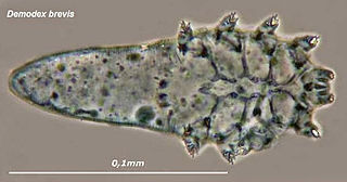
Demodex brevis is one of the two species of face mite that inhabit humans. They are about half as long, at 0.15 to 0.2 mm, as D. folliculorum, but otherwise have few differences. Most of the article on Demodex folliculorum applies equally to D. brevis.

<span class="mw-page-title-main">Mites of domestic animals</span> Type of parasite of domestic animals

Mites that infest and parasitize domestic animals cause disease and loss of production. Mites are small invertebrates, most of which are free living but some are parasitic. Mites are similar to ticks and both comprise the order Acari in the phylum Arthropoda. Mites are highly varied and their classification is complex; a simple grouping is used in this introductory article. Vernacular terms to describe diseases caused by mites include scab, mange, and scabies. Mites and ticks have substantially different biology from, and are classed separately from, insects. Mites of domestic animals cause important types of skin disease, and some mites infest other organs. Diagnosis of mite infestations can be difficult because of the small size of most mites, but understanding how mites are adapted to feed within the structure of the skin is useful.

Demodex bovis, also known as the cattle follicle mite, usually causes demodicosis, or demodectic mange, in cattle. This disease is common in tropical areas and is not usually found in temperate environments. Demodicosis is characterized by the formation of papules and nodules over the cattle's skin. These lesions most commonly occur on the neck, shoulders, and armpit of cattle; however, sometimes they also appear on the udder. This condition is often found in cattle with increased stress from pregnancy or lactation. Natural and acquired immunity can cause a decrease in the number of mites infesting a cow, as well as decreasing the severity of a cow's symptoms.

<span class="mw-page-title-main">Mites of livestock</span> Small crawling animals related to ticks and spiders

Mites are small crawling animals related to ticks and spiders. Most mites are free-living and harmless. Other mites are parasitic, and those that infest livestock animals cause many diseases that are widespread, reduce production and profit for farmers, and are expensive to control.

<span class="mw-page-title-main">Oclacitinib</span> Medication


Oclacitinib, sold under the brand name Apoquel among others, is a veterinary medication used in the control of atopic dermatitis and pruritus from allergic dermatitis in dogs at least 12 months of age. Chemically, it is a synthetic cyclohexylamino pyrrolopyrimidine janus kinase inhibitor that is relatively selective for JAK1. It inhibits signal transduction when the JAK is activated and thus helps downregulate expression of inflammatory cytokines.

<span class="mw-page-title-main">Notoedric mange</span> Skin disease caused by parasitic mites

Notoedric mange, also referred to as Feline scabies, is a highly contagious skin infestation caused by an ectoparasitic and skin burrowing mite Notoedres cati. N. cati is primarily a parasite of felids, but it can also infest rodents, lagomorphs, and occasionally also dogs and foxes. This skin disease also has zoonotic potential. Infestation is also called acariasis, which refers to a rash that is caused by mites.

References

  1. Owen, [Richard] (1843). "Lecture XIX. Arachnida". Lectures on Comparative Anatomy. London: Longman, Brown, Green, and Longmans. p. 252.
  2. Yong, Ed (August 31, 2012). "Everything you never wanted to know about the mites that eat, crawl, and have sex on your face". Not Exactly Rocket Science. Discover . Archived from the original on November 7, 2019. Retrieved April 24, 2013.
  3. Cassidy, Josh (21 May 2019). "Meet the Mites That Live on Your Face". NPR .
  4. δημός . Liddell, Henry George ; Scott, Robert ; An Intermediate Greek–English Lexicon at the Perseus Project.
  5. δήξ . Liddell, Henry George ; Scott, Robert ; A Greek–English Lexicon at the Perseus Project.
  6. "Demodex". Medical Dictionary (medicine.academic.ru).
  7. Simon, Gustav (1842). "Ueber eine in den kranken und normalen Haarsäcken des Menschen lebende Milbe" [On a Mite Living in the Diseased and Normal Hair Follicles of Humans]. In Müller, Johannes (ed.). Archiv für Anatomie, Physiologie und Wissenschaftliche Medicin [Archive for Anatomy, Physiology and Scientific Medicine] (in German). Berlin: Verlag von Veit & Comp. p. 221.
  8. Griffiths, Christopher E. M.; Barker, Jonathan; Bleiker, Tanya; Chalmers, Robert J. G.; Creamer, Daniel; Rook, Graham Arthur, eds. (2016). Rook's textbook of dermatology (9th ed.). Chichester, West Sussex Hoboken, NJ: John Wiley & Sons Inc. ISBN   978-1-118-44117-6.
  9. 1 2 Rufli, T.; Mumcuoglu, Y. (1981). "The hair follicle mites Demodex folliculorum and Demodex brevis: biology and medical importance. A review". Dermatologica . 162 (1): 1–11. doi:10.1159/000250228. PMID   6453029.
  10. Rush, Aisha (2000). "ADW: Demodex folliculorum". Animal Diversity Web . University of Michigan. Archived from the original on 2021-01-25. Retrieved 2022-02-10.
  11. Rather, Parvaiz Anwar; Hassan, Iffat (Jan–Feb 2014). "Human Demodex Mite: The Versatile Mite of Dermatological Importance". Indian Journal of Dermatology . 59 (1). Lippincott Williams & Wilkins: 60–66. doi: 10.4103/0019-5154.123498 . PMC   3884930 . PMID   24470662 . Retrieved 16 December 2023.
  12. Sengbusch, H. G.; Hauswirth, J. W. (1986). "Prevalence of hair follicle mites, Demodex folliculorum and D. brevis (Acari: Demodicidae), in a selected human population in western New York, USA". Journal of Medical Entomology . 23 (4): 384–388. doi:10.1093/jmedent/23.4.384. PMID   3735343.
  13. Thoemmes, Megan S.; Fergus, Daniel J.; Urban, Julie; Trautwein, Michelle; Dunn, Robert R.; Kolokotronis, Sergios-Orestis (27 August 2014). "Ubiquity and Diversity of Human-Associated Demodex Mites". PLoS ONE . 9 (8): e106265. Bibcode:2014PLoSO...9j6265T. doi: 10.1371/journal.pone.0106265 . PMC   4146604 . PMID   25162399.
  14. Liu, Jingbo; Sheha, Hosam; Tseng, Scheffer C. G. (October 2010). "Pathogenic role of Demodex mites in blepharitis". Current Opinion in Allergy and Clinical Immunology . 10 (5): 505–510. doi:10.1097/ACI.0b013e32833df9f4. PMC   2946818 . PMID   20689407.
  15. Zhao, Ya-e; Peng, Yan; Wang, Xiang-lan; Wu, Li-ping; Wang, Mei; Yan, Hu-ling; Xiao, Sheng-xiang (December 2011). "Facial dermatosis associated with Demodex: a case-control study". Journal of Zhejiang University Science B . 12 (8): 1008–1015. doi:10.1631/jzus.B1100179. PMC   3232434 . PMID   22135150.
  16. 1 2 Zhao, Ya-e; Hu, Li; Wu, Li-ping; Ma, Jun-xian (March 2012). "A meta-analysis of association between acne vulgaris and Demodex infestation". Journal of Zhejiang University Science B . 13 (3): 192–202. doi:10.1631/jzus.B1100285. PMC   3296070 . PMID   22374611.
  17. "2011-2012 Annual Evidence Update on Acne vulgaris" (PDF). University of Nottingham Centre of Evidence Based Dermatology. 2012. p. 10. Retrieved 23 September 2013.
  18. Douglas, Annyella; Zaenglein, Andrea L. (September 2019). "A case series of demodicosis in children". Pediatric Dermatology. 36 (5): 651–654. doi:10.1111/pde.13852. PMID   31197860. S2CID   189817759. Papulopustular lesions predominate, prompting the advice 'pustules on noses, think demodicosis!'
  19. MacKenzie, Debora (August 30, 2012). "Rosacea may be caused by mite faeces in your pores". New Scientist . Retrieved August 30, 2012.
  20. Jarmuda, Stanisław; O'Reilly, Niamh; Żaba, Ryszard; Jakubowicz, Oliwia; Szkaradkiewicz, Andrzej; Kavanagh, Kevin (1 November 2012). "Potential role of Demodex mites and bacteria in the induction of rosacea". Journal of Medical Microbiology. 61 (11): 1504–1510. doi: 10.1099/jmm.0.048090-0 . PMID   22933353.
  21. Savla, Keyur; Le, Jimmy T; Pucker, Andrew D (9 June 2019). "Tea tree oil for Demodex blepharitis". The Cochrane Database of Systematic Reviews. 2019 (6): CD013333. doi:10.1002/14651858.CD013333. PMC   6556368 .
  22. Liu, Jingbo; Sheha, Hosam; Tseng, Scheffer CG (2010). "Pathogenic role of Demodex mites in blepharitis". Current Opinion in Allergy and Clinical Immunology. 10 (5): 505–510. doi:10.1097/ACI.0b013e32833df9f4. ISSN   1528-4050. PMC   2946818 . PMID   20689407.
  23. Zhu, Minyi; Cheng, Chao; Yi, Haisu; Lin, Liping; Wu, Kaili (31 July 2018). "Quantitative Analysis of the Bacteria in Blepharitis With Demodex Infestation". Frontiers in Microbiology. 9: 1719. doi: 10.3389/fmicb.2018.01719 . PMC   6079233 . PMID   30108572.
  24. Palopoli, Michael F.; Fergus, Daniel J.; Minot, Samuel; Pei, Dorothy T.; Simison, W. Brian; Fernandez-Silva, Iria; Thoemmes, Megan S.; Dunn, Robert R.; Trautwein, Michelle (29 December 2015). "Global divergence of the human follicle mite Demodex folliculorum: Persistent associations between host ancestry and mite lineages". Proceedings of the National Academy of Sciences. 112 (52): 15958–15963. Bibcode:2015PNAS..11215958P. doi: 10.1073/pnas.1512609112 . PMC   4703014 . PMID   26668374.
  25. Bisbee, E; Rudnick, E; Loyd, A (October 2019). "Crusted demodicosis in an immunocompetent patient". Cutis. 104 (4): E9–E11. PMID   31774896.
  26. Yengil, Erhan; Cevik, Cengiz; Aycan Kaya, Özlem; Taner, Melis; Akkoca, Ayse; Ozer, Cahit (1 January 2014). "Relationship between demodex folliculorum and allergic rhinitis in adults". Acta Medica Mediterranea. 30: 27–31.
  27. Ramos-e-Silva M, Sampaio AL, Carneiro S (2014). "Red face revisited: Endogenous dermatitis in the form of atopic dermatitis and seborrheic dermatitis". Clin. Dermatol. 32 (1): 109–15. doi:10.1016/j.clindermatol.2013.05.032. PMID   24314384.
  28. Eran Galili; Aviv Barzilai; Gilad Twig; Tomm Caspi; Danny Daniely; Rony Shreberk-Hassidim; Nadav Astman (2020). "Allergic Rhinitis and Asthma Among Adolescents with Psoriasis: A Population-based Cross-sectional Study". Acta Dermato Venereologica. 100 (10): adv00133-5. doi:10.2340/00015555-3485. PMC   9137373 . PMID   32314795.
  29. Kolb L, Ferrer-Bruker SJ (2023). Atopic Dermatitis. Affiliations 1 VCOM/Orange Park Medical Center Copyright © 2022, StatPearls Publishing LLC. PMID   28846349.
  30. Belgrave, Danielle C M; Simpson, Angela; Buchan, Iain E; Custovic, Adnan (2015). "Atopic Dermatitis and Respiratory Allergy: What is the Link". Current Dermatology Reports. 4 (4): 221–227. doi:10.1007/s13671-015-0121-6. PMC   4635175 . PMID   26566461.
  31. Miller, William H. Jr.; Griffin, Craig E.; Campbell, Karen L. (2013). "Canine demodicosis". Muller & Kirk's small animal dermatology (7th ed.). St. Louis, Mo.: Elsevier. pp. 304–313. ISBN   978-1-4160-0028-0.
  32. Horvath, Christa; Neuber, Ariane (March 2007). "Pathogenesis of canine demodicosis". Companion Animal. 12 (2): 55–59. doi:10.1111/j.2044-3862.2007.tb00131.x.