Dihydrolipoyl transacetylase

Last updated
DLAT
PDB 1y8o EBI.jpg
Available structures
PDB Ortholog search: PDBe RCSB
Identifiers
Aliases DLAT , DLTA, PDC-E2, PDCE2, dihydrolipoamide S-acetyltransferase, Dihydrolipoyl transacetylase, E2
External IDs OMIM: 608770 MGI: 2385311 HomoloGene: 6814 GeneCards: DLAT
Orthologs
SpeciesHumanMouse
Entrez
Ensembl
UniProt
RefSeq (mRNA)

NM_001931

NM_145614

RefSeq (protein)

NP_663589

Location (UCSC) Chr 11: 112.02 – 112.06 Mb Chr 9: 50.55 – 50.57 Mb
PubMed search [3] [4]
Wikidata
View/Edit Human View/Edit Mouse

Dihydrolipoyl transacetylase (or dihydrolipoamide acetyltransferase) is an enzyme component of the multienzyme pyruvate dehydrogenase complex. The pyruvate dehydrogenase complex is responsible for the pyruvate decarboxylation step that links glycolysis to the citric acid cycle. This involves the transformation of pyruvate from glycolysis into acetyl-CoA which is then used in the citric acid cycle to carry out cellular respiration.

There are three different enzyme components in the pyruvate dehydrogenase complex. Pyruvate dehydrogenase (EC 1.2.4.1) is responsible for the oxidation of pyruvate, dihydrolipoyl transacetylase (this enzyme; EC 2.3.1.12) transfers the acetyl group to coenzyme A (CoA), and dihydrolipoyl dehydrogenase (EC 1.8.1.4) regenerates the lipoamide. Because dihydrolipoyl transacetylase is the second of the three enzyme components participating in the reaction mechanism for conversion of pyruvate into acetyl CoA, it is sometimes referred to as E2.

In humans, dihydrolipoyl transacetylase enzymatic activity resides in the pyruvate dehydrogenase complex component E2 (PDCE2) that is encoded by the DLAT (dihydrolipoamide S-acetyltransferase) gene. [5]

Nomenclature

The systematic name of this enzyme class is acetyl-CoA:enzyme N6-(dihydrolipoyl)lysine S-acetyltransferase.

Other names in common use include:

Structure

The cubic catalytic core structure made up of 24 dihydrolipoyl transacetylase subunits. Pyruvate dehydrogenase multienzyme complex.jpg
The cubic catalytic core structure made up of 24 dihydrolipoyl transacetylase subunits.
The dodecahedral catalytic core structure made up of 60 dihydrolipoyl transacetylase subunits from Geobacillus stearothermophilus: 3D electron microscopy map (left) and X-ray diffraction structure (right). 1B5S-crop.jpg
The dodecahedral catalytic core structure made up of 60 dihydrolipoyl transacetylase subunits from Geobacillus stearothermophilus : 3D electron microscopy map (left) and X-ray diffraction structure (right).

All dihydrolipoyl transacetylases have a unique multidomain structure consisting of (from N to C): 3 lipoyl domains, an interaction domain, and the catalytic domain (see the domain architecture at Pfam). All the domains are connected by disordered, low complexity linker regions.

Depending on the species, multiple subunits of dihydrolipoyl transacetylase enzymes can arrange together into either a cubic or dodecahedral shape. These structure then form the catalytic core of the pyruvate dehydrogenase complex which not only catalyzes the reaction that transfers an acetyl group to CoA, but also performs a crucial structural role in creating the architecture of the overall complex. [7]

Cube

The cubic core structure, found in species such as Azotobacter vinelandii , is made up of 24 subunits total. [8] [9] The catalytic domains are assembled into trimers with the active site located at the subunit interface. The topology of this trimer active site is identical to that of chloramphenicol acetyltransferase. Eight of these trimers are then arranged into a hollow truncated cube. The two main substrates, CoA and the lipoamide (Lip(SH)2), are found at two opposite entrances of a 30 Å long channel which runs between the subunits and forms the catalytic center. CoA enters from the inside of the cube, and the lipoamide enters from the outside. [10]

Dodecahedron

In many species, including bacteria such as Geobacillus stearothermophilus and Enterococcus faecalis [7] as well as mammals such as humans [11] and cows, [12] the dodecahedral core structure is made up of 60 subunits total. The subunits are arranged in sets of three, similar to the trimers in the cubic core shape, with each set making up one of the 20 dodecahedral vertices.

Function

dihydrolipoyllysine-residue acetyltransferase
Identifiers
EC no. 2.3.1.12
CAS no. 9032-29-5
Databases
IntEnz IntEnz view
BRENDA BRENDA entry
ExPASy NiceZyme view
KEGG KEGG entry
MetaCyc metabolic pathway
PRIAM profile
PDB structures RCSB PDB PDBe PDBsum
Gene Ontology AmiGO / QuickGO
Search
PMC articles
PubMed articles
NCBI proteins

Dihydrolipoyl transacetylase participates in the pyruvate decarboxylation reaction that links glycolysis to the citric acid cycle. These metabolic processes are important for cellular respiration—the conversion of biochemical energy from nutrients into adenosine triphosphate (ATP) which can then be used to carry out numerous biological reactions within a cell. The various parts of cellular respiration take place in different parts of the cell. In eukaryotes, glycolysis occurs in the cytoplasm, pyruvate decarboxylation in the mitochondria, the citric acid cycle within the mitochondrial matrix, and oxidative phosphorylation via the electron transport chain on the mitochondrial cristae. Thus pyruvate dehydrogenase complexes (containing the dihydrolipoyl transacetylase enzymes) are found in the mitochondria of eukaryotes (and simply in the cytosol of prokaryotes).

Mechanism

Dihydrolipoyl transacetylase mechanism Dihydrolipoyl transacetylase mechanism.png
Dihydrolipoyl transacetylase mechanism

Pyruvate decarboxylation requires a few cofactors in addition to the enzymes that make up the complex. The first is thiamine pyrophosphate (TPP), which is used by pyruvate dehydrogenase to oxidize pyruvate and to form a hydroxyethyl-TPP intermediate. This intermediate is taken up by dihydrolipoyl transacetylase and reacted with a second lipoamide cofactor to generate an acetyl-dihydrolipoyl intermediate, releasing TPP in the process. This second intermediate can then be attacked by the nucleophilic sulfur attached to Coenzyme A, and the dihydrolipoamide is released. This results in the production of acetyl CoA, which is the end goal of pyruvate decarboxylation. The dihydrolipoamide is taken up by dihydrolipoyl dehydrogenase, and with the additional cofactors FAD and NAD+, regenerates the original lipoamide (with NADH as a useful side product).

Interactive pathway map

Click on genes, proteins and metabolites below to link to respective articles. [§ 1]

[[File:
WP534.png go to articlego to articlego to articlego to articlego to articlego to articlego to articlego to articlego to articlego to articlego to articlego to articlego to articlego to articlego to articlego to articlego to articlego to articlego to articlego to articlego to articlego to articlego to articlego to articlego to articlego to articlego to articlego to articlego to articlego to articlego to articlego to articlego to articlego to articlego to articlego to articlego to articlego to articlego to articlego to articlego to articlego to articlego to articlego to articlego to articlego to articlego to articlego to articlego to articlego to articlego to articlego to articlego to articlego to articlego to articlego to articlego to articlego to articlego to articlego to articlego to articlego to articlego to articlego to articlego to articlego to articlego to articlego to articlego to articlego to articlego to WikiPathwaysgo to articlego to Entrezgo to article
[[
]]
[[
]]
[[
]]
[[
]]
[[
]]
[[
]]
[[
]]
[[
]]
[[
]]
[[
]]
[[
]]
[[
]]
[[
]]
[[
]]
[[
]]
[[
]]
[[
]]
[[
]]
[[
]]
[[
]]
[[
]]
[[
]]
[[
]]
[[
]]
[[
]]
[[
]]
[[
]]
[[
]]
[[
]]
[[
]]
[[
]]
[[
]]
[[
]]
[[
]]
[[
]]
[[
]]
[[
]]
[[
]]
[[
]]
[[
]]
[[
]]
[[
]]
[[
]]
[[
]]
[[
]]
[[
]]
[[
]]
[[
]]
[[
]]
[[
]]
[[
]]
[[
]]
[[
]]
[[
]]
[[
]]
[[
]]
[[
]]
[[
]]
[[
]]
[[
]]
[[
]]
[[
]]
[[
]]
[[
]]
[[
]]
[[
]]
[[
]]
[[
]]
[[
]]
[[
]]
[[
]]
[[
]]
[[
]]
[[
]]
[[
]]
WP534.png go to articlego to articlego to articlego to articlego to articlego to articlego to articlego to articlego to articlego to articlego to articlego to articlego to articlego to articlego to articlego to articlego to articlego to articlego to articlego to articlego to articlego to articlego to articlego to articlego to articlego to articlego to articlego to articlego to articlego to articlego to articlego to articlego to articlego to articlego to articlego to articlego to articlego to articlego to articlego to articlego to articlego to articlego to articlego to articlego to articlego to articlego to articlego to articlego to articlego to articlego to articlego to articlego to articlego to articlego to articlego to articlego to articlego to articlego to articlego to articlego to articlego to articlego to articlego to articlego to articlego to articlego to articlego to articlego to articlego to articlego to WikiPathwaysgo to articlego to Entrezgo to article
|alt=Glycolysis and Gluconeogenesis edit]]
Glycolysis and Gluconeogenesis edit
  1. The interactive pathway map can be edited at WikiPathways: "GlycolysisGluconeogenesis_WP534".

Clinical significance

Primary biliary cirrhosis

Primary biliary cirrhosis (PBC) is an autoimmune disease characterized by autoantibodies against mitochondrial and nuclear antigens. These are called anti-mitochondrial antibodies (AMA) and anti-nuclear antibodies (ANA), respectively. These antibodies are detectable in the sera of PBC patients and vary greatly with regards to epitope specificity from patient to patient. Of the mitochondrial antigens that can generate autoantibody reactivity in PBC patients, the E2 subunit of the pyruvate dehydrogenase complex, dihydrolipoyl transacetylase, is the most common epitope (other antigens include enzymes of the 2-oxoacid dehydrogenase complexes as well as the other enzymes of the pyruvate dehydrogenase complexes). [13] Recent evidence has suggested that peptides within the catalytic site may present the immunodominant epitopes recognized by the anti-PDC-E2 antibodies in PBC patients. [14] There is also evidence of anti-PDC-E2 antibodies in autoimmune hepatitis (AIH) patients. [15]

Pyruvate dehydrogenase deficiency

Pyruvate dehydrogenase deficiency (PDH) is a genetic disease resulting in lactic acidosis as well as neurological dysfunction in infancy and early childhood. Typically PDH is the result of a mutation in the X-linked gene for the E1 subunit of the pyruvate dehydrogenase complex. However, there have been a few rare cases in which a patient with PDH actually has a mutation in the autosomal gene for the E2 subunit instead. These patients have been reported to have much less severe symptoms, with the most prominent disease manifestation being episodic dystonia, though both hypotonia and ataxia were also present. [16]

Related Research Articles

<span class="mw-page-title-main">Pyruvate dehydrogenase complex</span> Three-enzyme complex

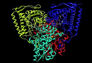
Pyruvate dehydrogenase complex (PDC) is a complex of three enzymes that converts pyruvate into acetyl-CoA by a process called pyruvate decarboxylation. Acetyl-CoA may then be used in the citric acid cycle to carry out cellular respiration, and this complex links the glycolysis metabolic pathway to the citric acid cycle. Pyruvate decarboxylation is also known as the "pyruvate dehydrogenase reaction" because it also involves the oxidation of pyruvate.

The oxoglutarate dehydrogenase complex (OGDC) or α-ketoglutarate dehydrogenase complex is an enzyme complex, most commonly known for its role in the citric acid cycle.

<span class="mw-page-title-main">Pyruvate dehydrogenase lipoamide kinase isozyme 1</span> Protein-coding gene in the species Homo sapiens

Pyruvate dehydrogenase lipoamide kinase isozyme 1, mitochondrial is an enzyme that in humans is encoded by the PDK1 gene. It codes for an isozyme of pyruvate dehydrogenase kinase (PDK).

The branched-chain α-ketoacid dehydrogenase complex is a multi-subunit complex of enzymes that is found on the mitochondrial inner membrane. This enzyme complex catalyzes the oxidative decarboxylation of branched, short-chain alpha-ketoacids. BCKDC is a member of the mitochondrial α-ketoacid dehydrogenase complex family, which also includes pyruvate dehydrogenase and alpha-ketoglutarate dehydrogenase, key enzymes that function in the Krebs cycle.

Pyruvate dehydrogenase deficiency is a rare neurodegenerative disorder associated with abnormal mitochondrial metabolism. PDCD is a genetic disease resulting from mutations in one of the components of the pyruvate dehydrogenase complex (PDC). The PDC is a multi-enzyme complex that plays a vital role as a key regulatory step in the central pathways of energy metabolism in the mitochondria. The disorder shows heterogeneous characteristics in both clinical presentation and biochemical abnormality.

<span class="mw-page-title-main">Anti-mitochondrial antibody</span> Autoantibodies against liver mitochondria, indicating primary biliary cholangitis

Anti-mitochondrial antibodies (AMA) are autoantibodies, consisting of immunoglobulins formed against mitochondria, primarily the mitochondria in cells of the liver.

Oxidative decarboxylation is a decarboxylation reaction caused by oxidation. Most are accompanied by α- Ketoglutarate α- Decarboxylation caused by dehydrogenation of hydroxyl carboxylic acids such as carbonyl carboxylic acid, malic acid, isocitric acid, etc.

<span class="mw-page-title-main">Pyruvate dehydrogenase</span> Class of enzymes

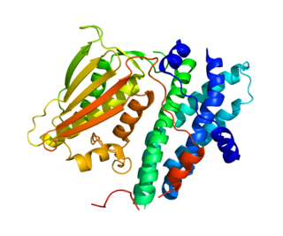
Pyruvate dehydrogenase is an enzyme that catalyzes the reaction of pyruvate and a lipoamide to give the acetylated dihydrolipoamide and carbon dioxide. The conversion requires the coenzyme thiamine pyrophosphate.

<span class="mw-page-title-main">Dihydrolipoamide dehydrogenase</span> Protein-coding gene in the species Homo sapiens

Dihydrolipoamide dehydrogenase (DLD), also known as dihydrolipoyl dehydrogenase, mitochondrial, is an enzyme that in humans is encoded by the DLD gene. DLD is a flavoprotein enzyme that oxidizes dihydrolipoamide to lipoamide.

<span class="mw-page-title-main">E3 binding protein</span> Protein-coding gene in the species Homo sapiens

E3 binding protein also known as pyruvate dehydrogenase protein X component, mitochondrial is a protein that in humans is encoded by the PDHX gene. The E3 binding protein is a component of the pyruvate dehydrogenase complex found only in eukaryotes. Defects in this gene are a cause of pyruvate dehydrogenase deficiency which results in neurological dysfunction and lactic acidosis in infancy and early childhood. This protein is also a minor antigen for antimitochondrial antibodies. These autoantibodies are present in nearly 95% of patients with primary biliary cholangitis, an autoimmune disease of the liver. In primary biliary cholangitis, activated T lymphocytes attack and destroy epithelial cells in the bile duct where this protein is abnormally distributed and overexpressed. Primary biliary cholangitis eventually leads to liver failure.

<span class="mw-page-title-main">Pyruvate dehydrogenase phosphatase</span> Protein-coding gene in the species Homo sapiens

Pyruvate dehydrogenase phosphatase catalytic subunit 1, also known as protein phosphatase 2C, is an enzyme that in humans is encoded by the PDP1 gene. PDPC 1 is an enzyme which serves to reverse the effects of pyruvate dehydrogenase kinase upon pyruvate dehydrogenase, activating pyruvate dehydrogenase.

<span class="mw-page-title-main">Pyruvate dehydrogenase (lipoamide) alpha 1</span> Protein-coding gene in the species Homo sapiens

Pyruvate dehydrogenase E1 component subunit alpha, somatic form, mitochondrial is an enzyme that in humans is encoded by the PDHA1 gene.The pyruvate dehydrogenase complex is a nuclear-encoded mitochondrial matrix multienzyme complex that provides the primary link between glycolysis and the tricarboxylic acid (TCA) cycle by catalyzing the irreversible conversion of pyruvate into acetyl-CoA. The PDH complex is composed of multiple copies of 3 enzymes: E1 (PDHA1); dihydrolipoyl transacetylase (DLAT) ; and dihydrolipoyl dehydrogenase (DLD). The E1 enzyme is a heterotetramer of 2 alpha and 2 beta subunits. The E1-alpha subunit contains the E1 active site and plays a key role in the function of the PDH complex.

<span class="mw-page-title-main">DBT (gene)</span> Mammalian protein found in Homo sapiens

Lipoamide acyltransferase component of branched-chain alpha-keto acid dehydrogenase complex, mitochondrial is an enzyme that in humans is encoded by the DBT gene.

<span class="mw-page-title-main">DLST</span> Protein-coding gene in the species Homo sapiens

Dihydrolipoyllysine-residue succinyltransferase component of 2-oxoglutarate dehydrogenase complex, mitochondrial is an enzyme that in humans is encoded by the DLST gene.

<span class="mw-page-title-main">PDK3</span> Protein-coding gene in the species Homo sapiens

Pyruvate dehydrogenase lipoamide kinase isozyme 3, mitochondrial is an enzyme that in humans is encoded by the PDK3 gene. It codes for an isozyme of pyruvate dehydrogenase kinase.The pyruvate dehydrogenase (PDH) complex is a nuclear-encoded mitochondrial multienzyme complex that catalyzes the overall conversion of pyruvate to acetyl-CoA and CO2. It provides the primary link between glycolysis and the tricarboxylic acid (TCA) cycle, and thus is one of the major enzymes responsible for the regulation of glucose metabolism. The enzymatic activity of PDH is regulated by a phosphorylation/dephosphorylation cycle, and phosphorylation results in inactivation of PDH. The protein encoded by this gene is one of the four pyruvate dehydrogenase kinases that inhibits the PDH complex by phosphorylation of the E1 alpha subunit. This gene is predominantly expressed in the heart and skeletal muscles. Alternatively spliced transcript variants encoding different isoforms have been found for this gene.

In chemistry, a compartment is a part of a protein that serves a specific function. They are essentially protein subunits with the added condition that a compartment has distinct functionality, rather than being just a structural component.

<span class="mw-page-title-main">OGDH</span> Enzyme involved in Krebs cycle

Alpha-ketoglutarate dehydrogenase also known as 2-oxoglutarate dehydrogenase E1 component, mitochondrial is an enzyme that in humans is encoded by the OGDH gene.

<span class="mw-page-title-main">Pyruvate dehydrogenase (lipoamide) alpha 2</span> Protein-coding gene in the species Homo sapiens

Pyruvate dehydrogenase (lipoamide) alpha 2, also known as pyruvate dehydrogenase E1 component subunit alpha, testis-specific form, mitochondrial or PDHE1-A type II, is an enzyme that in humans is encoded by the PDHA2 gene.

<span class="mw-page-title-main">Pyruvate dehydrogenase (lipoamide) beta</span> Protein-coding gene in the species Homo sapiens

Pyruvate dehydrogenase (lipoamide) beta, also known as pyruvate dehydrogenase E1 component subunit beta, mitochondrial or PDHE1-B is an enzyme that in humans is encoded by the PDHB gene. The pyruvate dehydrogenase (PDH) complex is a nuclear-encoded mitochondrial multienzyme complex that catalyzes the overall conversion of pyruvate to acetyl-CoA and CO2, and provides the primary link between glycolysis and the tricarboxylic acid (TCA) cycle. The PDH complex is composed of multiple copies of three enzymatic components: pyruvate dehydrogenase (E1), dihydrolipoamide acetyltransferase (E2) and lipoamide dehydrogenase (E3). The E1 enzyme is a heterotetramer of two alpha and two beta subunits. This gene encodes the E1 beta subunit. Mutations in this gene are associated with pyruvate dehydrogenase E1-beta deficiency.

<span class="mw-page-title-main">Pyruvate decarboxylation</span>

Pyruvate decarboxylation or pyruvate oxidation, also known as the link reaction, is the conversion of pyruvate into acetyl-CoA by the enzyme complex pyruvate dehydrogenase complex.

References

  1. 1 2 3 GRCh38: Ensembl release 89: ENSG00000150768 Ensembl, May 2017
  2. 1 2 3 GRCm38: Ensembl release 89: ENSMUSG00000000168 Ensembl, May 2017
  3. "Human PubMed Reference:". National Center for Biotechnology Information, U.S. National Library of Medicine.
  4. "Mouse PubMed Reference:". National Center for Biotechnology Information, U.S. National Library of Medicine.
  5. Leung PS, Watanabe Y, Munoz S, Teuber SS, Patel MS, Korenberg JR, Hara P, Coppel R, Gershwin ME (1993). "Chromosome localization and RFLP analysis of PDC-E2: the major autoantigen of primary biliary cirrhosis". Autoimmunity. 14 (4): 335–40. doi:10.3109/08916939309079237. PMID   8102256.
  6. Mattevi A, Obmolova G, Kalk KH, Teplyakov A, Hol WG (Apr 1993). "Crystallographic analysis of substrate binding and catalysis in dihydrolipoyl transacetylase (E2p)". Biochemistry. 32 (15): 3887–901. doi:10.1021/bi00066a007. PMID   8471601.
  7. 1 2 3 PDB: 1B5S ; Izard T, Aevarsson A, Allen MD, Westphal AH, Perham RN, de Kok A, Hol WG (February 1999). "Principles of quasi-equivalence and Euclidean geometry govern the assembly of cubic and dodecahedral cores of pyruvate dehydrogenase complexes". Proc. Natl. Acad. Sci. U.S.A. 96 (4): 1240–5. Bibcode:1999PNAS...96.1240I. doi: 10.1073/pnas.96.4.1240 . PMC   15447 . PMID   9990008.
  8. de Kok A, Hengeveld AF, Martin A, Westphal AH (Jun 1998). "The pyruvate dehydrogenase multi-enzyme complex from Gram-negative bacteria". Biochimica et Biophysica Acta (BBA) - Protein Structure and Molecular Enzymology. 1385 (2): 353–66. doi:10.1016/S0167-4838(98)00079-X. PMID   9655933.
  9. Hanemaaijer R, Westphal AH, Van Der Heiden T, De Kok A, Veeger C (Feb 1989). "The quaternary structure of the dihydrolipoyl transacetylase component of the pyruvate dehydrogenase complex from Azotobacter vinelandii. A reconsideration". European Journal of Biochemistry. 179 (2): 287–92. doi:10.1111/j.1432-1033.1989.tb14553.x. PMID   2917567.
  10. Mattevi A, Obmolova G, Schulze E, Kalk KH, Westphal AH, de Kok A, Hol WG (Mar 1992). "Atomic structure of the cubic core of the pyruvate dehydrogenase multienzyme complex". Science. 255 (5051): 1544–50. Bibcode:1992Sci...255.1544M. doi:10.1126/science.1549782. PMID   1549782.
  11. Brautigam CA, Wynn RM, Chuang JL, Chuang DT (May 2009). "Subunit and catalytic component stoichiometries of an in vitro reconstituted human pyruvate dehydrogenase complex". The Journal of Biological Chemistry. 284 (19): 13086–98. doi: 10.1074/jbc.M806563200 . PMC   2676041 . PMID   19240034.
  12. Zhou ZH, McCarthy DB, O'Connor CM, Reed LJ, Stoops JK (Dec 2001). "The remarkable structural and functional organization of the eukaryotic pyruvate dehydrogenase complexes". Proceedings of the National Academy of Sciences of the United States of America. 98 (26): 14802–7. Bibcode:2001PNAS...9814802Z. doi: 10.1073/pnas.011597698 . PMC   64939 . PMID   11752427.
  13. Mackay IR, Whittingham S, Fida S, Myers M, Ikuno N, Gershwin ME, Rowley MJ (Apr 2000). "The peculiar autoimmunity of primary biliary cirrhosis". Immunological Reviews. 174: 226–37. doi:10.1034/j.1600-0528.2002.017410.x. PMID   10807519. S2CID   596338. Archived from the original on 2013-01-05.
  14. Braun S, Berg C, Buck S, Gregor M, Klein R (Feb 2010). "Catalytic domain of PDC-E2 contains epitopes recognized by antimitochondrial antibodies in primary biliary cirrhosis". World Journal of Gastroenterology. 16 (8): 973–81. doi: 10.3748/wjg.v16.i8.973 . PMC   2828602 . PMID   20180236.
  15. O'Brien C, Joshi S, Feld JJ, Guindi M, Dienes HP, Heathcote EJ (Aug 2008). "Long-term follow-up of antimitochondrial antibody-positive autoimmune hepatitis". Hepatology. 48 (2): 550–6. doi: 10.1002/hep.22380 . PMID   18666262. S2CID   5400712.
  16. Head RA, Brown RM, Zolkipli Z, Shahdadpuri R, King MD, Clayton PT, Brown GK (Aug 2005). "Clinical and genetic spectrum of pyruvate dehydrogenase deficiency: dihydrolipoamide acetyltransferase (E2) deficiency". Annals of Neurology. 58 (2): 234–41. doi:10.1002/ana.20550. PMID   16049940. S2CID   38264402.

Further reading