Effects of sleep deprivation on cognitive performance

Last updated

The effects of sleep deprivation on cognitive performance encompass a broad range of impairments in various cognitive functions resulting from inadequate or insufficient sleep, impacting areas such as attention, memory, decision-making, and reaction time.

Contents

It has been estimated that over 20% of adults suffer from some form of sleep deprivation. [1] Insomnia and sleep deprivation are common symptoms of depression, and can be an indication of other mental disorders. [2] The consequences of not getting enough sleep could have dire results, not only to the health, cognition, energy level and the mood of the person, but also to those around them. Sleep deprivation increases the risk of human-error related accidents, especially with vigilance-based tasks involving technology. [3]

Attention

Neural substrates

Parietal lobe Parietal lobe.gif
Parietal lobe

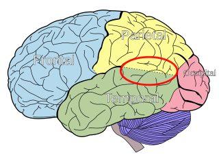
The parietal lobes of the brain are largely involved in attention. Lesions to this region of the brain in humans result in difficulty or inability to attend to events that are contralateral to the lesioned hemisphere. Those with lesions to the posterior parietal lobe have little to no difficulty shifting attention to and from stimuli appearing in the space ipsilateral to the lesioned hemisphere. However, they do display a slowed response in shifting their focus of current attention to events and stimuli appearing contralateral to the lesioned hemisphere. [4]

Studies involving single-unit recordings from the parietal lobes of monkeys have indicated that there are neurons solely involved in integrating visual spatial information with postural information. [5] Without this apparent combining of spatial information, it would be difficult or impossible to locate objects in external space (as information provided solely by the retina is insufficient). The position of the eyes, head and body must also be taken into consideration.

In addition, studies involving transcranial magnetic stimulation application over the parietal lobes as well as positron emission tomography (PET) analysis of the parietal lobes suggest that this region is involved in conjunction searches, but not in single-feature searches (see Visual search for supplementary information). [6]

Auditory attention

The primary auditory cortex is highlighted in magenta, and has been known to interact with all areas highlighted on this neural map. Brain Surface Gyri.SVG
The primary auditory cortex is highlighted in magenta, and has been known to interact with all areas highlighted on this neural map.

Auditory attention has been examined following sleep deprivation. In one study, researchers examined the auditory attention of twelve non-sleep-deprived subjects and twelve sleep-deprived subjects at various time intervals. Subjects were involved in an auditory attention task, which required the reproduction of the spatial relationships between four letters, using a graph composed of six squares, immediately following the presentation of an item from a tape recorder. It was found that auditory attention of sleep-deprived individuals is affected as the total amount of sleep-deprivation increases, possibly due to lowered perceptual vigilance. [7]

Divided attention

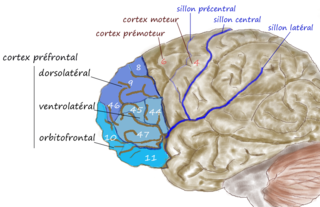
Functional magnetic resonance imaging (fMRI) scans of the brains of subjects exposed to thirty-five hours of sleep deprivation indicate that sleep deprivation is related to increases in prefrontal cortex and parietal lobe activation during tasks that combine verbal learning and arithmetic. This is particularly apparent in the right hemisphere. In non-sleep-deprived people involved in verbal learning and arithmetic tasks, the anterior cingulate cortex and the right prefrontal cortex are active. Following sleep deprivation, there is increased activation of the left inferior frontal gyrus and the bilateral parietal lobes. This information suggests that divided attention tasks require more attentional resources than normally required by a non-sleep-deprived person. [8]

Exogenous and endogenous attention

Studies using event-related potential (ERP) recordings have found that twenty-four hours of sleep deprivation decreases ERP response to signal inputs from endogenous, but not exogenous, sources. Therefore, it is suggested that sleep deprivation affects endogenously-driven selective attention to a greater extent than exogenously-driven selected attention. [9]

Selective attention

Twenty-four hours of sleep deprivation has been found to affect the functional connectivity between the inferior frontal parietal region (IPS) and the parahippocampal place area (PPA). However, sleep deprivation does not affect the attention modulation index of the PPA. With this information, researchers have concluded that the psychophysiological interaction (PPI) is more involved in selective attention than the IPS and PPA. [10]

Research has found that together, attention and sleep deprivation modulate the parahippocampal place area (PPA) activation and scene processing. Specifically, sleep deprivation was related to significant decreases in PPA activation while attending to scenes and when ignoring scenes. This is explained by the absence of change in the Attention Modulation Index (AMI). Face recognition is not affected by sleep deprivation. [10]

Sleep deprivation has been shown to negatively affect picture classification speed and accuracy, as well as recognition memory. [10] It results in an inability to avoid attending to irrelevant information displayed during attention-related tasks. (Norton) It also decreases activation in the ventral visual area and the frontal parietal control regions. [10]

Supervisory attention

Studies involving sleep deprived subjects’ performance on choice reaction time tests—in which response inhibition, task shifting skill and task strategy were involved—have been conducted and analyzed. These three cognitive processes are involved and critical in tasks involving supervisory attention, which is defined as behaviour that arises through the selection and implementation of schemas. [6] Following one night of total sleep deprivation, subjects showed no decline in task shifting or response inhibition performance. However, sleep deprivation does affect the ability to use preparatory bias to increase performance speed. It is suggested that the brain’s cognitive resources make an active effort to succeed in a challenging task when subjected to sleep deprivation, and that this deficit becomes apparent in tasks involving preparatory bias. [11] Similarly, partial sleep deprivation significantly influenced subjects simple reaction time, thus making it slower than subjects who were well rested [12]

Visuospatial attention

Deficits in cognitive performance due to continuous sleep restriction are not well understood. However, there have been studies looking into physiological arousal of the sleep-deprived brain. Participants, whose total amount of sleep had been restricted by 33% throughout one week, were subjected to reaction time tests. The results of these tests were analyzed using quantitative EEG analysis. The results indicate that the frontal regions of the brain are first to be affected, whereas the parietal regions remain active until the effects of sleep deprivation become more severe, which occurred towards the end of the week. In addition, EEG and ERP analysis reveals that activation deficits are more apparent in the non-dominant hemisphere—than in the dominant hemisphere. [13]

Thalamus. Brain chrischan thalamus.jpg
Thalamus .

The effects of sleep deprivation on cognitive performance have been studied through the use of parametric visual attention tasks. Functional magnetic resonance imaging of participants' brains who were involved in ball-tracking tasks of various difficulty levels were obtained. These images were taken during rested wakefulness and again after one night of sleep deprivation. The thalamus is more highly activated when accompanied by sleep deprivation—than when the subject is in a state of rested wakefulness. Contrarily, the thalamus is more highly activated during difficult tasks accompanied by rested wakefulness, but not during a state of sleep deprivation. Researchers propose that the thalamic resources, which are normally activated during difficult tasks, are being activated in an attempt to maintain alertness during states of sleep deprivation. In addition, an increase in thalamic activation is related to a decrease in the parietal, prefrontal and cingulate cortex activation, resulting in the overall impairment of attentional networks, which are necessary for visuospatial attention performance. [14]

Functional Magnetic Resonance Imaging (fMRI) studies indicate that the posterior cingulate (PCC) and medial prefrontal cortex are involved in the anticipatory allocation of spatial attention. When sleep-deprived, PCC activity decreases, impairing selective attention. Subjects were exposed to an attention-shifting task involving spatially informative, misleading and uninformative cues preceding the stimuli. When sleep-deprived, subjects showed increased activation in the left intraparietal sulcus. This region is activated when exposed to stimuli in unexpected locations. These findings suggest that sleep-deprived people may be impaired in their ability to anticipate the locations of upcoming events. In addition, inability to avoid attending to irrelevant events may also be induced by sleep-deprivation. [15]

By contrast, other studies have indicated that the effects of sleep deprivation on cognitive performance, specifically sustained visual attention, are more global and bilateral in nature (as opposed to more lateralized deficit explanations). In a study using the Choice Visual Perception Task, subjects were exposed to stimuli appearing in various locations in visual space. Results indicate that sleep deprivation results in a general decline in visual attention. It is also suggested that the sleep-deprived brain is able to maintain a certain level of cognitive performance during tasks requiring divided attention—by recruiting additional cortical regions that are not normally used for such tasks. [16]

Executive function

Executive functioning is "the ability to plan and coordinate a willful action in the face of alternatives, to monitor and update action as necessary and suppress distracting material by focusing attention on the task at hand". [17] The prefrontal cortex has been identified as the most important region involved in executive functioning. [18]

Researchers believe that three of the most 'basic' executive functions are: shifting, updating, and inhibition. [19] Shifting back and forth between different tasks is considered a very important mental behavior involved in executive functioning, as it involves active disengagement from the present task and engaging in a new task. [20] Updating is believed to be involved in working memory (closely associated with the function of the prefrontal cortex, [21] where the information that is active needs to be updated by replacing old, now irrelevant information with new, relevant information based on the objective. [22] [23] Inhibition involves controlled and deliberate impedance of automatic, predominant responses. [24]

The anterior cingulate cortex has been implemented in the process of inhibiting a habitual response or detecting possible conflicts in responses; [25] this is shown by the Stroop test. [26] Studies have found that as little as 36 hours of sleep deprivation can cause a performance reduction in tasks requiring these executive functions. [27]

Frontal lobe. Frontal lobe.gif
Frontal lobe.

The processes above illustrate a model of controlled versus automatic behavior that was hypothesized by Shallice et al. (1989), called the supervisory attentional system. This system is believed to come into play when intervention of habitual response is necessary. [28] Damage to the prefrontal cortex will cause a breakdown in this system, resulting in utilization behaviors. [28] These behaviors would include spontaneous sequences of action on irrelevant objects in the surroundings with no clear goal in mind. [28] This theory has helped to extend the current knowledge on executive functions.

Decision making

Decision making involves a range of executive functions that need to be combined and organized in order to respond in the most appropriate manner (i.e., respond with the most advantageous decision). [29] There are many aspects to the process of decision making, including those discussed above. Other processes involved that correlate to executive function will be discussed below.

Complexity

While most important decisions are made over a longer period of time involving more in-depth cognitive analysis, humans usually have limited time in which to assimilate a large amount of information into an informed decision. Lack of sleep appears to negatively affect one's ability to appreciate and respond to increasing complexity, as was found in performance deficits after 1 night of sleep deprivation on a simulated marketing game. [27]

The game involved subjects promoting a fictional product while getting feedback on the financial effects of their decisions. They would continuously have to take into account new variables to succeed which would increase the game's complexity. [29]

Other examples of inability to process complex information includes a decrease in ability to assess facial expressions, an increase in resolving to the use of stereotypes and racial biases in evaluations, and an increase in taking the easier solution to solving interpersonal problems. [30]

Innovation

Intuitively, because sleep deprivation had a negative effect on handling the complexity of the simulated marketing game, it also affected innovative processes as subjects failed to adopt a more innovative (and rewarding) style of play. [27] Innovative thinking involves the construction of new behaviors based on the assimilation of continuously changing or novel information. In a study of military personnel who had undergone two nights of sleep deprivation, results showed marked reductions in the ability to generate ideas about a given topic (categories test); this is known as ideational fluency. [31]

Approximate Location of the Orbitofrontal cortex. MRI of orbitofrontal cortex.jpg
Approximate Location of the Orbitofrontal cortex .

Risk

Risk versus reward analysis is an important part of decision making. Attempting to create a representation and response to a risky situation highly involves the orbitofrontal cortex. [32] In a study that involved risk taking analysis of drivers who had been driving for 12 hours straight, it was found that they were more willing to make hazardous maneuvers (and were reluctant to adopt any form of a cautious driving style). [33]

Some studies shed further light on this phenomenon. One study used real-life decision making scenarios involving choosing cards from 1 of 4 decks of cards. Different cards were considered as a reward while the others were a penalty. The sleep-deprived subjects failed to alter their selection methods, continuing to choose cards from decks that were producing a high amount of penalty cards, whereas the control subjects were able to change their choosing strategy by a cost-benefit analysis based on monitoring the outcomes they were getting as they went along. [34]

Planning

The process of planning would be done congruently with decision making in determining the outcome behavior. As has been shown so far, sleep deprivation has many detrimental effects on executive functions and planning is not spared. One study involved cadets who were required to complete simulated military operations under sleep deprived conditions. Results showed a decrease in the subjects ability to 'plan on the fly' and overall outcomes were less than those for well rested cadets. [35]

Another psychological test used to assess planning and decision making is the Tower of London test. This test has been widely used in the testing of executive functions—as well as studies of sleep-deprived subjects. In a study examining performance on this test after 45–50 hours of sleep deprivation, it was found that the sleep-deprived subjects not only took longer, but required more moves to complete the task than did the controls. [36]

Error correction

Being able to show insight into one's performance on different tasks and correct or modify those behaviors if they are incorrect is an important part of executive functioning. The problems that could be associated with being unable to learn from a mistake or adapt to a mistake could impair many behaviors.

A common test used to assess error correction and trouble shooting with regards to the frontal lobe is the Wisconsin Card Sorting Test. This test involves a change in the rules which requires a shift in strategy. In the same study discussed above, [36] detriments were also found on this task in the sleep deprived people.

Memory

Research evidence suggests that sleep is involved in the acquisition, maintenance and retrieval of memories [37] as well as memory consolidation. [38] Subsequently, sleep deprivation has been shown to affect both working memory and long-term memory processes.

Working memory

Sleep deprivation increases the number of errors made on working memory tasks. In one study, the working memory task involved illuminating a sequence of 3 or 4 coloured lights, then asking both sleep-deprived and non-sleep deprived people to memorize and repeat back the sequence. The sleep deprived performed the task much faster than those in the control condition (i.e., not sleep deprived), which initially appeared to be a positive effect. However, there was a significant difference in the number of errors made, with the fatigued group performing much worse. [39]

Evidence from imaging studies also demonstrates the influence of sleep deprivation on working memory. EEG studies have documented lower accuracy and slower reaction times among sleep deprived participants performing working memory tasks. Decreasing alertness and lack of focus triggered deficits in working memory that are accompanied by significant degradation of event-related potentials. [40]

PET scans shows global decrease in glucose metabolism in response to sleep deprivation. As subjects become increasingly impaired on working memory tasks, a more specific decrease of glucose occurs in the thalamus, prefrontal cortex and posterior parietal cortex. [41]

fMRI scans following brief sleep deprivation (24 hours or less) show increases in thalamic activation. Verbal working memory tasks normally cause increases in left temporal lobe activity. However, after 35 hours of deprivation, there are noted decreases in temporal lobe activation and increases in parietal lobe activation. [41]

Working memory model. Working memory model.svg
Working memory model.

The working memory span is also affected by sleep deprivation. When sleep deprived participants in a study were asked to remember a nonsense word and locate it among a number of similar words, the length of time they could hold it in their working memory decreased by 38% compared to rested persons. [42]

Long-term memory

One way sleep is involved in the creation of long-term memories is through memory consolidation—the process by which a new memory is changed into a more permanent form. This is believed to be accomplished by creating connections between the medial temporal lobes and neocortical areas. [37] NREM (non-REM) and REM sleep are both believed to be implicated, with current theories suggesting NREM is most particularly involved in procedural memory and REM with declarative memory. [37] [43]

Animal studies have partly validated these claims. For instance, one study conducted with rats showed that REM sleep deprivation after learning a new task disrupted their ability to perform the task again later. This was especially true if the task was complex (i.e., involved using unusual information or developing novel adaptive behaviours). [37]

There is similar evidence for the role of sleep in procedural memory in humans. Participants in one study were trained on a procedural memory skill involving perceptual-motor skills. Those who were NREM sleep deprived performed significantly worse on subsequent trials compared to those who were fully rested. [37] Another study using a visual-motor procedural memory task documented similar results. Participants who were sleep deprived following the initial training showed no improvement on trials the next day, while those who received sleep showed significant positive changes. [44] Studies such as these clearly demonstrate the disruptive influence sleep deprivation has on memory consolidation of procedural and declarative memories.

Sleep deprivation also has a documented effect on the ability to acquire new memories for subsequent consolidation. A study done on mice that were sleep deprived before learning a new skill but allowed to rest afterward displayed a similar number of errors on later trials as the mice that were sleep deprived only after the initial learning. [45] In this case, it is hypothesized that rather than preventing the memory from being consolidated, sleep deprivation interfered with the initial acquisition of the memory. Mice with pre-trial sleep deprivation also took significantly longer to learn a task than well-rested mice. [46]

Sleep deprivation is also implicated in impaired ability to retrieve stored long-term memories. When an aversive stimulus was included in a trial (i.e., a blowdryer blasting hot air and noise at a mouse), mice that were sleep deprived were less anxious on subsequent trials. This suggests they had not retrieved all of the memory related to the unpleasant experience. [46]

Explanations for the effect of sleep deprivation on memory

Several theories have been put forth to explain the effect sleep deprivation has on memory.

One early study into neurochemical influences on sleep and memory was conducted with cats and demonstrated that sleep deprivation increased brain protein synthesis. There is evidence that these altered levels of proteins could increase the excitability of the central nervous system, thus increasing the susceptibility of the brain to other neurochemical agents that can cause amnesia. [45] Further research has revealed that the protein kinase A (PKA) signaling pathway is crucial to long-term memory. If PKA or protein synthesis inhibition occurs at certain moments during sleep, memory consolidation can be disrupted[ citation needed ]. In addition, mice with genetic inhibition of PKA have been shown to have long-term memory deficits. [47] Thus, sleep deprivation may act through the inhibition of these protein synthesis pathways.

Acetylcholine (ACh) may also be involved in the effects of sleep deprivation, particularly with regards to spatial memory. Muscarinic antagonists, or chemicals that block ACh, impair spatial learning when administered prior to a training task among rats[ citation needed ]. ACh levels are also found to be lower when measured following a period of sleep deprivation. [47] ACh has also been shown to increase the activity of the PKA pathway, which is needed for memory consolidation. [47]

Serotonin levels (in the form of 5-HT) have been shown to decrease during REM and NREM sleep, leading some researchers to believe that it is also involved in memory consolidation during sleep[ citation needed ]. Mice lacking the receptor gene for 5-HT engage in more REM sleep and perform better on spatial memory tasks. [47] Researchers have hypothesized that sleep deprivation interferes with the normal reduction in levels of 5-HT, impairing the process of memory consolidation. [47]

Another theory suggests that the stress brought on by sleep deprivation affects memory consolidation by changing the concentration of corticosteroids in the body.[ citation needed ] This was simulated in one study by elevating the concentration of glucocorticoids during early sleep stages. The observed effects on memory retention the next day were similar to those obtained from people who had received no sleep. [48]

Sleep deprivation may affect memory by interfering with neuroplasticity as measured by long-term potentiation in the hippocampus. This reduced plasticity may be the root cause of impairments in both working memory among humans and spatial memory among rats. [49] Sleep deprivation may additionally affect memory by reducing the proliferation of cells in the hippocampus. [50]

Sleep deprivation has also been associated with decreased overall membrane excitability of neurons in the brain. Activation of these membranes is critical for the formation of memories. [51] Mitochondria play an essential role in modulating neuron excitability, and research has shown that sleep deprivation is involved in inhibiting mitochondrial metabolism. [51]

Practical effects

Risk of traffic collisions

Reduced duration of sleep, as well as an increase in time spent awake, are factors that highly contribute to the risk of traffic collisions, the severity and fatality rates of which are on the same level as driving under the influence of alcohol, [52] [53] with 19 hours of wakefulness corresponding to a BAC of 0.05%, and 24 hours of wakefulness corresponding to a BAC of 0.10%. [54] Compounding this issue is the proven dissociation between objective performance and subjective alertness; people vastly underestimate the effect that sleep deprivation has on their cognitive performance, particularly during the circadian night. [55] Much of the effect of acute sleep deprivation can be countered by napping, with longer naps giving more benefit than shorter naps. [56] Some industries, particularly the Fire Service, have traditionally allowed workers to sleep while on duty, between calls for service. In one study of EMS providers, 24-hour shifts were not associated with a higher frequency of negative safety outcomes when compared to shorter shifts. [57]

This is especially relevant for young adults as they require 8–9 hours of sleep at night to overcome excessive daytime sleepiness, [58] and are among the highest risk group for driving while feeling tired and sleep deprivation related crashes. [52] [59]

See also

Related Research Articles

Working memory is a cognitive system with a limited capacity that can hold information temporarily. It is important for reasoning and the guidance of decision-making and behavior. Working memory is often used synonymously with short-term memory, but some theorists consider the two forms of memory distinct, assuming that working memory allows for the manipulation of stored information, whereas short-term memory only refers to the short-term storage of information. Working memory is a theoretical concept central to cognitive psychology, neuropsychology, and neuroscience.

<span class="mw-page-title-main">Frontal lobe</span> Part of the brain

The frontal lobe is the largest of the four major lobes of the brain in mammals, and is located at the front of each cerebral hemisphere. It is parted from the parietal lobe by a groove between tissues called the central sulcus and from the temporal lobe by a deeper groove called the lateral sulcus. The most anterior rounded part of the frontal lobe is known as the frontal pole, one of the three poles of the cerebrum.

In the philosophy of mind, neuroscience, and cognitive science, a mental image is an experience that, on most occasions, significantly resembles the experience of "perceiving" some object, event, or scene but occurs when the relevant object, event, or scene is not actually present to the senses. There are sometimes episodes, particularly on falling asleep and waking up, when the mental imagery may be dynamic, phantasmagoric, and involuntary in character, repeatedly presenting identifiable objects or actions, spilling over from waking events, or defying perception, presenting a kaleidoscopic field, in which no distinct object can be discerned. Mental imagery can sometimes produce the same effects as would be produced by the behavior or experience imagined.

<span class="mw-page-title-main">Spatial memory</span> Memory about ones environment and spatial orientation

In cognitive psychology and neuroscience, spatial memory is a form of memory responsible for the recording and recovery of information needed to plan a course to a location and to recall the location of an object or the occurrence of an event. Spatial memory is necessary for orientation in space. Spatial memory can also be divided into egocentric and allocentric spatial memory. A person's spatial memory is required to navigate around a familiar city. A rat's spatial memory is needed to learn the location of food at the end of a maze. In both humans and animals, spatial memories are summarized as a cognitive map.

<span class="mw-page-title-main">Visual memory</span> Ability to process visual and spatial information

Visual memory describes the relationship between perceptual processing and the encoding, storage and retrieval of the resulting neural representations. Visual memory occurs over a broad time range spanning from eye movements to years in order to visually navigate to a previously visited location. Visual memory is a form of memory which preserves some characteristics of our senses pertaining to visual experience. We are able to place in memory visual information which resembles objects, places, animals or people in a mental image. The experience of visual memory is also referred to as the mind's eye through which we can retrieve from our memory a mental image of original objects, places, animals or people. Visual memory is one of several cognitive systems, which are all interconnected parts that combine to form the human memory. Types of palinopsia, the persistence or recurrence of a visual image after the stimulus has been removed, is a dysfunction of visual memory.

Explicit memory is one of the two main types of long-term human memory, the other of which is implicit memory. Explicit memory is the conscious, intentional recollection of factual information, previous experiences, and concepts. This type of memory is dependent upon three processes: acquisition, consolidation, and retrieval.

<span class="mw-page-title-main">Prefrontal cortex</span> Part of the brain responsible for personality, decision-making, and social behavior

In mammalian brain anatomy, the prefrontal cortex (PFC) covers the front part of the frontal lobe of the cerebral cortex. It is the association cortex in the frontal lobe. The PFC contains the Brodmann areas BA8, BA9, BA10, BA11, BA12, BA13, BA14, BA24, BA25, BA32, BA44, BA45, BA46, and BA47.

Affective neuroscience is the study of how the brain processes emotions. This field combines neuroscience with the psychological study of personality, emotion, and mood. The basis of emotions and what emotions are remains an issue of debate within the field of affective neuroscience.

<span class="mw-page-title-main">Executive functions</span> Cognitive processes necessary for control of behavior

In cognitive science and neuropsychology, executive functions are a set of cognitive processes that are necessary for the cognitive control of behavior: selecting and successfully monitoring behaviors that facilitate the attainment of chosen goals. Executive functions include basic cognitive processes such as attentional control, cognitive inhibition, inhibitory control, working memory, and cognitive flexibility. Higher-order executive functions require the simultaneous use of multiple basic executive functions and include planning and fluid intelligence.

<span class="mw-page-title-main">Posterior cingulate cortex</span> Caudal part of the cingulate cortex of the brain

The posterior cingulate cortex (PCC) is the caudal part of the cingulate cortex, located posterior to the anterior cingulate cortex. This is the upper part of the "limbic lobe". The cingulate cortex is made up of an area around the midline of the brain. Surrounding areas include the retrosplenial cortex and the precuneus.

<span class="mw-page-title-main">Posterior parietal cortex</span>

The posterior parietal cortex plays an important role in planned movements, spatial reasoning, and attention.

<span class="mw-page-title-main">Dorsolateral prefrontal cortex</span> Area of the prefrontal cortex of primates

The dorsolateral prefrontal cortex is an area in the prefrontal cortex of the primate brain. It is one of the most recently derived parts of the human brain. It undergoes a prolonged period of maturation which lasts into adulthood. The DLPFC is not an anatomical structure, but rather a functional one. It lies in the middle frontal gyrus of humans. In macaque monkeys, it is around the principal sulcus. Other sources consider that DLPFC is attributed anatomically to BA 9 and 46 and BA 8, 9 and 10.

<span class="mw-page-title-main">Temporoparietal junction</span> Area of the brain where the temporal and parietal lobes meet

The temporoparietal junction (TPJ) is an area of the brain where the temporal and parietal lobes meet, at the posterior end of the lateral sulcus. The TPJ incorporates information from the thalamus and the limbic system as well as from the visual, auditory, and somatosensory systems. The TPJ also integrates information from both the external environment as well as from within the body. The TPJ is responsible for collecting all of this information and then processing it.

<span class="mw-page-title-main">Default mode network</span> Large-scale brain network active when not focusing on an external task

In neuroscience, the default mode network (DMN), also known as the default network, default state network, or anatomically the medial frontoparietal network (M-FPN), is a large-scale brain network primarily composed of the dorsal medial prefrontal cortex, posterior cingulate cortex, precuneus and angular gyrus. It is best known for being active when a person is not focused on the outside world and the brain is at wakeful rest, such as during daydreaming and mind-wandering. It can also be active during detailed thoughts related to external task performance. Other times that the DMN is active include when the individual is thinking about others, thinking about themselves, remembering the past, and planning for the future.

The neuroanatomy of memory encompasses a wide variety of anatomical structures in the brain.

Recognition memory, a subcategory of explicit memory, is the ability to recognize previously encountered events, objects, or people. When the previously experienced event is reexperienced, this environmental content is matched to stored memory representations, eliciting matching signals. As first established by psychology experiments in the 1970s, recognition memory for pictures is quite remarkable: humans can remember thousands of images at high accuracy after seeing each only once and only for a few seconds.

<span class="mw-page-title-main">Inhibitory control</span> Cognitive process

Inhibitory control, also known as response inhibition, is a cognitive process – and, more specifically, an executive function – that permits an individual to inhibit their impulses and natural, habitual, or dominant behavioral responses to stimuli in order to select a more appropriate behavior that is consistent with completing their goals. Self-control is an important aspect of inhibitory control. For example, successfully suppressing the natural behavioral response to eat cake when one is craving it while dieting requires the use of inhibitory control.

<span class="mw-page-title-main">Sleep and memory</span> Relationship between sleep and memory

The relationship between sleep and memory has been studied since at least the early 19th century. Memory, the cognitive process of storing and retrieving past experiences, learning and recognition, is a product of brain plasticity, the structural changes within synapses that create associations between stimuli. Stimuli are encoded within milliseconds; however, the long-term maintenance of memories can take additional minutes, days, or even years to fully consolidate and become a stable memory that is accessible. Therefore, the formation of a specific memory occurs rapidly, but the evolution of a memory is often an ongoing process.

<span class="mw-page-title-main">Fronto-cerebellar dissociation</span>

Fronto-cerebellar dissociation is the disconnection and independent function of frontal and cerebellar regions of the brain. It is characterized by inhibited communication between the two regions, and is notably observed in cases of ADHD, schizophrenia, alcohol use disorder, and heroin use. The frontal and cerebellar regions make distinctive contributions to cognitive performance, with the left-frontal activations being responsible for selecting a response to a stimulus, while the right-cerebellar activation is responsible for the search for a given response to a stimulus. Left-frontal activation increases when there are many appropriate responses to a stimulus, and right-cerebellar activation increases when there is a single appropriate response to a stimulus. A person with dissociated frontal and cerebellar regions may have difficulties with selecting a response to a stimuli, or difficulties with response initiation. Fronto-cerebellar dissociation can often result in either the frontal lobe or the cerebellum becoming more active in place of the less active region as a compensatory effect.

The neurocircuitry that underlies executive function processes and emotional and motivational processes are known to be distinct in the brain. However, there are brain regions that show overlap in function between the two cognitive systems. Brain regions that exist in both systems are interesting mainly for studies on how one system affects the other. Examples of such cross-modal functions are emotional regulation strategies such as emotional suppression and emotional reappraisal, the effect of mood on cognitive tasks, and the effect of emotional stimulation of cognitive tasks.

References

  1. Hublin C, Kaprio J, Partinen M, Koskenvuo M (June 2001). "Insufficient sleep--a population-based study in adults". Sleep. 24 (4): 392–400. doi: 10.1093/sleep/24.4.392 . PMID   11403523.
  2. Diagnostic and statistical manual of mental disorders: DSM-IV. American Psychiatric Association, Washington, DC: 1994
  3. Dinges DF (December 1995). "An overview of sleepiness and accidents". Journal of Sleep Research. 4 (S2): 4–14. doi:10.1111/j.1365-2869.1995.tb00220.x. PMID   10607205.
  4. Cohen A, Rafal RD (1991). "Attention and Feature Integration". Psychological Science. 2 (2): 106–110. doi:10.1111/j.1467-9280.1991.tb00109.x. S2CID   145171384.
  5. Promsote W, Chumpolkulwong K, Musich T, Corley MJ, Ndhlovu LC, Sopanaporn J, et al. (November 2023). "Impact of sleep deprivation on neurocognition and inflammation in rhesus macaques". Brain, Behavior, & Immunity - Health. 33: 100683. doi:10.1016/j.bbih.2023.100683. PMC   10493883 . PMID   37701789.
  6. 1 2 Ward J (2006). "Space, Attention and the Parietal Lobes.". The Student's Guide to Cognitive Neuroscience. New York, NY: Psychology Press.
  7. Linde L, Edland A, Bergström M (May 1999). "Auditory attention and multiattribute decision-making during a 33 h sleep-deprivation period: mean performance and between-subject dispersions". Ergonomics. 42 (5): 696–713. doi:10.1080/001401399185397. PMID   10327892.
  8. Drummond SP, Gillin JC, Brown GG (June 2001). "Increased cerebral response during a divided attention task following sleep deprivation". Journal of Sleep Research. 10 (2): 85–92. doi:10.1046/j.1365-2869.2001.00245.x. PMID   11422722.
  9. Trujillo LT, Kornguth S, Schnyer DM (October 2009). "An ERP examination of the different effects of sleep deprivation on exogenously cued and endogenously cued attention". Sleep. 32 (10): 1285–1297. doi:10.1093/sleep/32.10.1285. PMC   2753807 . PMID   19848358.
  10. 1 2 3 4 Chee MW, Tan JC, Parimal S, Zagorodnov V (January 2010). "Sleep deprivation and its effects on object-selective attention". NeuroImage. 49 (2): 1903–10. CiteSeerX   10.1.1.493.2042 . doi:10.1016/j.neuroimage.2009.08.067. PMID   19761853.
  11. Jennings JR, Monk TH, van der Molen MW (September 2003). "Sleep deprivation influences some but not all processes of supervisory attention". Psychological Science. 14 (5): 473–479. doi:10.1111/1467-9280.02456. PMID   12930479. S2CID   29269260.
  12. van den Berg J, Neely G (April 2006). "Performance on a simple reaction time task while sleep deprived". Perceptual and Motor Skills. 102 (2): 589–599. doi:10.2466/pms.102.2.589-599. PMID   16826680. S2CID   1623919.
  13. Cote KA, Milner CE, Osip SL, Baker ML, Cuthbert BP (October 2008). "Physiological arousal and attention during a week of continuous sleep restriction". Physiology & Behavior. 95 (3): 353–364. doi:10.1016/j.physbeh.2008.06.016. PMID   18655799. S2CID   24230233.
  14. Tomasi D, Wang RL, Telang F, Boronikolas V, Jayne MC, Wang GJ, et al. (January 2009). "Impairment of attentional networks after 1 night of sleep deprivation". Cerebral Cortex. 19 (1): 233–240. doi:10.1093/cercor/bhn073. PMC   2638746 . PMID   18483003.
  15. Mander BA, Reid KJ, Davuluri VK, Small DM, Parrish TB, Mesulam MM, et al. (June 2008). "Sleep deprivation alters functioning within the neural network underlying the covert orienting of attention". Brain Research. 1217: 148–156. doi:10.1016/j.brainres.2008.04.030. PMC   2528837 . PMID   18511023.
  16. Kendall AP, Kautz MA, Russo MB, Killgore WD (October 2006). "Effects of sleep deprivation on lateral visual attention". The International Journal of Neuroscience. 116 (10): 1125–1138. doi:10.1080/00207450500513922. PMID   16923682. S2CID   26456267.
  17. Jones K, Harrison Y (December 2001). "Frontal lobe function, sleep loss and fragmented sleep". Sleep Medicine Reviews. 5 (6): 463–475. doi:10.1053/smrv.2001.0203. PMID   12531154.
  18. Koechlin E, Ody C, Kouneiher F (November 2003). "The architecture of cognitive control in the human prefrontal cortex". Science. 302 (5648): 1181–1185. Bibcode:2003Sci...302.1181K. CiteSeerX   10.1.1.71.8826 . doi:10.1126/science.1088545. PMID   14615530. S2CID   18585619.
  19. Miyake A, Friedman NP, Emerson MJ, Witzki AH, Howerter A, Wager TD (August 2000). "The unity and diversity of executive functions and their contributions to complex "Frontal Lobe" tasks: a latent variable analysis". Cognitive Psychology. 41 (1): 49–100. CiteSeerX   10.1.1.485.1953 . doi:10.1006/cogp.1999.0734. PMID   10945922. S2CID   10096387.
  20. Norman DA, Shallice T (1986). "Attention to action: Willed and automatic control of behavior.". In Davidson RJ, Schwartz GE, Shapiro D (eds.). Consciousness and self-regulation: Advances in research and theory. Vol. 4. New York: Plenum. pp. 1–18. doi:10.1007/978-1-4757-0629-1_1. ISBN   978-1-4757-0629-1.
  21. Jonides J, Smith EE (1997). "The architecture of working memory.". In Rugg MD (ed.). Cognitive Neuroscience. Cambridge, MA: MIT Press. pp. 243–276. doi:10.4324/9780203765401. ISBN   978-0-203-76540-1.
  22. Morris N, Jones DM (1990). "Memory updating in working memory: The role of the central executive". British Journal of Psychology. 81 (2): 111–121. doi:10.1111/j.2044-8295.1990.tb02349.x.
  23. Szmalec A, Verbruggen F, Vandierendonck A, Kemps E (February 2011). "Control of interference during working memory updating". Journal of Experimental Psychology. Human Perception and Performance. 37 (1): 137–151. doi:10.1037/a0020365. PMID   20731517.
  24. Gambin M, Swiecicka M (November 2009). "Relation between response inhibition and symptoms of inattention and hyperactivity-impulsivity in children". The British Journal of Clinical Psychology. 48 (Pt 4): 425–430. doi:10.1348/014466509X449765. PMID   19523279.
  25. Braver TS, Barch DM, Gray JR, Molfese DL, Snyder A (September 2001). "Anterior cingulate cortex and response conflict: effects of frequency, inhibition and errors". Cerebral Cortex. 11 (9): 825–836. doi: 10.1093/cercor/11.9.825 . PMID   11532888.
  26. Sagaspe P, Sanchez-Ortuno M, Charles A, Taillard J, Valtat C, Bioulac B, et al. (February 2006). "Effects of sleep deprivation on Color-Word, Emotional, and Specific Stroop interference and on self-reported anxiety". Brain and Cognition. 60 (1): 76–87. doi:10.1016/j.bandc.2005.10.001. PMID   16314019. S2CID   33994666.
  27. 1 2 3 Harrison Y, Horne JA (September 2000). "The impact of sleep deprivation on decision making: a review". Journal of Experimental Psychology. Applied. 6 (3): 236–249. doi:10.1037/1076-898x.6.3.236. PMID   11014055.
  28. 1 2 3 Shallice T, Burgess PW, Schon F, Baxter DM (December 1989). "The origins of utilization behaviour". Brain. 112 (6): 1587–1598. doi:10.1093/brain/112.6.1587. PMID   2597999.
  29. 1 2 Harrison Y, Horne JA (May 1999). "One night of sleep loss impairs innovative thinking and flexible decision making". Organizational Behavior and Human Decision Processes. 78 (2): 128–145. doi:10.1006/obhd.1999.2827. PMID   10329298.
  30. Gordon AM, Mendes WB, Prather AA (October 2017). "The social side of sleep: Elucidating the links between sleep and social processes". Current Directions in Psychological Science. 26 (5): 470–475. doi:10.1177/0963721417712269. PMC   5791747 . PMID   29398789.
  31. May J, Kline P (November 1987). "Measuring the effects upon cognitive abilities of sleep loss during continuous operations". British Journal of Psychology. 78 (4): 443–455. doi:10.1111/j.2044-8295.1987.tb02261.x. PMID   3427310.
  32. Pais-Vieira M, Lima D, Galhardo V (March 2007). "Orbitofrontal cortex lesions disrupt risk assessment in a novel serial decision-making task for rats". Neuroscience. 145 (1): 225–231. doi:10.1016/j.neuroscience.2006.11.058. PMID   17204373. S2CID   29401671.
  33. Brown ID, Tickner AH, Simmonds DC (March 1970). "Effect of prolonged driving on overtaking criteria". Ergonomics. 13 (2): 239–242. doi:10.1080/00140137008931137. PMID   5432365.
  34. Harrison Y, Horne JA (June 1998). "Sleep loss impairs short and novel language tasks having a prefrontal focus". Journal of Sleep Research. 7 (2): 95–100. doi:10.1046/j.1365-2869.1998.00104.x. PMID   9682180. S2CID   34980267.
  35. McCann, C., Pointing, T. (1995) The effect of alerting drugs on planning performance during sustained operations. Defence and Civil Institute of Environmental Medicine, North York, Ontario, Canada. Retrieved onon November 19, 2009 from http://cradpdf.drdc.gc.ca/PDFS/zbb55/p506693.pdf/%5B%5D
  36. 1 2 Killgore WD, Kahn-Greene ET, Grugle NL, Killgore DB, Balkin TJ (February 2009). "Sustaining executive functions during sleep deprivation: A comparison of caffeine, dextroamphetamine, and modafinil". Sleep. 32 (2): 205–216. doi:10.1093/sleep/32.2.205. PMC   2635585 . PMID   19238808.
  37. 1 2 3 4 5 Rauchs G, Desgranges B, Foret J, Eustache F (June 2005). "The relationships between memory systems and sleep stages". Journal of Sleep Research. 14 (2): 123–140. doi:10.1111/j.1365-2869.2005.00450.x. PMID   15910510.
  38. Saxvig IW, Lundervold AJ, Grønli J, Ursin R, Bjorvatn B, Portas CM (March 2008). "The effect of a REM sleep deprivation procedure on different aspects of memory function in humans". Psychophysiology. 45 (2): 309–317. doi:10.1111/j.1469-8986.2007.00623.x. PMID   17995908.
  39. Kahol K, Leyba MJ, Deka M, Deka V, Mayes S, Smith M, et al. (February 2008). "Effect of fatigue on psychomotor and cognitive skills". American Journal of Surgery. 195 (2): 195–204. doi:10.1016/j.amjsurg.2007.10.004. PMID   18194679.
  40. Smith ME, McEvoy LK, Gevins A (November 2002). "The impact of moderate sleep loss on neurophysiologic signals during working-memory task performance". Sleep. 25 (7): 784–794. doi: 10.1093/sleep/25.7.56 . PMC   1626388 . PMID   12405615.
  41. 1 2 Durmer JS, Dinges DF (March 2005). "Neurocognitive consequences of sleep deprivation". Seminars in Neurology. 25 (1): 117–129. doi:10.1055/s-2005-867080. PMC   3564638 . PMID   15798944.
  42. Turner TH, Drummond SP, Salamat JS, Brown GG (November 2007). "Effects of 42 hr of total sleep deprivation on component processes of verbal working memory". Neuropsychology. 21 (6): 787–795. doi:10.1037/0894-4105.21.6.787. PMID   17983292.
  43. Zohuri B, McDaniel PJ (2019-08-27). Electrical brain stimulation for the treatment of neurological disorders. Oakville, ON: Apple Academic Press. ISBN   978-0-429-32563-2. OCLC   1108812410.
  44. Gais S, Köster S, Sprenger A, Bethke J, Heide W, Kimmig H (November 2008). "Sleep is required for improving reaction times after training on a procedural visuo-motor task". Neurobiology of Learning and Memory. 90 (4): 610–615. doi:10.1016/j.nlm.2008.07.016. PMID   18723102. S2CID   31790114.
  45. 1 2 Linden ER, Bern D, Fishbein W (April 1975). "Retrograde amnesia: prolonging the fixation phase of memory consolidation by paradoxical sleep deprivation". Physiology & Behavior. 14 (4): 409–412. doi:10.1016/0031-9384(75)90004-9. PMID   166396. S2CID   9895768.
  46. 1 2 Alvarenga TA, Patti CL, Andersen ML, Silva RH, Calzavara MB, Lopez GB, et al. (November 2008). "Paradoxical sleep deprivation impairs acquisition, consolidation, and retrieval of a discriminative avoidance task in rats". Neurobiology of Learning and Memory. 90 (4): 624–632. doi:10.1016/j.nlm.2008.07.013. PMID   18707010. S2CID   465398.
  47. 1 2 3 4 5 Graves L, Pack A, Abel T (April 2001). "Sleep and memory: a molecular perspective". Trends in Neurosciences. 24 (4): 237–243. doi:10.1016/s0166-2236(00)01744-6. PMID   11250009. S2CID   32046011.
  48. Plihal W, Born J (September 1999). "Memory consolidation in human sleep depends on inhibition of glucocorticoid release". NeuroReport. 10 (13): 2741–2747. doi:10.1097/00001756-199909090-00009. PMID   10511433.
  49. Campbell IG, Guinan MJ, Horowitz JM (August 2002). "Sleep deprivation impairs long-term potentiation in rat hippocampal slices". Journal of Neurophysiology. 88 (2): 1073–1076. doi:10.1152/jn.2002.88.2.1073. PMID   12163556. S2CID   8621057.
  50. McEwen BS (October 2006). "Sleep deprivation as a neurobiologic and physiologic stressor: Allostasis and allostatic load". Metabolism. 55 (10 Suppl 2): S20–S23. doi:10.1016/j.metabol.2006.07.008. PMID   16979422.
  51. 1 2 Yang RH, Hu SJ, Wang Y, Zhang WB, Luo WJ, Chen JY (September 2008). "Paradoxical sleep deprivation impairs spatial learning and affects membrane excitability and mitochondrial protein in the hippocampus". Brain Research. 1230: 224–232. doi:10.1016/j.brainres.2008.07.033. PMID   18674519. S2CID   915190.
  52. 1 2 Pack AI, Pack AM, Rodgman E, Cucchiara A, Dinges DF, Schwab CW (December 1995). "Characteristics of crashes attributed to the driver having fallen asleep". Accident; Analysis and Prevention. 27 (6): 769–775. doi: 10.1016/0001-4575(95)00034-8 . PMID   8749280.
  53. Stutts JC, Wilkins JW, Scott Osberg J, Vaughn BV (May 2003). "Driver risk factors for sleep-related crashes". Accident; Analysis and Prevention. 35 (3): 321–331. doi:10.1016/s0001-4575(02)00007-6. PMID   12643949.
  54. Dawson D, Reid K (July 1997). "Fatigue, alcohol and performance impairment". Nature. 388 (6639): 235. Bibcode:1997Natur.388..235D. doi: 10.1038/40775 . PMID   9230429.
  55. Bermudez EB, Klerman EB, Czeisler CA, Cohen DA, Wyatt JK, Phillips AJ (2016). "Prediction of Vigilant Attention and Cognitive Performance Using Self-Reported Alertness, Circadian Phase, Hours since Awakening, and Accumulated Sleep Loss". PLOS ONE. 11 (3): e0151770. Bibcode:2016PLoSO..1151770B. doi: 10.1371/journal.pone.0151770 . PMC   4809494 . PMID   27019198.
  56. Mollicone DJ, Van Dongen HP, Dinges DF (2007). "Optimizing sleep/wake schedules in space: Sleep during chronic nocturnal sleep restriction with and without diurnal naps". Acta Astronautica. 60 (4–7): 354–361. Bibcode:2007AcAau..60..354M. doi:10.1016/j.actaastro.2006.09.022.
  57. Patterson PD, Weaver MD, Frank RC, Warner CW, Martin-Gill C, Guyette FX, et al. (2012). "Association between poor sleep, fatigue, and safety outcomes in emergency medical services providers". Prehospital Emergency Care. 16 (1): 86–97. doi:10.3109/10903127.2011.616261. PMC   3228875 . PMID   22023164.
  58. Roehrs T, Timms V, Zwyghuizen-Doorenbos A, Roth T (October 1989). "Sleep extension in sleepy and alert normals". Sleep. 12 (5): 449–457. doi: 10.1093/sleep/12.5.449 . PMID   2799218.
  59. Carskadon MA (1989). "Adolescent sleepiness: increased risk in a high-risk population". Alcohol, Drugs and Driving. 5–6: 317–328.