Endoscopy

Last updated
Endoscopy
Endoscopy start.jpg
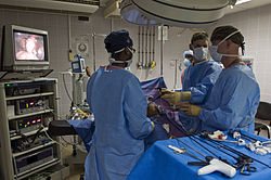
An example of an endoscopic procedure
MeSH D004724
OPS-301 code 1-40...1-49, 1-61...1-69
MedlinePlus 003338

An endoscopy is a procedure used in medicine to look inside the body. [1] The endoscopy procedure uses an endoscope to examine the interior of a hollow organ or cavity of the body. Unlike many other medical imaging techniques, endoscopes are inserted directly into the organ.

Contents

There are many types of endoscopies. Depending on the site in the body and type of procedure, an endoscopy may be performed by either a doctor or a surgeon. A patient may be fully conscious or anaesthetised during the procedure. Most often, the term endoscopy is used to refer to an examination of the upper part of the gastrointestinal tract, known as an esophagogastroduodenoscopy. [2]

For nonmedical use, similar instruments are called borescopes.

History

Adolf Kussmaul was fascinated by sword swallowers who would insert a sword down their throat without gagging. This drew inspiration to insert a hollow tube for observation; the next problem to solve was how to shine light through the tube, as they were still relying on candles and oil lamps as light sources. [3]

The term endoscope was first used on February 7, 1855, by engineer-optician Charles Chevalier, in reference to the uréthroscope of Désormeaux, who himself began using the former term a month later. [4] The self-illuminated endoscope was developed at Glasgow Royal Infirmary in Scotland (one of the first hospitals to have mains electricity) in 1894/5 by John Macintyre as part of his specialization in the investigation of the larynx. [5] [ failed verification ]

Medical uses

Operation part of the endoscope PENTAX Colonoscope002.jpg
Operation part of the endoscope
Insertion tip of an endoscope Insertion tip of endoscope.jpg
Insertion tip of an endoscope

Endoscopy may be used to investigate symptoms in the digestive system including nausea, vomiting, abdominal pain, difficulty swallowing, and gastrointestinal bleeding. [6] It is also used in diagnosis, most commonly by performing a biopsy to check for conditions such as anemia, bleeding, inflammation, and cancers of the digestive system. [6] The procedure may also be used for treatment such as cauterization of a bleeding vessel, widening a narrow esophagus, clipping off a polyp or removing a foreign object. [6]

Specialty professional organizations that specialize in digestive problems advise that many patients with Barrett's esophagus receive endoscopies too frequently. [7] Such societies recommend that patients with Barrett's esophagus and no cancer symptoms after two biopsies receive biopsies as indicated and no more often than the recommended rate. [8] [9]

Applications

An anoscope, a proctoscope, and a rectoscope with approximate lengths Anoscope, proctoscope and rectoscope.svg
An anoscope, a proctoscope, and a rectoscope with approximate lengths
Endoscopy surgery US Navy 081117-N-7526R-568 Cmdr. Thomas Nelson and Lt. Robert Roadfuss discuss proper procedures while performing a laparoscopic cholecystectomy surgery.jpg
Endoscopy surgery
Esophageal Bougie Dilator Esophageal Bougie Dilator.jpg
Esophageal Bougie Dilator

Health care providers can use endoscopy to review any of the following body parts:

Endoscopy is used for many procedures:

An endoscopy is a simple procedure that allows a doctor to look inside human bodies using an instrument called an endoscope. A cutting tool can be attached to the end of the endoscope, and the apparatus can then be used to perform minor procedures such as tissue biopsies, banding of oesophageal varices or removal of polyps.

Application in other fields

Risks

The main risks are infection, over-sedation, perforation, or a tear of the stomach or esophagus lining and bleeding. [10] Although perforation generally requires surgery, certain cases may be treated with antibiotics and intravenous fluids. Bleeding may occur at the site of a biopsy or polyp removal. Such typically minor bleeding may simply stop on its own or be controlled by cauterisation. Seldom does surgery become necessary. Perforation and bleeding are rare during gastroscopy. Other minor risks include drug reactions and complications related to other diseases the patient may have. Consequently, patients should inform their doctor of all allergic tendencies and medical problems. Occasionally, the site of the sedative injection may become inflamed and tender for a short time. This is usually not serious and warm compresses for a few days are usually helpful. While any of these complications may possibly occur, each of them occurs quite infrequently. A doctor can further discuss risks with the patient with regard to the particular need for gastroscopy.

After the endoscopy

After the procedure, the patient will be observed and monitored by a qualified individual in the endoscopy room, or a recovery area, until a significant portion of the medication has worn off. Occasionally the patient is left with a mild sore throat, which may respond to saline gargles, or chamomile tea. It may last for weeks or not happen at all. The patient may have a feeling of distention from the insufflated air that was used during the procedure. Both problems are mild and fleeting. When fully recovered, the patient will be instructed when to resume their usual diet (probably within a few hours) and will be allowed to be taken home. Where sedation has been used, most facilities mandate that the patient be taken home by another person and that they not drive or handle machinery for the remainder of the day. Patients who have had an endoscopy without sedation are able to leave unassisted.

Related Research Articles

<span class="mw-page-title-main">Gastroenterology</span> Branch of medicine focused on the digestive system and its disorders

Gastroenterology is the branch of medicine focused on the digestive system and its disorders. The digestive system consists of the gastrointestinal tract, sometimes referred to as the GI tract, which includes the esophagus, stomach, small intestine and large intestine as well as the accessory organs of digestion which include the pancreas, gallbladder, and liver. The digestive system functions to move material through the GI tract via peristalsis, break down that material via digestion, absorb nutrients for use throughout the body, and remove waste from the body via defecation. Physicians who specialize in the medical specialty of gastroenterology are called gastroenterologists or sometimes GI doctors. Some of the most common conditions managed by gastroenterologists include gastroesophageal reflux disease, gastrointestinal bleeding, irritable bowel syndrome, inflammatory bowel disease (IBD) which includes Crohn's disease and ulcerative colitis, peptic ulcer disease, gallbladder and biliary tract disease, hepatitis, pancreatitis, colitis, colon polyps and cancer, nutritional problems, and many more.

<span class="mw-page-title-main">Colonoscopy</span> Examination of the bowel

Colonoscopy or coloscopy is a medical procedure involving the endoscopic examination of the large bowel (colon) and the distal portion of the small bowel. This examination is performed using either a CCD camera or a fiber optic camera, which is mounted on a flexible tube and passed through the anus.

<span class="mw-page-title-main">Endoscope</span> Instrument to visually examine the interior of a hollow space

An endoscope is an inspection instrument composed of image sensor, optical lens, light source and mechanical device, which is used to look deep into the body by way of openings such as the mouth or anus. A typical endoscope applies several modern technologies including optics, ergonomics, precision mechanics, electronics, and software engineering. With an endoscope, it is possible to observe lesions that cannot be detected by X-ray, making it useful in medical diagnosis. Endoscopes use tubes which are only a few millimeters thick to transfer illumination in one direction and high-resolution images in real time in the other direction, resulting in minimally invasive surgeries. It is used to examine the internal organs like the throat or esophagus. Specialized instruments are named after their target organ. Examples include the cystoscope (bladder), nephroscope (kidney), bronchoscope (bronchus), arthroscope (joints) and colonoscope (colon), and laparoscope. They can be used to examine visually and diagnose, or assist in surgery such as an arthroscopy.

<span class="mw-page-title-main">Esophagogastroduodenoscopy</span> Diagnostic endoscopic procedure

Esophagogastroduodenoscopy (EGD) or oesophagogastroduodenoscopy (OGD), also called by various other names, is a diagnostic endoscopic procedure that visualizes the upper part of the gastrointestinal tract down to the duodenum. It is considered a minimally invasive procedure since it does not require an incision into one of the major body cavities and does not require any significant recovery after the procedure. However, a sore throat is common.

<span class="mw-page-title-main">Endoscopic retrograde cholangiopancreatography</span> Use of endoscopy and fluoroscopy to treat and diagnose digestive issues.

Endoscopic retrograde cholangiopancreatography (ERCP) is a technique that combines the use of endoscopy and fluoroscopy to diagnose and treat certain problems of the biliary or pancreatic ductal systems. It is primarily performed by highly skilled and specialty trained gastroenterologists. Through the endoscope, the physician can see the inside of the stomach and duodenum, and inject a contrast medium into the ducts in the biliary tree and pancreas so they can be seen on radiographs.

<span class="mw-page-title-main">Mallory–Weiss syndrome</span> Bleeding from a laceration in the mucosa at the junction of the stomach and esophagus

Mallory–Weiss syndrome or gastro-esophageal laceration syndrome refers to bleeding from a laceration in the mucosa at the junction of the stomach and esophagus. This is usually caused by severe vomiting because of alcoholism or bulimia, but can be caused by any condition which causes violent vomiting and retching such as food poisoning. The syndrome presents with hematemesis. The laceration is sometimes referred to as a Mallory–Weiss tear.

<span class="mw-page-title-main">Percutaneous endoscopic gastrostomy</span> Feeding tube going into the stomach through the abdominal wall

Percutaneous endoscopic gastrostomy (PEG) is an endoscopic medical procedure in which a tube is passed into a patient's stomach through the abdominal wall, most commonly to provide a means of feeding when oral intake is not adequate. This provides enteral nutrition despite bypassing the mouth; enteral nutrition is generally preferable to parenteral nutrition. The PEG procedure is an alternative to open surgical gastrostomy insertion, and does not require a general anesthetic; mild sedation is typically used. PEG tubes may also be extended into the small intestine by passing a jejunal extension tube through the PEG tube and into the jejunum via the pylorus.

<span class="mw-page-title-main">Endoscopic ultrasound</span> Medical imaging procedure

Endoscopic ultrasound (EUS) or echo-endoscopy is a medical procedure in which endoscopy is combined with ultrasound to obtain images of the internal organs in the chest, abdomen and colon. It can be used to visualize the walls of these organs, or to look at adjacent structures. Combined with Doppler imaging, nearby blood vessels can also be evaluated.

<span class="mw-page-title-main">Endoscopic foreign body retrieval</span>

Endoscopic foreign body retrieval refers to the removal of ingested objects from the esophagus, stomach and duodenum by endoscopic techniques. It does not involve surgery, but rather encompasses a variety of techniques employed through the gastroscope for grasping foreign bodies, manipulating them, and removing them while protecting the esophagus and trachea. It is of particular importance with children, people with mental illness, and prison inmates as these groups have a high rate of foreign body ingestion.

<span class="mw-page-title-main">Double-balloon enteroscopy</span>

Double-balloon enteroscopy, also known as push-and-pull enteroscopy, is an endoscopic technique for visualization of the small bowel. It was developed by Hironori Yamamoto in 2001. It is novel in the field of diagnostic gastroenterology as it is the first endoscopic technique that allows for the entire gastrointestinal tract to be visualized in real time.

<span class="mw-page-title-main">Colonic polypectomy</span> Removal of colorectal polyps to prevent them turning cancerous

Colonic polypectomy is the removal of colorectal polyps in order to prevent them from turning cancerous.

<span class="mw-page-title-main">Endoclip</span>

An endoclip is a metallic mechanical device used in endoscopy in order to close two mucosal surfaces without the need for surgery and suturing. Its function is similar to a suture in gross surgical applications, as it is used to join together two disjointed surfaces, but, can be applied through the channel of an endoscope under direct visualization. Endoclips have found use in treating gastrointestinal bleeding, in preventing bleeding after therapeutic procedures such as polypectomy, and in closing gastrointestinal perforations. Many forms of endoclips exist of different shapes and sizes, including two and three prong devices, which can be administered using single use and reloadable systems, and may or may not open and close to facilitate placement.

<span class="mw-page-title-main">Capsule endoscopy</span> Medical imaging procedure

Capsule endoscopy is a medical procedure used to record internal images of the gastrointestinal tract for use in disease diagnosis. Newer developments are also able to take biopsies and release medication at specific locations of the entire gastrointestinal tract. Unlike the more widely used endoscope, capsule endoscopy provides the ability to see the middle portion of the small intestine. It can be applied to the detection of various gastrointestinal cancers, digestive diseases, ulcers, unexplained bleedings, and general abdominal pains. After a patient swallows the capsule, it passes along the gastrointestinal tract, taking a number of images per second which are transmitted wirelessly to an array of receivers connected to a portable recording device carried by the patient. General advantages of capsule endoscopy over standard endoscopy include the minimally invasive procedure setup, ability to visualize more of the gastrointestinal tract, and lower cost of the procedure.

<span class="mw-page-title-main">Esophageal food bolus obstruction</span> Medical condition

An esophageal food bolus obstruction is a medical emergency caused by the obstruction of the esophagus by an ingested foreign body.

<span class="mw-page-title-main">Esophageal stent</span>

An esophageal stent is a stent (tube) placed in the esophagus to keep a blocked area open so the patient can swallow soft food and liquids. They are effective in the treatment of conditions causing intrinsic esophageal obstruction or external esophageal compression. For the palliative treatment of esophageal cancer most esophageal stents are self-expandable metallic stents. For benign esophageal disease such as refractory esophageal strictures, plastic stents are available. Common complications include chest pain, overgrowth of tissue around the stent and stent migration. Esophageal stents may also be used to staunch the bleeding of esophageal varices.

Therapeutic endoscopy is the medical term for an endoscopic procedure during which treatment is carried out via the endoscope. This contrasts with diagnostic endoscopy, where the aim of the procedure is purely to visualize a part of the gastrointestinal, respiratory or urinary tract in order to aid diagnosis. In practice, a procedure which starts as a diagnostic endoscopy may become a therapeutic endoscopy depending on the findings, such as in cases of upper gastrointestinal bleeding, or the finding of polyps during colonoscopy.

<span class="mw-page-title-main">Endoscopy unit</span>

An endoscopy unit refers to a dedicated area where medical procedures are performed with endoscopes, which are cameras used to visualize structures within the body, such as the digestive tract and genitourinary system. Endoscopy units may be located within a hospital, incorporated within other medical care centres, or may be stand-alone in nature.

<span class="mw-page-title-main">Chromoendoscopy</span>

Chromoendoscopy is a medical procedure wherein dyes are instilled into the gastrointestinal tract at the time of visualization with fibre-optic endoscopy. The purposes of chromoendoscopy is chiefly enhance the characterization of tissues, although dyes may be used for other functional purposes. The detail achieved with chromoendoscopy can often allow for identification of the tissue type or pathology based upon the pattern uncovered.

Narrow-band imaging is an imaging technique for endoscopic diagnostic medical tests, where light of specific blue and green wavelengths is used to enhance the detail of certain aspects of the surface of the mucosa. A special filter is electronically activated by a switch in the endoscope leading to the use of ambient light of wavelengths of 415 nm (blue) and 540 nm (green). Because the peak light absorption of hemoglobin occurs at these wavelengths, blood vessels will appear very dark, allowing for their improved visibility and in the improved identification of other surface structures.

Endoscopic mucosal resection is a technique used to remove cancerous or other abnormal lesions found in the digestive tract. It is one method of performing a mucosectomy.

References

  1. "Endoscopy". British Medical Association Complete Family Health Encyclopedia. Dorling Kindersley Limited. 1990. ISBN   978-0-751-30161-8.
  2. "Endoscopy". Cancer Research UK. Archived from the original on 1 February 2017. Retrieved 5 November 2015.
  3. "The pioneers of endoscopy and the sword swallowers". Archived from the original on 2022-01-27. Retrieved 2022-01-26.
  4. Janssen, Diederik F (2021-05-17). "Who named and built the Désormeaux endoscope? The case of unacknowledged opticians Charles and Arthur Chevalier". Journal of Medical Biography. 29 (3): 176–179. doi:10.1177/09677720211018975. ISSN   0967-7720. PMID   33998906. S2CID   234747817. Archived from the original on 2023-02-04. Retrieved 2021-05-17.
  5. "The Scottish Society of the History of Medicine" (PDF). Archived (PDF) from the original on 2016-12-20. Retrieved 2017-07-11.
  6. 1 2 3 Staff (2012). "Upper endoscopy". Mayo Clinic. Archived from the original on 25 May 2013. Retrieved 24 September 2012.
  7. American Gastroenterological Association, "Five Things Physicians and Patients Should Question" (PDF), Choosing Wisely: an initiative of the ABIM Foundation , American Gastroenterological Association, archived from the original (PDF) on August 9, 2012, retrieved August 17, 2012
  8. Spechler SJ, Sharma P, Souza RF, Inadomi JM, Shaheen NJ (March 2011). "American Gastroenterological Association medical position statement on the management of Barrett's esophagus". Gastroenterology. 140 (3): 1084–91. doi: 10.1053/j.gastro.2011.01.030 . PMID   21376940.
  9. Wang KK, Sampliner RE (March 2008). "Updated guidelines 2008 for the diagnosis, surveillance and therapy of Barrett's esophagus". The American Journal of Gastroenterology. 103 (3): 788–97. doi: 10.1111/j.1572-0241.2008.01835.x . PMID   18341497. S2CID   8443847.
  10. "Endoscopy". NHS Choices. NHS Gov.UK. Archived from the original on May 2, 2017. Retrieved April 20, 2017.