Erythroferrone

Last updated
ERFE
Identifiers
Aliases ERFE , C1QTNF15, CTRP15, FAM132B, Erythroferrone, family with sequence similarity 132 member B
External IDs OMIM: 615099 MGI: 3606476 HomoloGene: 87245 GeneCards: ERFE
Orthologs
SpeciesHumanMouse
Entrez
Ensembl
UniProt
RefSeq (mRNA)

NM_152521
NM_001291832

NM_173395

RefSeq (protein)

NP_001278761

NP_775571

Location (UCSC) Chr 2: 238.16 – 238.17 Mb Chr 1: 91.29 – 91.3 Mb
PubMed search [3] [4]
Wikidata
View/Edit Human View/Edit Mouse
Erythroferrone
Identifiers
SymbolERFE
NCBI gene 151176
HGNC 26727
OMIM 615099
RefSeq NM_001291832.1
UniProt Q4G0M1
Other data
Locus Chr. 2 q37.3
Search for
Structures Swiss-model
Domains InterPro

Erythroferrone is a protein hormone encoded in humans by the ERFE gene. Erythroferrone is produced by erythroblasts, inhibits the production of hepcidin in the liver, and so increases the amount of iron available for hemoglobin synthesis. [5] [6] Skeletal muscle secreted ERFE has been shown to maintain systemic metabolic homeostasis. [7]

Contents

Discovery

It was identified in 2014 in mice where the transcript was found in bone marrow, encoded by the mouse Fam132b gene. [6] The homologous gene in humans is FAM132B and the sequence is conserved in other species. The protein is synthesized by erythroblasts and secreted. [6] This sequence had previously been found expressed in mouse skeletal muscle, called myonectin (CTRP15), and linked to lipid homeostasis. [8]

Seldin and his colleagues have written: "Myonectin is expressed and secreted predominantly by skeletal muscle.... (Our) results suggest that myonectin is a nutrient-responsive metabolic regulator secreted by skeletal muscle in response to changes in cellular energy state resulting from glucose or fatty acid fluxes. Many metabolically relevant secreted proteins (e.g. adiponectin, leptin, resistin, and RBP) and the signaling pathways they regulate in tissues are known to be dysregulated in the condition of obesity. The reduction in expression and circulating levels of myonectin in the obese state may represent yet another component of the complex metabolic circuitry dysregulated by excess caloric intake. Although exercise has long been known to have profound positive impacts on systemic insulin sensitivity and energy balance, the underlying mechanisms remain incompletely understood. That voluntary exercise dramatically increases the expression and circulating levels of myonectin to promote fatty acid uptake into cells may underlie one of the beneficial effects of physical exercise." [9]

Myonectin was shown in 2015 to be identical to erythroferrone, a hormone produced in erythroblasts that is involved in iron metabolism. [5] > [6]

Structure

Erythroferrone in humans is transcribed as a precursor of 354 amino acids, with a signal peptide of 28 amino acids. The mouse gene encodes a 340 amino acid protein which is 71% identical. [6] Homology is greater at the C-terminal where there is a TNF-alpha-like domain. [10] As a member of the C1q/TNF-Related Protein (CTRP) family, erythroferrone has a 4-domain structure with a unique N-terminus. The two larger domains are connected by a short, proline-rich, collagenous linker that is thought to promote protein multimerization. Erythroferrone is predicted to contain two PCSK3/furin recognition sites. The protein hormone weighs approximately 35-40 kDa. [11]

Function

Erythroferrone is a hormone that regulates iron metabolism through its actions on hepcidin. [5] As shown in mice and humans, it is produced in erythroblasts, which proliferate when new red cells are synthesized, such as after hemorrhage when more iron is needed (so-called stress erythropoiesis). [12] This process is governed by the renal hormone, erythropoietin. [6]

Its mechanism of action is to inhibit the expression of the liver hormone, hepcidin. [12] This process is governed by the renal hormone, erythropoietin. [6] By suppressing hepcidin, ERFE increases the function of the cellular iron export channel, ferroportin. This then results in increased iron absorption from the intestine and mobilization of iron from stores, which can then be used in the synthesis of hemoglobin in new red blood cells. [6] Erythroferrone inhibits hepcidin synthesis by binding bone morphogenetic proteins and thereby inhibiting the bone morphogenetic protein pathway that controls hepcidin expression. [13] [14]

Mice deficient in the gene encoding erythroferrone have transient maturational hemoglobin deficits and impaired hepcidin suppression in response to phlebotomy with a delayed recovery from anemia. [6]

In its role as myonectin, it also promotes lipid uptake into adipocytes and hepatocytes. [8]

Regulation

Synthesis of erythroferrone is stimulated by erythropoietin binding to its receptor and activating the Jak2/Stat5 signaling pathway. [6]

Clinical significance

The clinical significance in humans is becoming clear. [15] From parallels in the mouse studies, there may be diseases where its function could be relevant. In a mouse model of thalassemia, its expression is increased, resulting in iron overload, which is also a feature of the human disease. [16] A role in the recovery from the anemia of inflammation in mice has been shown [17] and involvement in inherited anemias with ineffective erythropoiesis, anemia of chronic kidney diseases and iron-refractory iron-deficiency anemia has been suggested. [6] [15]

Erythroferrone levels in blood have been shown by immunoassay to be higher after blood loss or erythropoetin administration. Patients with beta-thalassemia have very high levels, and these decrease after blood transfusion. [18]

Related Research Articles

<span class="mw-page-title-main">Erythropoietin</span> Protein that stimulates red blood cell production

Erythropoietin, also known as erythropoetin, haematopoietin, or haemopoietin, is a glycoprotein cytokine secreted mainly by the kidneys in response to cellular hypoxia; it stimulates red blood cell production (erythropoiesis) in the bone marrow. Low levels of EPO are constantly secreted in sufficient quantities to compensate for normal red blood cell turnover. Common causes of cellular hypoxia resulting in elevated levels of EPO include any anemia, and hypoxemia due to chronic lung disease and mouth disease.

<span class="mw-page-title-main">Erythropoiesis</span> Process which produces red blood cells

Erythropoiesis is the process which produces red blood cells (erythrocytes), which is the development from erythropoietic stem cell to mature red blood cell.

Anemia of chronic disease (ACD) or anemia of chronic inflammation is a form of anemia seen in chronic infection, chronic immune activation, and malignancy. These conditions all produce elevation of interleukin-6, which stimulates hepcidin production and release from the liver. Hepcidin production and release shuts down ferroportin, a protein that controls export of iron from the gut and from iron storing cells. As a consequence, circulating iron levels are reduced. Other mechanisms may also play a role, such as reduced erythropoiesis. It is also known as anemia of inflammation, or anemia of inflammatory response.

<span class="mw-page-title-main">Human iron metabolism</span> Iron metabolism in the body

Human iron metabolism is the set of chemical reactions that maintain human homeostasis of iron at the systemic and cellular level. Iron is both necessary to the body and potentially toxic. Controlling iron levels in the body is a critically important part of many aspects of human health and disease. Hematologists have been especially interested in systemic iron metabolism, because iron is essential for red blood cells, where most of the human body's iron is contained. Understanding iron metabolism is also important for understanding diseases of iron overload, such as hereditary hemochromatosis, and iron deficiency, such as iron-deficiency anemia.

<span class="mw-page-title-main">Hepcidin</span> Protein-coding gene in the species Homo sapiens

Hepcidin is a protein that in humans is encoded by the HAMP gene. Hepcidin is a key regulator of the entry of iron into the circulation in mammals.

<span class="mw-page-title-main">Ferroportin</span> Protein

Ferroportin-1, also known as solute carrier family 40 member 1 (SLC40A1) or iron-regulated transporter 1 (IREG1), is a protein that in humans is encoded by the SLC40A1 gene. Ferroportin is a transmembrane protein that transports iron from the inside of a cell to the outside of the cell. Ferroportin is the only known iron exporter.

<span class="mw-page-title-main">GATA1</span> Protein-coding gene in humans

GATA-binding factor 1 or GATA-1 is the founding member of the GATA family of transcription factors. This protein is widely expressed throughout vertebrate species. In humans and mice, it is encoded by the GATA1 and Gata1 genes, respectively. These genes are located on the X chromosome in both species.

<span class="mw-page-title-main">Beta thalassemia</span> Thalassemia characterized by the reduced or absent synthesis of the beta globin chains of hemoglobin

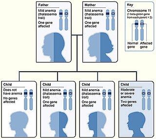
Beta thalassemias are a group of inherited blood disorders. They are forms of thalassemia caused by reduced or absent synthesis of the beta chains of hemoglobin that result in variable outcomes ranging from severe anemia to clinically asymptomatic individuals. Global annual incidence is estimated at one in 100,000. Beta thalassemias occur due to malfunctions in the hemoglobin subunit beta or HBB. The severity of the disease depends on the nature of the mutation.

<span class="mw-page-title-main">Hephaestin</span> Mammalian protein found in Homo sapiens

Hephaestin, also known as HEPH, is a protein which in humans is encoded by the HEPH gene.

<span class="mw-page-title-main">Hemojuvelin</span>

Hemojuvelin (HJV), also known as repulsive guidance molecule C (RGMc) or hemochromatosis type 2 protein (HFE2), is a membrane-bound and soluble protein in mammals that is responsible for the iron overload condition known as juvenile hemochromatosis in humans, a severe form of hemochromatosis. In humans, the hemojuvelin protein is encoded by the HFE2 gene. Hemojuvelin is a member of the repulsive guidance molecule family of proteins. Both RGMa and RGMb are found in the nervous system, while hemojuvelin is found in skeletal muscle and the liver.

<span class="mw-page-title-main">Iron in biology</span> Use of Iron by organisms

Iron is an important biological element. It is used in both the ubiquitous iron-sulfur proteins and in vertebrates it is used in hemoglobin which is essential for blood and oxygen transport.

<span class="mw-page-title-main">KLF2</span> Protein-coding gene in the species Homo sapiens

Krüppel-like Factor 2 (KLF2), also known as lung Krüppel-like Factor (LKLF), is a protein that in humans is encoded by the KLF2 gene on chromosome 19. It is in the Krüppel-like factor family of zinc finger transcription factors, and it has been implicated in a variety of biochemical processes in the human body, including lung development, embryonic erythropoiesis, epithelial integrity, T-cell viability, and adipogenesis.

Congenital hemolytic anemia (CHA) is a diverse group of rare hereditary conditions marked by decreased life expectancy and premature removal of erythrocytes from blood flow. Defects in erythrocyte membrane proteins and red cell enzyme metabolism, as well as changes at the level of erythrocyte precursors, lead to impaired bone marrow erythropoiesis. CAH is distinguished by variable anemia, chronic extravascular hemolysis, decreased erythrocyte life span, splenomegaly, jaundice, biliary lithiasis, and iron overload. Immune-mediated mechanisms may play a role in the pathogenesis of these uncommon diseases, despite the paucity of data regarding the immune system's involvement in CHAs.

<span class="mw-page-title-main">GLRX5</span> Protein-coding gene in the species Homo sapiens

Glutaredoxin 5, also known as GLRX5, is a protein which in humans is encoded by the GLRX5 gene located on chromosome 14. This gene encodes a mitochondrial protein, which is evolutionarily conserved. It is involved in the biogenesis of iron- sulfur clusters, which are required for normal iron homeostasis. Mutations in this gene are associated with autosomal recessive pyridoxine-refractory sideroblastic anemia.

Ineffective erythropoiesis is defined by the expansion of early-stage erythroid precursors driven by erythropoietin, accompanied by the apoptosis of late-stage precursors. This mechanism is principally responsible for the anemia seen in acquired conditions such as certain subtypes of myelodysplastic syndrome (MDS) and inherited disorders such as β-thalassemia, inherited sideroblastic anemias, as well as congenital dyserythropoietic anemias.

A myokine is one of several hundred cytokines or other small proteins and proteoglycan peptides that are produced and released by skeletal muscle cells in response to muscular contractions. They have autocrine, paracrine and/or endocrine effects; their systemic effects occur at picomolar concentrations.

Elizabeta Nemeth is an American physiologist who has made many contributions to the understanding of inflammatory disorders, thalassemias, and iron overload diseases.

Hemochromatosis type 4 is a hereditary iron overload disorder that affects ferroportin, an iron transport protein needed to export iron from cells into circulation. Although the disease is rare, it is found throughout the world and affects people from various ethnic groups. While the majority of individuals with type 4 hemochromatosis have a relatively mild form of the disease, some affected individuals have a more severe form. As the disease progresses, iron may accumulate in the tissues of affected individuals over time, potentially resulting in organ damage.

<span class="mw-page-title-main">Adropin</span> Peptide hormone

Adropin is a peptide encoded by the energy homeostasis-associated gene ENHO, which is highly conserved across mammals.

Hepatokines are proteins produced by liver cells (hepatocytes) that are secreted into the circulation and function as hormones across the organism. Research is mostly focused on hepatokines that play a role in the regulation of metabolic diseases such as diabetes and fatty liver and include: Adropin, ANGPTL4, Fetuin-A, Fetuin-B, FGF-21, Hepassocin, LECT2, RBP4,Selenoprotein P, Sex hormone-binding globulin.

References

  1. 1 2 3 GRCh38: Ensembl release 89: ENSG00000178752 - Ensembl, May 2017
  2. 1 2 3 GRCm38: Ensembl release 89: ENSMUSG00000047443 - Ensembl, May 2017
  3. "Human PubMed Reference:". National Center for Biotechnology Information, U.S. National Library of Medicine.
  4. "Mouse PubMed Reference:". National Center for Biotechnology Information, U.S. National Library of Medicine.
  5. 1 2 3 Koury MJ. "Erythroferrone: A Missing Link in Iron Regulation". The Hematologist. American Society of Hematology. Archived from the original on 28 January 2019. Retrieved 26 August 2015.
  6. 1 2 3 4 5 6 7 8 9 10 11 Kautz L, Jung G, Valore EV, Rivella S, Nemeth E, Ganz T (July 2014). "Identification of erythroferrone as an erythroid regulator of iron metabolism". Nature Genetics. 46 (7): 678–84. doi:10.1038/ng.2996. PMC   4104984 . PMID   24880340.
  7. Wang H (2017). "Skeletal muscle secreted myonectin maintains systemic metabolic homeostasis". The FASEB Journal. 31 (S1): 1036.17. doi: 10.1096/fasebj.31.1_supplement.1036.17 . ISSN   1530-6860. S2CID   232543402.
  8. 1 2 Seldin MM, Peterson JM, Byerly MS, Wei Z, Wong GW (April 2012). "Myonectin (CTRP15), a novel myokine that links skeletal muscle to systemic lipid homeostasis". The Journal of Biological Chemistry. 287 (15): 11968–80. doi: 10.1074/jbc.M111.336834 . PMC   3320944 . PMID   22351773.
  9. Marcus M. Seldin, Jonathan M. Peterson, Mardi S. Byerly, Zhikui Wei, and G. William Wong. "Myonectin (CTRP15), a Novel Myokine That Links Skeletal Muscle to Systemic Lipid Homeostasis." THE JOURNAL OF BIOLOGICAL CHEMISTRY VOL. 287, NO. 15, pp. 11968–11980, April 6, 2012, doi: 10.1074/jbc.M111.336834 originally published online February 17, 2012.
  10. Srole DN, Jung G, Waring AJ, Nemeth E, Ganz T (December 2023). "Characterization of erythroferrone structural domains relevant to its iron-regulatory function". Journal of Biological Chemistry. 299 (12): 105374. doi: 10.1016/j.jbc.2023.105374 . PMC   10692919 . PMID   37866631.
  11. Srole DN, Ganz T (July 2021). "Erythroferrone structure, function, and physiology: Iron homeostasis and beyond". Journal of Cellular Physiology. 236 (7): 4888–4901. doi:10.1002/jcp.30247. PMC   8026552 . PMID   33372284.
  12. 1 2 Kim A, Nemeth E (May 2015). "New insights into iron regulation and erythropoiesis". Current Opinion in Hematology. 22 (3): 199–205. doi:10.1097/MOH.0000000000000132. PMC   4509743 . PMID   25710710.
  13. Arezes J, Foy N, McHugh K, Sawant A, Quinkert D, Terraube V, et al. (October 2018). "Erythroferrone inhibits the induction of hepcidin by BMP6". Blood. 132 (14): 1473–1477. doi:10.1182/blood-2018-06-857995. PMC   6238155 . PMID   30097509.
  14. Arezes J, Foy N, McHugh K, Quinkert D, Benard S, Sawant A, et al. (February 2020). "Antibodies against the erythroferrone N-terminal domain prevent hepcidin suppression and ameliorate murine thalassemia". Blood. 135 (8): 547–557. doi: 10.1182/blood.2019003140 . PMC   7046598 . PMID   31899794.
  15. 1 2 Pasricha SR, McHugh K, Drakesmith H (July 2016). "Regulation of Hepcidin by Erythropoiesis: The Story So Far". Annual Review of Nutrition. 36: 417–34. doi:10.1146/annurev-nutr-071715-050731. PMID   27146013.
  16. Kautz L, Jung G, Du X, Gabayan V, Chapman J, Nasoff M, et al. (October 2015). "Erythroferrone contributes to hepcidin suppression and iron overload in a mouse model of β-thalassemia". Blood. 126 (17): 2031–7. doi:10.1182/blood-2015-07-658419. PMC   4616236 . PMID   26276665.
  17. Kautz L, Jung G, Nemeth E, Ganz T (October 2014). "Erythroferrone contributes to recovery from anemia of inflammation". Blood. 124 (16): 2569–74. doi:10.1182/blood-2014-06-584607. PMC   199959 . PMID   25193872.
  18. Ganz T, Jung G, Naeim A, Ginzburg Y, Pakbaz Z, Walter PB, et al. (September 2017). "Immunoassay for human serum erythroferrone". Blood. 130 (10): 1243–1246. doi:10.1182/blood-2017-04-777987. PMC   5606005 . PMID   28739636.