Face and neck development of the human embryo

Last updated

The face and neck development of the human embryo refers to the development of the structures from the third to eighth week that give rise to the future head and neck. They consist of three layers, the ectoderm, mesoderm and endoderm, which form the mesenchyme (derived form the lateral plate mesoderm and paraxial mesoderm), neural crest and neural placodes (from the ectoderm). [1] The paraxial mesoderm forms structures named somites and somitomeres that contribute to the development of the floor of the brain and voluntary muscles of the craniofacial region. [2] The lateral plate mesoderm consists of the laryngeal cartilages (arytenoid and cricoid). The three tissue layers give rise to the pharyngeal apparatus, [3] formed by six pairs of pharyngeal arches, a set of pharyngeal pouches and pharyngeal grooves, which are the most typical feature in development of the head and neck. The formation of each region of the face and neck is due to the migration of the neural crest cells which come from the ectoderm. These cells determine the future structure to develop in each pharyngeal arch. Eventually, they also form the neurectoderm, which forms the forebrain, midbrain and hindbrain, cartilage, bone, dentin, tendon, dermis, pia mater and arachnoid mater, sensory neurons, and glandular stroma.

Contents

Pharyngeal arches

Pharyngeal arches are formed during the fourth week. Each arch consists of a mesenchymal tissue covered on the outside by ectoderm and on the inside by epithelium of endodermal origin. In human embryology, there are six arches which are separated by pharyngeal grooves externally and pharyngeal pouches internally. These arches contribute to the physical appearance of the embryo because they are the main components that build the face and neck. In addition, the muscular components of each arch have their own cranial nerve, and wherever the muscle cells migrate, they carry their nerve component with them. Plus, each arch has its own arterial component. When neural cells migrate to the arches and surround them, they begin to increase in size. [4] The six pharyngeal arches give rise to much of the skeletal and muscular tissue in the head and neck region. When the embryo is 42 days old, the mesenchymal arches can be recognized with its corresponding cranial nerve. [1]

The first pharyngeal arch forms maxillary and mandibular processes. It is innervated by the trigeminal nerve and molds muscles related to mastication such as temporal, masseter, medial, lateral, pterygoid bones, tensor palati, and tensor tympani. This arch originates maxillar and mandibular prominences, part of the temporal bone and Meckel's cartilage (malleus and incus) as skeletal structures. The second pharyngeal arch is innervated by the facial cranial nerve. Muscles that arise from the arch are those involved with facial expression and the posterior digastric muscle. Skeletal structures that originate here are the cervical sinus, Reichert cartilage (stape) the styloid process of the temporal bone, the lesser cornu and the hyoid bone. [4] The third pharyngeal arch is innervated by glossopharyngeal nerve. It molds the stylopharyngeus muscle and forms the skeletal structures of the greater horn and lower portion of body hyoid bone. [5] The fourth and sixth arches are innervated by the vagus cranial nerve. Both arches will fuse to form the laryngeal cartilages. The fifth cartilage does not appear to have any contribution to adult anatomy and disappears. [2] [6]

Pharyngeal pouches

Pharyngeal pouches develop into future parts in face and head. The pouches penetrate the surrounding mesenchyme but do not establish communication with the pharyngeal grooves. They appear simultaneously with the development of the arches. [7] The first pharyngeal pouch is characterized by narrowing at its final segment. However, it does not disappear and eventually forms the eustachian tube. Second pharyngeal pouch develops differently from the first one mainly because most of it disappears, leaving the tonsillar fossa (Rohen). At the end of the fifth month, the palatine tonsil as a protective structure is completed. This structure covers the tonsillar fossa. The third pharyngeal pouch will give rise to the inferior parathyroid gland and thymus. [2] The fourth and fifth pouches develop as a unique structure that molds the superior parathyroid and parafolicular cells of thyroid gland. [1]

Pharyngeal grooves

Initially, pharyngeal grooves consist of four bars of mesenchymal tissue that separate pharyngeal nerves. Most of these structures obliterate, only the ear canal remains. [2]

Development of the tongue

In the fourth week of the pregnancy the structures that develop the tongue appear. These structures develop from the first pharyngeal arch are two lingual lateral prominences and one in the middle that does not develop and disappears. A second prominence, the hypobranchial eminence, comes from the second, third and fourth pharyngeal arches. A third prominence that comes from the fourth arch develops the epiglottis. The laryngeal orifice is behind the third prominence, which is surrounded by the arytenoid prominences. [1] Later, the lateral and middle prominences join forming the first of the three parts of the tongue. The terminal sulcus is a V-shaped line that separates the body of the tongue from the posterior part. [1] The corresponding nerve for the three prominences of the anterior tongue is the trigeminal nerve. The posterior tongue is innervated by the glossopharyngeal nerve. The muscles of the tongue are innervated by the hypoglossal nerve. [1]

Development of the thyroid

The thyroid appears as an epithelial proliferation in the pharynx floor between the copula linguae and the tuberculum impar. This point later will be the foramen cecum. [1] Later, the thyroid descends in front of the pharyngeal gut when it already has a belobed diverticulum shape. The thyroglossal duct keeps the thyroid joined to the tongue until it disappears. [1] The thyroid keeps descending in front of the hyoid bone until finally it affixes to the front of the trachea in the seventh week. The thyroid starts working in the third month when the first follicles are visible and start producing colloid. The parafollicular cells come from the ultimobranchial body and produce calcitonin. [8]

Development of the face

The facial prominences are five swellings that appear in the fourth week and come from the first and second pharyngeal arch. They are basically made of mesenchyme that comes from the neural crest. [4] [9]

The frontonasal prominence is a single structure that is ventral to the forebrain. It is derived from neural crest cells, which have an ectodermal origin. These neural crest cells migrate from the ectoderm as the forebrain closes, invading the space that will form the frontonasal prominence. The maxillary and mandibular prominences are derived from the first arch. The maxillary prominence is initially located superior/lateral to the stomodeum while the mandibular prominence is located inferior to it and will fuse early on.

Nasal placodes originate on the frontonasal prominence from the ectoderm. They thicken and sink in to form Nasal Pits, which deepen to form the Nasal Sacs. At the same time, mesodermal cells proliferate around the placodes, and the sides of these swellings form the medial and lateral nasal prominences. The lateral nasal prominence is separated from the maxillary prominence by the nasolacrimal groove. [4] [9]

As the maxillary prominences continue growing they merge laterally with the mandibular prominences to form the cheeks. Their growth compresses the medial nasal prominences and causes them to fuse around the 10th week of development. This establishes the bridge of the nose and the intermaxillary segment, which is the region of the medial nasal prominence located inferior to the bridge of the nose and superior to the mandibular prominence. The intermaxillary segment yields the portion of the upper lip containing the philtrum, the upper jaw with 4 incisors, and the primary palate. The medial prominence fuses with the maxillary prominence, giving rise to a smooth upper lip while fusing the primary and secondary palate. Meanwhile, the lateral nasal prominence gives rise to the alae of the nose and fuses with the maxillary prominence, forming the Nasolacrimal duct. This duct is formed when the ectoderm thickens into a cord and sinks into the underlying mesenchyme. [4] [9]

Development of the nasal cavity

The formation of the lateral and medial nasal prominences makes the nasal placodes lie in the floor of the depression, called nasal pits. The nasal pits deepen and develop the nasal sacs in the sixth week. These new structures grow dorsocaudally in front of the forming brain. In the beginning, the nasal sacs are separated from the oral cavity by the oronasal membrane. This membrane disappears in the seventh week leaving a connection between the nasal cavities and the oral cavity, called the primitive choanae. Later, when the development of the secondary palate occurs, the choanae changes its position and locates at the junction of the nasal cavity and the pharynx. The nasal septum grows as a downgrowth from the merged nasal prominences and fuses with the palatine process between the ninth and eleventh week. Finally, the superior, middle and inferior conchae develop the lateral wall of each nasal cavity. [4] [9]

See also

Related Research Articles

<span class="mw-page-title-main">Mesoderm</span> Middle germ layer of embryonic development

The mesoderm is the middle layer of the three germ layers that develops during gastrulation in the very early development of the embryo of most animals. The outer layer is the ectoderm, and the inner layer is the endoderm.

<span class="mw-page-title-main">Temporomandibular joint</span> Joints connecting the jawbone to the skull

In anatomy, the temporomandibular joints (TMJ) are the two joints connecting the jawbone to the skull. It is a bilateral synovial articulation between the temporal bone of the skull above and the mandible below; it is from these bones that its name is derived. This joint is unique in that it is a bilateral joint that functions as one unit. Since the TMJ is connected to the mandible, the right and left joints must function together and therefore are not independent of each other.

<span class="mw-page-title-main">Facial nerve</span> Cranial nerve VII, for the face and tasting

The facial nerve, also known as the seventh cranial nerve, cranial nerve VII, or simply CN VII, is a cranial nerve that emerges from the pons of the brainstem, controls the muscles of facial expression, and functions in the conveyance of taste sensations from the anterior two-thirds of the tongue. The nerve typically travels from the pons through the facial canal in the temporal bone and exits the skull at the stylomastoid foramen. It arises from the brainstem from an area posterior to the cranial nerve VI and anterior to cranial nerve VIII.

Articles related to anatomy include:

<span class="mw-page-title-main">External carotid artery</span> Major artery of the head and neck

The external carotid artery is a major artery of the head and neck. It arises from the common carotid artery when it splits into the external and internal carotid artery. The external carotid artery supplies blood to the face, brain and neck.

<span class="mw-page-title-main">Muscles of mastication</span> Muscles that aid chewing

The four classical muscles of mastication elevate the mandible and move it forward/backward and laterally, facilitating biting and chewing. Other muscles are responsible for opening the jaw, namely the geniohyoid, mylohyoid, and digastric muscles.

<span class="mw-page-title-main">Somite</span> Each of several blocks of mesoderm that flank the neural tube on either side in embryogenesis

The somites are a set of bilaterally paired blocks of paraxial mesoderm that form in the embryonic stage of somitogenesis, along the head-to-tail axis in segmented animals. In vertebrates, somites subdivide into the dermatomes, myotomes, sclerotomes and syndetomes that give rise to the vertebrae of the vertebral column, rib cage, part of the occipital bone, skeletal muscle, cartilage, tendons, and skin.

<span class="mw-page-title-main">Stylopharyngeus muscle</span> Muscle

The stylopharyngeus muscle is a muscle in the head. It originates from the temporal styloid process. Some of its fibres insert onto the thyroid cartilage, while others end by intermingling with proximal structures. It is innervated by the glossopharyngeal nerve. It acts to elevate the larynx and pharynx, and dilate the pharynx, thus facilitating swallowing.

<span class="mw-page-title-main">Pharyngeal arch</span> Embryonic precursor structures in vertebrates

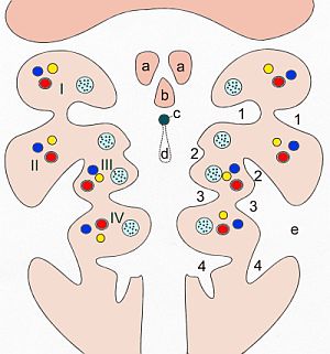
The pharyngeal arches, also known as visceral arches, are structures seen in the embryonic development of vertebrates that are recognisable precursors for many structures. In fish, the arches are known as the branchial arches, or gill arches.

<span class="mw-page-title-main">Pharyngeal pouch (embryology)</span>

In the embryonic development of vertebrates, pharyngeal pouches form on the endodermal side between the pharyngeal arches. The pharyngeal grooves form the lateral ectodermal surface of the neck region to separate the arches.

In embryology, Carnegie stages are a standardized system of 23 stages used to provide a unified developmental chronology of the vertebrate embryo.

<span class="mw-page-title-main">Maxillary prominence</span>

Continuous with the dorsal end of the first pharyngeal arch, and growing forward from its cephalic border, is a triangular process, the maxillary prominence, the ventral extremity of which is separated from the mandibular arch by a ">"-shaped notch.

<span class="mw-page-title-main">Frontonasal process</span>

The frontonasal process, or frontonasal prominence is one of the five swellings that develop to form the face. The frontonasal process is unpaired, and the others are the paired maxillary prominences, and the paired mandibular prominences. During the fourth week of embryonic development, an area of thickened ectoderm develops, on each side of the frontonasal process called the nasal placodes or olfactory placodes, and appear immediately under the forebrain.

<span class="mw-page-title-main">Human nose</span> Feature of the human face

The human nose is the first organ of the respiratory system. It is also the principal organ in the olfactory system. The shape of the nose is determined by the nasal bones and the nasal cartilages, including the nasal septum which separates the nostrils and divides the nasal cavity into two.

<span class="mw-page-title-main">Human embryonic development</span> Development and formation of the human embryo

Human embryonic development or human embryogenesis is the development and formation of the human embryo. It is characterised by the processes of cell division and cellular differentiation of the embryo that occurs during the early stages of development. In biological terms, the development of the human body entails growth from a one-celled zygote to an adult human being. Fertilization occurs when the sperm cell successfully enters and fuses with an egg cell (ovum). The genetic material of the sperm and egg then combine to form the single cell zygote and the germinal stage of development commences. Embryonic development in the human, covers the first eight weeks of development; at the beginning of the ninth week the embryo is termed a fetus. The eight weeks has 23 stages.

<span class="mw-page-title-main">Outline of human anatomy</span> Overview of and topical guide to human anatomy

The following outline is provided as an overview of and topical guide to human anatomy:

<span class="mw-page-title-main">Pharynx</span> Part of the throat that is behind the mouth and nasal cavity

The pharynx is the part of the throat behind the mouth and nasal cavity, and above the esophagus and trachea. It is found in vertebrates and invertebrates, though its structure varies across species. The pharynx carries food to the esophagus and air to the larynx. The flap of cartilage called the epiglottis stops food from entering the larynx.

The cranial neural crest is one of the four regions of the neural crest.

<span class="mw-page-title-main">Trabecular cartilage</span> Embryonic cartilage type

Trabecular cartilages are paired, rod-shaped cartilages, which develop in the head of the vertebrate embryo. They are the primordia of the anterior part of the cranial base, and are derived from the cranial neural crest cells.

<span class="mw-page-title-main">Outline of the human nervous system</span> Overview of and topical guide to the human nervous system

The following diagram is provided as an overview of and topical guide to the human nervous system:

References

  1. 1 2 3 4 5 6 7 8 Sadler, T.W. (January 1, 2009). "Capítulo 16: Cabeza y cuello Family". Langman Embriología Médica. Editorial Médica Panamericana. pp. 267–293. ISBN   978-950-06-0077-4.
  2. 1 2 3 4 Al-Yawer, Malak (2012). Head and Neck (PDF) (1 ed.). Retrieved April 23, 2013.
  3. Baylis, Allison (2009). Head and Neck Embryology: An Overview of Developmente, Growth and Defect in the Human Fetus (1 ed.). University of Connecticut. Digital Commons. Retrieved April 17, 2013.[ permanent dead link ]
  4. 1 2 3 4 5 6 Moore, K.; Persaud, T. (January 1, 2008). "Capítulo 6: Cabeza y cuello". Embriología Clínica. Editorial Elsevier. pp. 160–188. ISBN   978-0-7216-9412-2.
  5. Rohen, Johannes (2006). "Chapter 4: Desarrollo de la Cabeza". Embriología Funcional. Editorial Médica Panamericana. pp. 111–113. ISBN   978-84-98-35-155-2.
  6. Carlson, Bruce (2004). "Chapter 14: Cabeza y Cuello". Embriología Humana y Biología del Desarrollo. Mosby. pp. 317–325. ISBN   84-8174-785-8.
  7. Patel, Pravin (2012). Head and Neck Embryology (1st ed.). Archived from the original on July 31, 2013. Retrieved April 17, 2013.
  8. Tortora, Gerard J. (January 1, 2005). "Capítulo 23: The Endocryne System". Principles of Human Anatomy. Bergen Community College. pp. 711–714. ISBN   0-471-42081-6.
  9. 1 2 3 4 Moore, K.; Persaud, T.; Shiota, K. (January 1, 1996). "Capítulo 3: De la tercera a la octava semana del desarrollo humano". Atlas de Embriología Clínica. Editorial Médica Panamericana. pp. 25–32. ISBN   84-7903-240-5.