Fetal warfarin syndrome

Last updated
Fetal warfarin syndrome
Warfarin.svg
Warfarin
Specialty Teratology
Usual onsetEmbryo
CausesMaternal warfarin administration
Diagnostic method Observation of key symptoms
PreventionAvoid administration of warfarin during pregnancy
TreatmentAdminister Vitamin K and plasma with clotting factors. Surgical correction

Fetal warfarin syndrome is a disorder of the embryo which occurs in a child whose mother took the medication warfarin (brand name: Coumadin) during pregnancy. Resulting abnormalities include low birth weight, slower growth, intellectual disability, deafness, small head size, and malformed bones, cartilage, and joints. [1]

Contents

Warfarin is an oral anticoagulant drug (blood thinner) used to reduce blood clots, deep vein thrombosis, and embolism in people with prosthetic heart valves, atrial fibrillation, or those who have had ischemic stroke. [2] Warfarin blocks the action of vitamin K, causing an inhibition of blood clotting factors and the pro-bone-building hormone osteocalcin.

Warfarin is a teratogen which can cross from the mother to the developing fetus. The inhibition of clotting factors can lead to internal bleeding of the fetus while the inhibition of osteocalcin causes lower bone growth. As well as birth defects, warfarin can induce spontaneous abortion or stillbirth. [3] Because of this, warfarin is contraindicated during pregnancy.

Signs and symptoms

The key symptoms, visible at birth, vary between individuals exposed to warfarin in utero . The severity and occurrence of symptoms is dose dependent with higher doses (>5 mg warfarin daily) more likely to cause immediately noticeable defects. [4]

The period of pregnancy in which warfarin is administered can affect the category of defects which develop. Warfarin taken in the first trimester of pregnancy is more likely to result in physical abnormalities while warfarin taken in the second and third trimester more commonly causes abnormalities of the central nervous system. The more extreme symptoms such as severe intellectual disability, blindness and deafness occur more often when warfarin is used throughout all three trimesters. [3]

Growth

Babies born with fetal warfarin syndrome may have a below-average birth weight and do continue to grow at a reduced rate. [5]

Facial features

Children with fetal warfarin syndrome show many otolaryngological abnormalities consistent with abnormal bone and cartilage growth. Children may present with hypoplasia of the nasal ridge and a deep groove at the midline of the nose, [3] thinned or absent nasal septum, [6] choanal atresia; a narrowing the airway at the posterior nasal cavity, cleft lip and laryngomalacia; [3] large soft protrusions into the larynx. These facial defects and narrowing of the airways often lead to respiratory distress, noisy breathing and later; speech defects. Narrow airways often widen with age and allow for easier breathing. [3] Dental problems are also seen with abnormally large dental buds and late eruption of deciduous teeth. [6]

Development of the eyes is also affected by warfarin. Microphthalmia; abnormally small eyes, telecanthus; abnormally far apart eyes and strabismus; misaligned or crossed eyes are common signs of fetal warfarin syndrome. [6] The appearance of an ectopic lacrimal duct, where the tear duct protrudes laterally onto the eye has also been noted. [6]

Bodily features

Whole body skeletal abnormalities are common in fetal warfarin syndrome. A generalized reduction in bone size causes rhizomelia; disproportionally short limbs, brachydactyly; short fingers and toes, [3] a shorter neck, [6] short trunk, scoliosis; abnormal curvature of the spine and stippled epiphyses; malformation of joints. Abnormalities of the chest: either pectus carinatum; [3] a protruding sternum, or pectus excavatum; [6] a sunken sternum form an immediately recognizable sign of fetal warfarin syndrome.

Congenital heart defects such as a thinned atrial septum, coarctation of the aorta, patent ductus arteriosus; a connection between the pulmonary artery and aorta occur in 8% of fetal warfarin syndrome patients. Situs inversus totalis, the complete left-right mirroring of thoracic organs, has also been observed

CNS

Defects of the central nervous system can lead to profound intellectual disabilities. Fetal warfarin syndrome can lead to microcephaly; an abnormally small head, hydrocephaly; increased ventricle size and CSF volume, and agenesis of the corpus callosum. These defects contribute to the appearance of significant intellectual disability in 31% of fetal warfarin syndrome cases. [3] Hypotonia, whole body muscle relaxation, can appear in newborns with severe nervous deficits. Atrophy of the optic nerve can also cause blindness in fetal warfarin syndrome. [7]

Physiological

Inhibition of coagulation and resultant internal bleeding can cause too few red blood cells to be present in the bloodstream and low blood pressure in newborns with fetal warfarin syndrome. [5] Low hemoglobin levels can lead to partial oxygen starvation, a high level of lactic acid in the bloodstream, and acidosis. Prolonged oozing of fluid from the stump of the cut umbilical cord is common.

Cause

Fetal warfarin syndrome appears in greater than 6% of children whose mothers took warfarin during pregnancy. [3] Warfarin has a low molecular weight so can pass from the maternal to fetal bloodstream through the tight filter-like junctions of the placental barrier.

As the teratogenic effects of warfarin are well known, the medication is rarely prescribed to pregnant women. However, for some patients, the risks associated with discontinuing warfarin use may outweigh the risk of embryopathy. Patients with prosthetic heart valves carry a particularly high risk of thrombus formation due to the inorganic surface and turbulent blood flow generated by a mechanical prosthesis. The risk of blood clotting is further increased by generalized hypercoagulability as concentrations of clotting factors rise during pregnancy. [8] This increased chance of blood clots leads to an increased risk of potentially fatal pulmonary or systemic emboli cutting off blood flow and oxygen to critical organs. Thus, some patients may continue taking warfarin throughout the pregnancy despite the risks to the developing child.

Mechanism

Warfarin's ability to cause fetal warfarin syndrome in utero stems from its ability to limit vitamin K activation. [3] Warfarin binds to and blocks the enzyme Vitamin K epoxide reductase which is usually responsible for activating vitamin K during vitamin K recycling. Vitamin K, once activated, is able to add a carboxylic acid group to glutamate residues of certain proteins which assists in correct protein folding. [9] Without active vitamin K, a fetus exposed to warfarin is unable to produce large quantities of clotting and bone growth factors.

Without vitamin K, clotting factors II, VII, IX and X are unable to be produced. Without these vital parts of the coagulation cascade a durable fibrin plug cannot form to block fluid escaping from damaged or permeable vasculature. [2] Anemia is common in fetuses exposed to warfarin as blood constantly seeps into the interstitial fluid or amniotic cavity. [5] High doses of warfarin and heavy bleeding lead to abortion and stillbirth.

Osteocalcin is another protein dependent on vitamin K for correct folding and function. Osteocalcin is normally secreted by osteoblast cells and plays a role in aiding correct bone mineralization and bone maturation. [10] In the presence of warfarin and subsequent absence of vitamin K and active osteocalcin, bone mineralization and growth are stunted.

Prevention

Fetal warfarin syndrome is prevented by withholding prescription to pregnant women or those trying to conceive. As warfarin can remain in the mother's body for up to five days, [11] warfarin should not be administered in the days leading up to conception. Doctors must take care to ensure women of reproductive age are aware of the risks to the baby should they get pregnant, before prescribing warfarin.

For some women, such as those with prosthetic heart valves, anticoagulation medication cannot be suspended during pregnancy as the risk of thrombus and emboli is too high. In such cases an alternate anticoagulant, which cannot pass through the placental barrier to the fetus, is proscribed in place of warfarin. Heparin is one such anticoagulant medication, although its efficacy in patients with prosthetic heart valves is not well established. [12]

Management

Medication

As well as the routine dose of vitamin K given to newborns after birth, babies born with fetal warfarin syndrome are given additional doses intramuscularly to overcome any remaining warfarin in the circulation and prevent further bleeding. Fresh frozen plasma is also administered to raise concentrations of active blood clotting factors. If the child is anemic from extensive bleeding in-utero, red blood cell concentrate is given to restore oxygen carrying capacity. [5]

Surgical correction

Surgical interventions can be given to improve functionality and correct cosmetic abnormalities. Osteotomy (bone cutting) and zetaplasty surgeries are used to cut away abnormal tissue growths at the piriform aperture around and pharynx to reduce airway obstruction. [6] Rhinoplasty surgery is used to restore normal appearance and function of the nose. [6] Heart surgery may also be required to close a patent ductus arteriosus.

Related Research Articles

<span class="mw-page-title-main">Vitamin K</span> Fat-soluble vitamers

Vitamin K is a family of structurally similar, fat-soluble vitamers found in foods and marketed as dietary supplements. The human body requires vitamin K for post-synthesis modification of certain proteins that are required for blood coagulation or for controlling binding of calcium in bones and other tissues. The complete synthesis involves final modification of these so-called "Gla proteins" by the enzyme gamma-glutamyl carboxylase that uses vitamin K as a cofactor.

<span class="mw-page-title-main">Bleeding</span> Loss of blood escaping from the circulatory system

Bleeding, hemorrhage, haemorrhage or blood loss, is blood escaping from the circulatory system from damaged blood vessels. Bleeding can occur internally, or externally either through a natural opening such as the mouth, nose, ear, urethra, vagina or anus, or through a puncture in the skin. Hypovolemia is a massive decrease in blood volume, and death by excessive loss of blood is referred to as exsanguination. Typically, a healthy person can endure a loss of 10–15% of the total blood volume without serious medical difficulties. The stopping or controlling of bleeding is called hemostasis and is an important part of both first aid and surgery.

<span class="mw-page-title-main">Anticoagulant</span> Class of drugs

Anticoagulants, commonly known as blood thinners, are chemical substances that prevent or reduce coagulation of blood, prolonging the clotting time. Some of them occur naturally in blood-eating animals such as leeches and mosquitoes, where they help keep the bite area unclotted long enough for the animal to obtain some blood. As a class of medications, anticoagulants are used in therapy for thrombotic disorders. Oral anticoagulants (OACs) are taken by many people in pill or tablet form, and various intravenous anticoagulant dosage forms are used in hospitals. Some anticoagulants are used in medical equipment, such as sample tubes, blood transfusion bags, heart–lung machines, and dialysis equipment. One of the first anticoagulants, warfarin, was initially approved as a rodenticide.

<span class="mw-page-title-main">Warfarin</span> Medication

Warfarin is an anticoagulant used as a medication under several brand names including Coumadin. While the drug is described as a "blood thinner", it does not reduce viscosity but rather inhibits coagulation. Accordingly, it is commonly used to prevent blood clots in the circulatory system such as deep vein thrombosis and pulmonary embolism, and to protect against stroke in people who have atrial fibrillation, valvular heart disease, or artificial heart valves. Less commonly, it is used following ST-segment elevation myocardial infarction and orthopedic surgery. It is usually taken by mouth, but may also be administered intravenously.

<span class="mw-page-title-main">Antiphospholipid syndrome</span> Medical condition

Antiphospholipid syndrome, or antiphospholipid antibody syndrome, is an autoimmune, hypercoagulable state caused by antiphospholipid antibodies. APS provokes blood clots (thrombosis) in both arteries and veins as well as pregnancy-related complications such as miscarriage, stillbirth, preterm delivery, and severe preeclampsia. Although the exact etiology of APS is still not clear, genetics is believed to play a key role in the development of the disease. The diagnostic criteria require one clinical event and two positive blood test results spaced at least three months apart that detect lupus anticoagulant, anti-apolipoprotein antibodies, or anti-cardiolipin antibodies.

<span class="mw-page-title-main">Prothrombin time</span> Assay for evaluating the extrinsic pathway & common pathway of coagulation

The prothrombin time (PT) – along with its derived measures of prothrombin ratio (PR) and international normalized ratio (INR) – is an assay for evaluating the extrinsic pathway and common pathway of coagulation. This blood test is also called protime INR and PT/INR. They are used to determine the clotting tendency of blood, in such things as the measure of warfarin dosage, liver damage, and vitamin K status. PT measures the following coagulation factors: I (fibrinogen), II (prothrombin), V (proaccelerin), VII (proconvertin), and X.

<span class="mw-page-title-main">Thrombophilia</span> Abnormality of blood coagulation

Thrombophilia is an abnormality of blood coagulation that increases the risk of thrombosis. Such abnormalities can be identified in 50% of people who have an episode of thrombosis that was not provoked by other causes. A significant proportion of the population has a detectable thrombophilic abnormality, but most of these develop thrombosis only in the presence of an additional risk factor.

<span class="mw-page-title-main">Placental abruption</span> Medical condition

Placental abruption is when the placenta separates early from the uterus, in other words separates before childbirth. It occurs most commonly around 25 weeks of pregnancy. Symptoms may include vaginal bleeding, lower abdominal pain, and dangerously low blood pressure. Complications for the mother can include disseminated intravascular coagulopathy and kidney failure. Complications for the baby can include fetal distress, low birthweight, preterm delivery, and stillbirth.

<span class="mw-page-title-main">Renal vein thrombosis</span> Medical condition

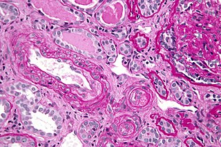
Renal vein thrombosis (RVT) is the formation of a clot in the vein that drains blood from the kidneys, ultimately leading to a reduction in the drainage of one or both kidneys and the possible migration of the clot to other parts of the body. First described by German pathologist Friedrich Daniel von Recklinghausen in 1861, RVT most commonly affects two subpopulations: newly born infants with blood clotting abnormalities or dehydration and adults with nephrotic syndrome.

<span class="mw-page-title-main">Hypoprothrombinemia</span> Medical condition

Hypoprothrombinemia is a rare blood disorder in which a deficiency in immunoreactive prothrombin, produced in the liver, results in an impaired blood clotting reaction, leading to an increased physiological risk for spontaneous bleeding. This condition can be observed in the gastrointestinal system, cranial vault, and superficial integumentary system, affecting both the male and female population. Prothrombin is a critical protein that is involved in the process of hemostasis, as well as illustrating procoagulant activities. This condition is characterized as an autosomal recessive inheritance congenital coagulation disorder affecting 1 per 2,000,000 of the population, worldwide, but is also attributed as acquired.

<span class="mw-page-title-main">Phenprocoumon</span> Drug

Phenprocoumon is a long-acting blood thinner drug to be taken by mouth, and a derivative of coumarin. It acts as a vitamin K antagonist and inhibits blood clotting (coagulation) by blocking synthesis of coagulation factors II, VII, IX and X. It is used for the prophylaxis and treatment of thromboembolic disorders such as heart attacks and pulmonary (lung) embolism. The most common adverse effect is bleeding. The drug interacts with a large number of other medications, including aspirin and St John's Wort. It is the standard coumarin used in Germany, Austria, and other European countries.

<span class="mw-page-title-main">Dabigatran</span> Anticoagulant medication

Dabigatran, sold under the brand name Pradaxa among others, is an anticoagulant used to treat and prevent blood clots and to prevent stroke in people with atrial fibrillation. Specifically it is used to prevent blood clots following hip or knee replacement and in those with a history of prior clots. It is used as an alternative to warfarin and does not require monitoring by blood tests. In a meta analysis of 7 different studies, there was no benefit of dabigatran over warfarin in preventing ischemic stroke; however, dabigatran were associated with a lower hazard for intracranial bleeding compared with warfarin, but also had a higher risk of gastrointestinal bleeding relative to warfarin. It is taken by mouth.

Hypercoagulability in pregnancy is the propensity of pregnant women to develop thrombosis. Pregnancy itself is a factor of hypercoagulability, as a physiologically adaptive mechanism to prevent post partum bleeding. However, when combined with an additional underlying hypercoagulable states, the risk of thrombosis or embolism may become substantial.

<span class="mw-page-title-main">Percutaneous umbilical cord blood sampling</span>

Percutaneous umbilical cord blood sampling (PUBS), also called cordocentesis, fetal blood sampling, or umbilical vein sampling is a diagnostic genetic test that examines blood from the fetal umbilical cord to detect fetal abnormalities. Fetal and maternal blood supply are typically connected in utero with one vein and two arteries to the fetus. The umbilical vein is responsible for delivering oxygen rich blood to the fetus from the mother; the umbilical arteries are responsible for removing oxygen poor blood from the fetus. This allows for the fetus’ tissues to properly perfuse. PUBS provides a means of rapid chromosome analysis and is useful when information cannot be obtained through amniocentesis, chorionic villus sampling, or ultrasound ; this test carries a significant risk of complication and is typically reserved for pregnancies determined to be at high risk for genetic defect. It has been used with mothers with immune thrombocytopenic purpura.

<span class="mw-page-title-main">VKORC1</span> Protein-coding gene in the species Homo sapiens

The human gene VKORC1 encodes for the enzyme, Vitamin K epOxide Reductase Complex (VKORC) subunit 1. This enzymatic protein complex is responsible for reducing vitamin K 2,3-epoxide to its active form, which is important for effective clotting (coagulation). In humans, mutations in this gene can be associated with deficiencies in vitamin-K-dependent clotting factors.

<span class="mw-page-title-main">Vitamin K antagonist</span>

Vitamin K antagonists (VKA) are a group of substances that reduce blood clotting by reducing the action of vitamin K. The term "vitamin K antagonist" is technically a misnomer, as the drugs do not directly antagonize the action of vitamin K in the pharmacological sense, but rather the recycling of vitamin K. Vitamin K antagonists (VKAs) have been the mainstay of anticoagulation therapy for more than 50 years.

<span class="mw-page-title-main">Vitamin K reaction</span> Medical condition

Vitamin K reactions are adverse side effects that may occur after injection with vitamin K. The liver utilizes vitamin K to produce coagulation factors that help the body form blood clots which prevent excessive bleeding. Vitamin K injections are administered to newborns as a preventative measure to reduce the risk of hemorrhagic disease of the newborn (HDN).

Women should speak to their doctor or healthcare professional before starting or stopping any medications while pregnant. Non-essential drugs and medications should be avoided while pregnant. Tobacco, alcohol, marijuana, and illicit drug use while pregnant may be dangerous for the unborn baby and may lead to severe health problems and/or birth defects. Even small amounts of alcohol, tobacco, and marijuana have not been proven to be safe when taken while pregnant. In some cases, for example, if the mother has epilepsy or diabetes, the risk of stopping a medication may be worse than risks associated with taking the medication while pregnant. The mother's healthcare professional will help make these decisions about the safest way to protect the health of both the mother and unborn child. In addition to medications and substances, some dietary supplements are important for a healthy pregnancy, however, others may cause harm to the unborn child.

<span class="mw-page-title-main">Circumvallate placenta</span> Medical condition

Circumvallate placenta is a rare condition affecting about 1-2% of pregnancies, in which the amnion and chorion fetal membranes essentially "double back" on the fetal side around the edges of the placenta. After delivery, a circumvallate placenta has a thick ring of membranes on its fetal surface. Circumvallate placenta is a placental morphological abnormality associated with increased fetal morbidity and mortality due to the restricted availability of nutrients and oxygen to the developing fetus.

A pre-existing disease in pregnancy is a disease that is not directly caused by the pregnancy, in contrast to various complications of pregnancy, but which may become worse or be a potential risk to the pregnancy. A major component of this risk can result from necessary use of drugs in pregnancy to manage the disease.

References

  1. Yurdakök, M. 2012, "Fetal and neonatal effects of anticoagulants used in pregnancy: a review", The Turkish journal of pediatrics, vol. 54, no. 3, pp. 207.
  2. 1 2 Reid, A., Forrester, C. & Shwe, K. 2009, "Warfarin", Student BMJ, vol. 17.
  3. 1 2 3 4 5 6 7 8 9 10 Hou, J. 2004, "Fetal warfarin syndrome", Chang Gung medical journal, vol. 27, no. 9, pp. 691.
  4. Vitale, N., De Feo, M., De Santo, L.S., Pollice, A., Tedesco, N. & Cotrufo, M. 1999, "Dose-dependent fetal complications of warfarin in pregnant women with mechanical heart valves", Journal of the American College of Cardiology, vol. 33, no. 6, pp. 1637-1641.
  5. 1 2 3 4 Starling, L.D., Sinha, A., Boyd, D. & Furck, A. 2012, "Fetal warfarin syndrome", BMJ case reports, vol. 2012, no. 1, pp. 1-4
  6. 1 2 3 4 5 6 7 8 Silveira, D.B., da Rosa, E.B., de Mattos, V.F., Goetze, T.B., Sleifer, P., Santa Maria, F.D., Rosa, R.C.M., Rosa, R.F.M. & Zen, P.R.G. 2015, "Importance of a multidisciplinary approach and monitoring in fetal warfarin syndrome", American Journal of Medical Genetics Part A, vol. 167, no. 6, pp. 1294-1299.
  7. Raghav, S. & Reutens, D. 2006;2007;, "Neurological sequelae of intrauterine warfarin exposure", Journal of Clinical Neuroscience, vol. 14, no. 2, pp. 99-103.
  8. Maiello, M., Torella, M., Caserta, L., Caserta, R., Sessa, M., Tagliaferri, A., Bernacchi, M., Napolitano, M., Nappo, C., De Lucia, D. & Panariello, S. 2006, "Hypercoagulability during pregnancy: evidences for a thrombophilic state", Minerva ginecologica, vol. 58, no. 5, pp. 417.
  9. Danziger, J. 2008, "Vitamin K-dependent Proteins, Warfarin, and Vascular Calcification", Clinical Journal of the American Society of Nephrology, vol. 3, no. 5, pp. 1504-1510.
  10. Yamauchi, M., Yamaguchi, T., Nawata, K., Takaoka, S. & Sugimoto, T. 2010, "Relationships between undercarboxylated osteocalcin and vitamin K intakes, bone turnover, and bone mineral density in healthy women", Clinical Nutrition, vol. 29, no. 6, pp. 761-765.
  11. Walfisch, A. & Koren, G. 2010, "The "Warfarin Window" in Pregnancy: The Importance of Half-life", Journal of obstetrics and gynaecology Canada, vol. 32, no. 10, pp. 988.
  12. Loftus, C. (1996). Neurosurgical aspects of pregnancy. Park Ridge, Ill.: American Association of Neurological Surgeons.